. 2015 Nov 3;16(11):26318-32.
doi: 10.3390/ijms161125955.

Aβ1-25-Derived Sphingolipid-Domain Tracer Peptide SBD Interacts with Membrane Ganglioside Clusters via a Coil-Helix-Coil Motif

Affiliations

Aβ1-25-Derived Sphingolipid-Domain Tracer Peptide SBD Interacts with Membrane Ganglioside Clusters via a Coil-Helix-Coil Motif

Yaofeng Wang et al. Int J Mol Sci. .

Abstract

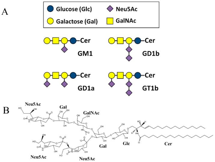
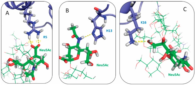
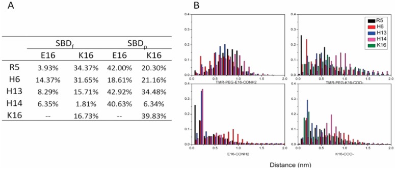
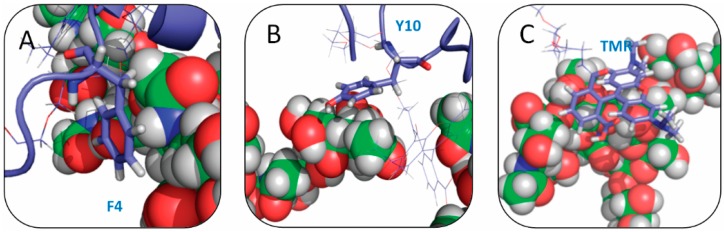
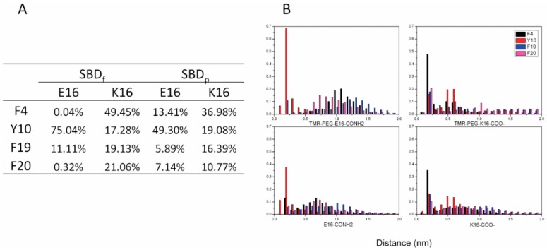
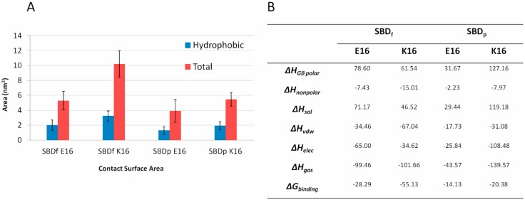
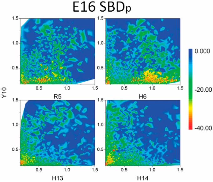
The Amyloid-β (Aβ)-derived, sphingolipid binding domain (SBD) peptide is a fluorescently tagged probe used to trace the diffusion behavior of sphingolipid-containing microdomains in cell membranes through binding to a constellation of glycosphingolipids, sphingomyelin, and cholesterol. However, the molecular details of the binding mechanism between SBD and plasma membrane domains remain unclear. Here, to investigate how the peptide recognizes the lipid surface at an atomically detailed level, SBD peptides in the environment of raft-like bilayers were examined in micro-seconds-long molecular dynamics simulations. We found that SBD adopted a coil-helix-coil structural motif, which binds to multiple GT1b gangliosides via salt bridges and CH-π interactions. Our simulation results demonstrate that the CH-π and electrostatic forces between SBD monomers and GT1b gangliosides clusters are the main driving forces in the binding process. The presence of the fluorescent dye and linker molecules do not change the binding mechanism of SBD probes with gangliosides, which involves the helix-turn-helix structural motif that was suggested to constitute a glycolipid binding domain common to some sphingolipid interacting proteins, including HIV gp120, prion, and Aβ.

Keywords: Sphingolipid binding domain; lipid rafts; molecular dynamics simulation.


Figures

Figure 1
Figure 1
(A) Structure of the major brain gangliosides; (B) Detailed chemical structure of gangliosides GT1b. Cer, ceramide.
Figure 2
Figure 2
(A) Superimpositions of SBD configuration binding with GT1b during the simulations, one frame per 2 ns; (B) Secondary structure distributions of SBDs in the binding modes with GT1b. E16 SBDf; K16 SBDf; E16 SBDp; K16 SBDp
Figure 3
Figure 3
Binding modes of SBDf with GT1b: (A) E16 variant; and (B) E16 variant. GT1b molecules are shown as green balls and the SBD molecule is represented by blue sticks.
Figure 4
Figure 4
Three most frequent binding modes of SBDp with GT1b: (AC) E16 variant and (DF) K16 variant.
Figure 5
Figure 5
Snapshots of electrostatic interactions between positively charged residues of SBD in (A) arginine; (B) histidine; and (C) lysine, to Neu5Ac of GT1b gangliosides.
Figure 6
Figure 6
Salt bridge distances between SBD and GT1b during the last 100 ns of simulation: (A) probabilities of strong salt bridges (<0.35 nm); and (B) distance distributions of salt bridges.
Figure 7
Figure 7
Snapshots of CH–π interactions between SBD at (A) phenylalanine 4; (B) tyrosine 10; and (C) TMR (tetra methyl rhodamine) and GT1b gangliosides.
Figure 8
Figure 8
Distances of CH–π interactions between SBD and GT1b during last the 100 ns of simulation: (A) probabilities of CH–π interactions (<0.35 nm); and (B) distance distributions of CH–π interactions.
Figure 9
Figure 9
(A) Contact surface areas between SBD and GT1b; (B) Average MM-GBSA binding energies between SBD and GT1b (Kcal/mol).
Figure 10
Figure 10
MM-GBSA binding energies (Kcal/mol) between SBD and GT1b gangliosides. Distances of salt bridge and CH–π interaction were set as two coefficients of variation. Distances of salt bridges between positive residues (R5, H6, H13 and H14) and Neu5Ac were set as the horizontal axis, and distances of CH–π interactions between aromatic rings of Y10 and CH groups in sugar rings were set as the vertical axis.

References

    1. Simons K., Ikonen E. Functional rafts in cell membranes. Nature. 1997;387:569–572. doi: 10.1038/42408. - DOI - PubMed
    1. Pike L.J. Rafts defined: A report on the keystone symposium on lipid rafts and cell function. J. Lipid Res. 2006;47:1597–1598. doi: 10.1194/jlr.E600002-JLR200. - DOI - PubMed
    1. Mattner J., DeBord K.L., Ismail N., Goff R.D., Cantu C., Zhou D.P., Saint-Mezard P., Wang V., Gao Y., Yin N., et al. Exogenous and endogenous glycolipid antigens activate nkt cells during microbial infections. Nature. 2005;434:525–529. doi: 10.1038/nature03408. - DOI - PubMed
    1. Brown D.A., London E. Structure and function of sphingolipid- and cholesterol-rich membrane rafts. J. Biol. Chem. 2000;275:17221–17224. doi: 10.1074/jbc.R000005200. - DOI - PubMed
    1. Lingwood C.A. Glycolipid receptors for verotoxin and helicobacter pylori: Role in pathology. Biochim. Biophys. Acta. 1999;1455:375–386. doi: 10.1016/S0925-4439(99)00062-9. - DOI - PubMed

Publication types