. 2015 Nov 24;6(37):39457-68.
doi: 10.18632/oncotarget.6277.

Changes in autophagy, proteasome activity and metabolism to determine a specific signature for acute and chronic senescent mesenchymal stromal cells

Affiliations

Changes in autophagy, proteasome activity and metabolism to determine a specific signature for acute and chronic senescent mesenchymal stromal cells

Stefania Capasso et al. Oncotarget. .

Abstract

A sharp definition of what a senescent cell is still lacking since we do not have in depth understanding of mechanisms that induce cellular senescence. In addition, senescent cells are heterogeneous, in that not all of them express the same genes and present the same phenotype. To further clarify the classification of senescent cells, hints may be derived by the study of cellular metabolism, autophagy and proteasome activity. In this scenario, we decided to study these biological features in senescence of Mesenchymal Stromal Cells (MSC). These cells contain a subpopulation of stem cells that are able to differentiate in mesodermal derivatives (adipocytes, chondrocytes, osteocytes). In addition, they can also contribute to the homeostatic maintenance of many organs, hence, their senescence could be very deleterious for human body functions. We induced MSC senescence by oxidative stress, doxorubicin treatment, X-ray irradiation and replicative exhaustion. The first three are considered inducers of acute senescence while extensive proliferation triggers replicative senescence also named as chronic senescence. In all conditions, but replicative and high IR dose senescence, we detected a reduction of the autophagic flux, while proteasome activity was impaired in peroxide-treated and irradiated cells. Differences were observed also in metabolic status. In general, all senescent cells evidenced metabolic inflexibility and prefer to use glucose as energy fuel. Irradiated cells with low dose of X-ray and replicative senescent cells show a residual capacity to use fatty acids and glutamine as alternative fuels, respectively. Our study may be useful to discriminate among different senescent phenotypes.

Keywords: Gerotarget; autophagy; mesenchymal stem cells; metabolism; proteasome; senescence.


Conflict of interest statement

CONFLICTS OF INTERESTS

None of the authors have competing interests.

Figures

Figure 1
Figure 1. Workflow for metabolic assays
MSC were in growth medium (D0). After 24h stress-induced senescence was triggered as described in methods (D1). Chronic senescent cells were plated in growth medium (D0). After 24 hours, we performed medium change without use of stressors (D1). The day after (D2), we replaced the growth medium with the substrate-limited medium. After 24 hours (T0), we changed the substrate-limited medium with the assay medium. After 30 min (T30) in a group of samples, we added the metabolic pathway inhibitor drugs (2D-glucose, etomoxir and BPTES). After 60 min (T90), we added the relative substrates (glucose, palmitate and glutamine) in all samples. Following 60 min incubation (T150), we performed the lactate, ATP and O2 assays.
Figure 2
Figure 2. Induction of senescence in MSC cultures
Representative microscopic fields of acid beta-galactosidase (blue) in treated and control cells are shown. The graph shows mean percentage value of senescent cells (± SD, n = 3, *p < 0.05).
Figure 3
Figure 3. Cellular energetic pathways
Glucose, fatty acids and amino acids (glutamine in the example) are the main energy fuels for the cells. Glucose is metabolized to pyruvate through glycolysis into cytoplasm. This can be converted to lactate or enter mitochondria to be converted in acetyl-CoA. This is combined with oxaloacetate to give citrate and undergo further oxidation in trycarboxylic acid (TCA) cycle. Fatty acids can enter mitochondria as acyl-CoA by mean of carnitinepalmitoyltransferase 1 (CPT1) and here can be oxidized to acetyl-CoA and fuel the TCA cycle. Glutamine can be converted to glutamate by glutaminase (GLS) and further metabolized to α-ketoglutarate and then oxaloacetate to fuel TCA cycle. 2-deoxyglucose inhibits glycolysis; etomoxir impair acyl-CoA transfer into mitochondria; BPTES inhibits GLS activity.
Figure 4
Figure 4. Evaluation of metabolic activity in senescent cells
A. ATP level measurement. Intracellular ATP level was determined using the ATP Colorimetric/Fluorometric Assay. Cells were incubated in medium containing glucose (left graph) or palmitate (middle graph) or glutamine (right graph). The ATP level was measured in absence of energy substrate (white bars), in presence of specific substrate (gray bars) and in the presence of substrate and its inhibitor (black bar). For every condition, the asterisk (*) denotes significant differences (p < 0.05) between ATP level in a medium with a specific substrate (gray bar) and a medium without substrate plus its inhibitor (black bar). Data are expressed in arbitrary units (± SD, n = 3). B. Basal oxygen consumption rate. We used MitoXpress® Xtra assay for measurement of extracellular oxygen consumption rates (OCR). Cells were incubated in medium containing glucose (left graph) or palmitate (middle graph) or glutamine (right graph). For every condition, the asterisk (*) denotes significant differences (p < 0.05) between OCR in a medium with a specific substrate (black bar) and a medium without energy substrates (white bar). The hash symbol (#) denotes differences between ATP in control healthy cells and senescent cells (D, H, IRL, IRH, Rep). Data are expressed in arbitrary units (± SD, n = 3). C. Maximal respiration rate. OCR was measured following sequential addition of FCCP, that is, a protonophore and uncoupler of oxidative phosphorylation in mitochondria. Cells were incubated in medium containing glucose (left graph) or palmitate (middle graph) or glutamine (right graph). For every condition the asterisk (*) denotes significant differences (p < 0.05) between maximal OCR in a medium with a specific substrate (black bar) and a medium without energy substrates (white bar). The hash symbol (#) denotes differences between OCR in control healthy cells and senescent cells (D, H, IRL, IRH, Rep). Data are expressed in arbitrary units (± SD, n = 3). C. Oxygen for mitochondrial ATP production. OCR was measured following sequential addition of olygomycin that is inhibitor of the F0 part of H+/ATP-synthase in mitochondria. Cells were incubated in medium containing glucose (left graph) or palmitate (middle graph) or glutamine (right graph). For every condition the asterisk (*) denotes significant differences (p < 0.05) between OCR in a medium with a specific substrate (black bar) and a medium without energy substrates (white bar). The hash symbol (#) denotes differences between OCR in control healthy cells and senescent cells (D, H, IRL, IRH, Rep). Data are expressed in arbitrary units (± SD, n = 3).
Figure 5
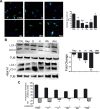
Figure 5. Autophagy and proteasome activity assays
A. Cyto-ID assay. Representative microscopic fields of cells with active autophagy (green) are shown. Nuclei were counterstained with Hoechst 33342 (blue). The graph shows mean percentage value of Cyto-ID-positive cells (± SD, n = 3, *p < 0.05, **p < 0.01). B. Autophagic flux evaluation – The picture shows western blot detection of LC3-I and LC3-II in senescent and control MSC. Following induction of senescence with different stressors cells were incubated for six hours and then harvested for western blot analysis. Two hours before the end of cell sample preparation, senescent and control MSC cultures were incubated with 100 nMB afilomycin A1 (inhibitor of lysosomal degradation) or PBS to detect autophagic flux. We used Gel Doc 2000 Gel Documentation Systems (Bio-Rad, CA, USA) to measure LC3-I and II band intensities that were normalized with tubulin (TUB). We determined autophagic flux (AF) for LC3 II as follows: IR-treated MSC AF = (IR-treated MSC + Bafylomycin A1) - (IR treated MSC + PBS); Control MSCs AF = (Control MSC + Bafylomycin A1) - (Control MSC + PBS). Change in autophagic flux (ΔAF) between IR-treated and control MSC was calculated as ΔAF = IR-treated MSC AF - Control MSC AF. The graph shows AF changes in IR-treated MSC compared to control cultures. Data are expressed in change folds (n = 3; *p < 0.05, **p < 0.01). C. Proteasome activity. Following induction of senescence with different stressors, cells were incubated for six hours and then harvested for fluorimetric assay determination of proteasome activity. Two hours before the end of cell sample preparation, senescent and control MSC cultures were incubated with 25 μM lactacystin (a specific and potent inhibitor of proteasomes) or PBS. The graph shows proteasome activity in control and senescent cells both in the presence and absence of lactacystin. Data are expressed in arbitrary units (n = 3; *p < 0.05).

References

    1. Campisi J, d'Adda di Fagagna F. Cellular senescence: when bad things happen to good cells. Nat Rev Mol Cell Biol. 2007;8(9):729–740. - PubMed
    1. van Deursen JM. The role of senescent cells in ageing. Nature. 2014;509(7501):439–446. - PMC - PubMed
    1. Campisi J. Aging, cellular senescence, and cancer. Annu Rev Physiol. 2013;75:685–705. - PMC - PubMed
    1. Galderisi U, Giordano A. The gap between the physiological and therapeutic roles of mesenchymal stem cells. Medicinal research reviews. 2014;34(5):1100–1126. - PubMed
    1. Giordano A, Galderisi U, Marino IR. From the laboratory bench to the patient's bedside: an update on clinical trials with mesenchymal stem cells. Journal of cellular physiology. 2007;211(1):27–35. - PubMed

Publication types

MeSH terms

LinkOut - more resources