. 2015 Nov;56(12):7085-99.
doi: 10.1167/iovs.14-16246.

Human Adult Retinal Pigment Epithelial Stem Cell-Derived RPE Monolayers Exhibit Key Physiological Characteristics of Native Tissue

Affiliations

Human Adult Retinal Pigment Epithelial Stem Cell-Derived RPE Monolayers Exhibit Key Physiological Characteristics of Native Tissue

Timothy A Blenkinsop et al. Invest Ophthalmol Vis Sci. 2015 Nov.

Abstract

Purpose: We tested what native features have been preserved with a new culture protocol for adult human RPE.

Methods: We cultured RPE from adult human eyes. Standard protocols for immunohistochemistry, electron microscopy, electrophysiology, fluid transport, and ELISA were used.

Results: Confluent monolayers of adult human RPE cultures exhibit characteristics of native RPE. Immunohistochemistry demonstrated polarized expression of RPE markers. Electron microscopy illustrated characteristics of native RPE. The mean transepithelial potential (TEP) was 1.19 ± 0.24 mV (mean ± SEM, n = 31), apical positive, and the mean transepithelial resistance (RT) was 178.7 ± 9.9 Ω·cm2 (mean ± SEM, n = 31). Application of 100 μM adenosine triphosphate (ATP) apically increased net fluid absorption (Jv) by 6.11 ± 0.53 μL·cm2·h-1 (mean ± SEM, n = 6) and TEP by 0.33 ± 0.048 mV (mean ± SEM, n = 25). Gene expression of cultured RPE was comparable to native adult RPE (n = 5); however, native RPE RNA was harvested between 24 and 40 hours after death and, therefore, may not accurately reflect healthy native RPE. Vascular endothelial growth factor secreted preferentially basally 2582 ± 146 pg/mL/d, compared to an apical secretion of 1548 ± 162 pg/mL/d (n = 14, P < 0.01), while PEDF preferentially secreted apically 1487 ± 280 ng/mL/d compared to a basolateral secretion of 864 ± 132 ng/mL/d (n = 14, P < 0.01).

Conclusions: The new culture model preserves native RPE morphology, electrophysiology, and gene and protein expression patterns, and may be a useful model to study RPE physiology, disease, and transplantation.


Figures

Figure 1
Figure 1
Schematic illustrating the method to culture ahRPE. Human globes are received in sterile moist chambers on ice. After rinsing, a circumferential incision is made posterior to the ora serrata and the vitreous and retina are removed. The eye cup is rinsed, then Dispase I is put into the cup and incubated. The RPE layer is scraped gently from the Bruch's membrane using an angled, double-beveled spoon blade to minimize damage. The RPE are collected and run on a sucrose gradient to separate RPE sheets from single RPE cells. The RPE sheets are plated on ECM-coated plates and typically attach within 48 hours. Confluence is reached after 2 months, with routine media changes.
Figure 2
Figure 2
Adult human RPE cultures express markers typical of native RPE. Nuclei are labeled with 4′,6-diamidino-2-phenylendole (DAPI; cyan), whereas all other immunofluorescence is red or yellow. Claudin 19, Phalloidin (F-actin), Ezrin, ZO1, and MCT1 are preferentially located on the apical side, as observed by looking at the Z-plane located to the right of the 2D image. RPE65, Cytokeratin 8, CRALBP are cytoplasmic. Bestrophin is found perinuclear and on the basal side. CA IX and Na+K+-ATPase are located on the apical and basolateral membranes. MCT3 is observed basally. Scale bars: 10 μM.
Figure 3
Figure 3
In electron microscopy, ahRPE cultures exhibit multiple features of native tissue. (A) Scanning electron microscopy (SEM) images of the surface of an ahRPE culture at multiple magnifications. (B) A 6-month culture of ahRPE. (C) Time course of ahRPE polarization: 1 week (Ci), 2 weeks (Cii), and 2 months (Cii). Tight junctions (arrow 1), apical microvilli (arrow 2), polarized apical pigmentation (arrow 3), basally localized nucleus (arrow 4), basement membrane (arrow 5) are identified.
Figure 4
Figure 4
Steady-state and TER of ahRPE primary cultures. Retinal pigment epithelium cultivated on polyester membrane Transwells maintain stable TEP and TER for over 1 hour. (A) TEP (solid line) and RT (dotted line) plotted as a function of time. (B) Box plots of the average baseline TEP (mV) and TER (Ω·cm2) of 31 experiments.
Figure 5
Figure 5
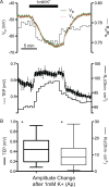
Effect of reducing apical bath [K+]o from 5 to 1 mM on VA, VB, TER, and TEP in ahRPE cultures. (A) Reduction of apical K+ from 5 to 1mM (solid bar) depolarized VA (dotted orange line), VB (solid green line) each from −48.4 to −66.7, while TEP increased (solid line) by 0.1 mV and RT increased by 7Ω·cm (dotted line). (B) Box plots of the average change in TEP and TER upon application of 1 mM K+ apically in 16 experiments. *P < 0.001.
Figure 6
Figure 6
Effect of a 5-fold increase in basal side [K+]o on TEP and RT in ahRPE. (A) Changing the basal bath [K+]o from 5 to 25 mM (solid bar) reversibly increased TEP by 0.91 (solid line) and decreased RT by 39 Ω·cm (dotted line). (B) Box plots of TEP and TER changes (n = 16). Ba, Basal. *P < 0.001.
Figure 7
Figure 7
Epinephrine-induced electrical responses (TEP, RT) in ahRPE. (A) Transepithelial potential (solid line) and RT (dotted line) plotted as a function of time. Apical epinephrine (10 nM, solid bar) increased TEP by 0.41 mV and RT by 14 Ω·cm2. (B) Box plots of the change in amplitude of TEP and RT upon 10 nM epinephrine administration to apical bath (n = 5). *P < 0.001.
Figure 8
Figure 8
Electrical responses of ahRPE following addition of 100 μM ATP (solid bar) to the apical bath. (A) Transepithelial potential (solid line) and RT (dotted line) plotted as a function of time. The ATP elicited a triphasic response indicated as I, II, III, and by vertical dotted lines. Phase I is characterized by a rapid 0.63 mV increase in TEP concomitant with a drop in RT of 39 Ω·cm. This increase was followed in phase II by a drop in TEP. In phase III, TEP again increased by 0.46 mV at a slower rate. (B) Box plots of the TEP and RT changes in amplitude following addition of apical ATP (100 μM, n = 25). Ap, Apical. *P < 0.01.
Figure 9
Figure 9
Adenosine triphosphate–induced fluid absorption (Jv) by ahRPE. (A) Jv (solid circles) plotted as a function of time (absorption indicated by positive values). (B) Concomitant TEP (solid line) and RT (dotted line) measurements. Solid bar indicates addition of Ringer's solution containing 100 μM ATP which increased Jv from 2.6 to 5.6 μL·cm2·h−2 and decreased RT by 10.3 Ω·cm; EP increased by 0.95 mV. (C) Summary of six experiments as plotted. *P < 0.05 from previous condition.
Figure 10
Figure 10
Gene expression comparing ahRPE to nhRPE and fhRPE. Complementary DNA from freshly isolated RPE from adult cadaver donors was compared to their genetically-matched cultured RPE counterparts as well as to nongenetically matched cultured fetal RPE by quantitative PCR. Adult cultured RPE was normalized to nhRPE (A) and fhRPE (B) data and plotted in LOG10 scale. *Significant difference, with a P < 0.05, n = 5.
Figure 11
Figure 11
Polarized expression of PEDF and VEGF. Adult human RPE was passaged onto Transwell inserts and cultured for at least 2 months. Media then was taken from the top and bottom wells 24 hours after media change, and assayed for concentrations of VEGF and PEDF proteins via ELISA. In the 14 donors assayed, mean VEGF secretion was 1548 pg/mL/d in the apical side and 2582 pg/mL/d in the basal side. Mean PEDF secretion was 1487 ng/mL/d in the apical side and 863.8 ng/mL/d in the basal side. *Significant difference, with a P < 0.01, n = 14.

References

    1. Curcio CA,, Johnson M,, Rudolf M,, Huang JD. The oil spill in ageing Bruch membrane. Br J Ophthalmol. 2011; 95: 1638–1645. - PMC - PubMed
    1. Zarbin MA,, Rosenfeld PJ. Pathway-based therapies for age-related macular degeneration: an integrated survey of emerging treatment alternatives. Retina. 2010; 30: 1350–1367. - PubMed
    1. Kolomeyer AM,, Zarbin MA. Trophic factors in the pathogenesis and therapy for retinal degenerative diseases. Surv Ophthalmol. 2014; 59: 134–165. - PubMed
    1. Gallemore RP,, Hughes BA,, Miller SS. Light-induced responses of the retinal pigment epithelium. : Marmor MF,, Wolfensburger TJ, Retinal Pigment Epithelium: Current Aspects of Function and Disease. Oxford: Oxford University Press; 1998: 103–134.
    1. Maminishkis A,, Jalickee S,, Blaug SA,, et al. The P2Y(2) receptor agonist INS37217 stimulates RPE fluid transport in vitro and retinal reattachment in rat. Invest Ophthalmol Vis Sci. 2002; 43: 3555–3566. - PubMed

Publication types