. 2015 Nov;56(12):7214-23.
doi: 10.1167/iovs.15-17107.

Treatment of Inherited Eye Defects by Systemic Hematopoietic Stem Cell Transplantation

Affiliations

Treatment of Inherited Eye Defects by Systemic Hematopoietic Stem Cell Transplantation

Celine J Rocca et al. Invest Ophthalmol Vis Sci. 2015 Nov.

Abstract

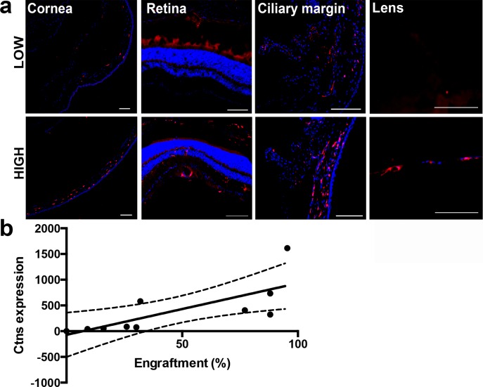
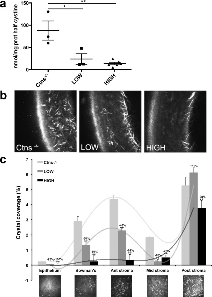
Purpose: Cystinosis is caused by a deficiency in the lysosomal cystine transporter, cystinosin (CTNS gene), resulting in cystine crystal accumulation in tissues. In eyes, crystals accumulate in the cornea causing photophobia and eventually blindness. Hematopoietic stem progenitor cells (HSPCs) rescue the kidney in a mouse model of cystinosis. We investigated the potential for HSPC transplantation to treat corneal defects in cystinosis.

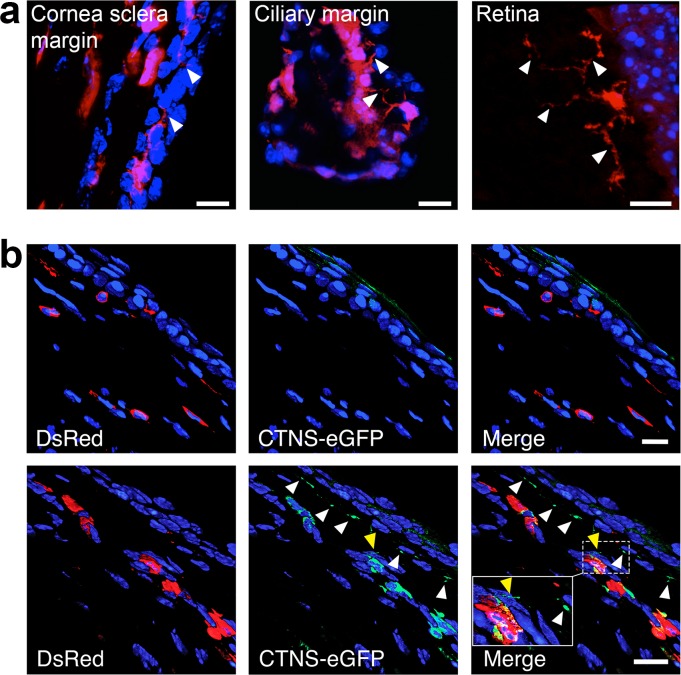
Methods: We isolated HSPCs from transgenic DsRed mice and systemically transplanted irradiated Ctns-/- mice. A year posttransplantation, we investigated the fate and function of HSPCs by in vivo confocal and fluorescence microscopy (IVCM), quantitative RT-PCR (RT-qPCR), mass spectrometry, histology, and by measuring the IOP. To determine the mechanism by which HSPCs may rescue disease cells, we transplanted Ctns-/- mice with Ctns-/- DsRed HSPCs virally transduced to express functional CTNS-eGFP fusion protein.

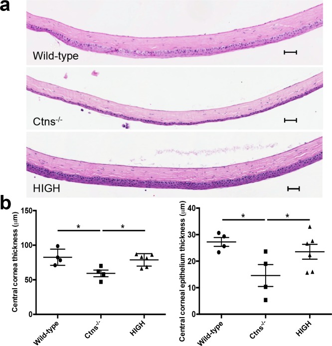
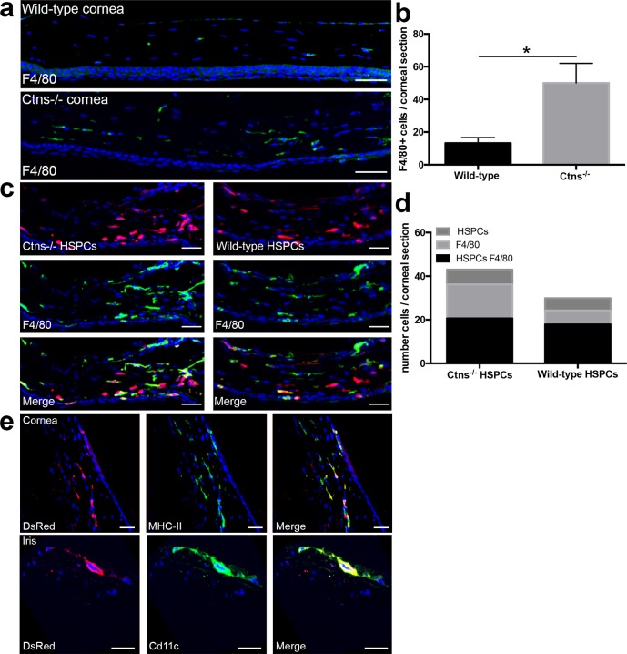
Results: We found that a single systemic transplantation of wild-type HSPCs prevented ocular pathology in the Ctns-/- mice. Engraftment-derived HSPCs were detected within the cornea, and also in the sclera, ciliary body, retina, choroid, and lens. Transplantation of HSPC led to substantial decreases in corneal cystine crystals, restoration of normal corneal thickness, and lowered IOP in mice with high levels of donor-derived cell engraftment. Finally, we found that HSPC-derived progeny differentiated into macrophages, which displayed tunneling nanotubes capable of transferring cystinosin-bearing lysosomes to diseased cells.

Conclusions: To our knowledge, this is the first demonstration that HSPCs can rescue hereditary corneal defects, and supports a new potential therapeutic strategy for treating ocular pathologies.


Figures

Figure 1
Figure 1
Ocular engraftment of transplanted HSPCs depends on donor-derived blood cell engraftment. (a) Systemic DsRed-expressing HSPC transplantation resulted in the presence of bone marrow–derived cells (seen in red) in the whole eye as shown in sagittal sections of the eyes of mice with donor-derived blood cell engraftment <50% (Low) compared to >50% (High). More abundant HSPC-derived cells were seen when donor-derived blood cell engraftment in High than in Low mice in the different eye compartments. Scale bars: 100 μm. (b) Linear regression analysis of Ctns expression in the whole eye showing that Ctns expression increases with the level of transplanted DsRed-HSPCs. The linear regression interrupted lines correspond to 95% confidence.
Figure 2
Figure 2
Transplantation of HSPC leads to ocular cystine and corneal cystine crystal clearance. (a) Cystine levels measured in the whole eye were significantly reduced in the HSPC-transplanted mice, Low and High, compared to the Ctns−/− controls. Error bars: SEM (*P < 0.05, **P < 0.005). (b) Lateral cornea IVCM representations of Ctns−/− controls and Low and High HSPC-transplanted mice. Abundant cystine crystals were observed in the whole cornea in Ctns−/− mice in contrast to Ctns−/− with high level of engraftment where residual crystals can be seen only in the posterior stroma. (c) Surface crystal quantification within each layer of the full IVCM cornea scans from both eyes of Ctns−/− controls and transplanted (Low and High) mice. Transplantation of HSPC with high level of engraftment prevents almost completely cystine crystal formation within the superior layers of the cornea (epithelium to middle stroma). The mice with low level of engraftment exhibit significant but more modest crystal reduction. Error bars: SEM (*P < 0.05, **P < 0.005).
Figure 3
Figure 3
Corneal structure is preserved in HSPC-transplanted Ctns−/− mice. (a) Representative sagittal hematoxylin and eosin sections of the central cornea of wild-type mice, nontreated Ctns−/−, and highly engrafted Ctns−/− (High) mice. The Ctns−/− mice exhibit markedly reduced CCT compared to wild-type animal, while treated Ctns−/− mice presented with CCT comparable wild-type. Scale bars: 200 μm. (b) Thickness measurement of the central cornea and central cornea epithelium of wild-type, Ctns−/−, and transplanted (High) Ctns−/− mice showing that HSPC transplantation prevents corneal reduction. Error bars: SEM (*P < 0.05).
Figure 4
Figure 4
Hematopoietic stem and progenitor cells differentiate into phagocytic cells within the cornea. (a) Representative confocal pictures of corneal sections from wild-type and Ctns−/− mice. Nuclei were stained with DAPI are seen in blue and F4/80+ cells are seen in green. (b) Quantification of F4/80-positive cells within corneas of wild-type (n = 4) and Ctns−/− (n = 4) mice. (c) Representative confocal pictures of corneal sections from Ctns−/− mice transplanted with either DsRed Ctns−/− HSPCs or DsRed wild-type HSPCs. Nuclei were stained with DAPI are seen in blue, HSPCs-derived cells are seen in red, and F4/80-positive cells are seen in green. (d) Quantification of DsRed-positive HSPC-derived cells (HSPCs), host F4/80-positive cells (F4/80), and F4/80-positive and DsRed-positive HSPC-derived cells (HSPCs F4/80) within the corneas of Ctns−/− mice transplanted with Ctns−/− DsRed HSPCs (n = 5) or wild-type DsRed HSPCs (n = 7) mice. (e) All the DsRed-expressing bone marrow–derived colocalize with the antigen presenting cells (APC) marker MHC-II class II (seen in green, upper). Few of them expressed CD11c (seen in green, lower) indicating that some of them differentiated into dendritic cells. Error bars: SEM (*P < 0.05). Scale bars: (a, c) 50 μm and (e) 30 μm.
Figure 5
Figure 5
HSPCs-derived macrophages transfer cystinosin-bearing lysosomes via TNTs to the adjacent disease cells. (a) Tunneling nanotubes generated from the DsRed bone marrow–derived cells were observed in the cornea (sclera margin and ciliary margin) and retina (arrows). (b) Representative confocal pictures of the cornea of Ctns−/− mice transplanted with Ctns−/− DsRed HSPCs as control (upper) or with Ctns−/− DsRed HSPCs stably expressing the fusion protein CTNS-eGFP (lower). Green vesicles are observed within the DsRed-bone marrow–derived cells in the mice treated with CTNS-eGFP-HSPCs but also in the no color host cells (white arrowheads) showing that lysosomes containing CTNS-eGFP could be transferred. Inset shows a TNT containing cystinosin-bearing vesicles (yellow arrowhead). Scale bars: (a) 10 μm, (b) 15 μm.

References

    1. Oliva MS,, Schottman T,, Gulati M. Turning the tide of corneal blindness. Indian J Ophthalmol. 2012; 60: 423–427. - PMC - PubMed
    1. Haddadin RI,, Chodosh J. Corneal transplantation and glaucoma. Semin Ophthalmol. 2014; 29: 380–396. - PubMed
    1. He H,, Yiu SC. Stem cell-based therapy for treating limbal stem cells deficiency: a review of different strategies. Saudi J Ophthalmol. 2014; 28: 188–194. - PMC - PubMed
    1. Li F,, Zhao SZ. Mesenchymal stem cells: potential role in corneal wound repair and transplantation. World J Stem Cells. 2014; 6: 296–304. - PMC - PubMed
    1. Town M,, Jean G,, Cherqui S,, et al. A novel gene encoding an integral membrane protein is mutated in nephropathic cystinosis. Nat Genet. 1998; 18: 319–324. - PubMed

Publication types