. 2015 Nov 6:5:16320.
doi: 10.1038/srep16320.

Mycobacterium tuberculosis Inhibits RAB7 Recruitment to Selectively Modulate Autophagy Flux in Macrophages

Affiliations

Mycobacterium tuberculosis Inhibits RAB7 Recruitment to Selectively Modulate Autophagy Flux in Macrophages

Pallavi Chandra et al. Sci Rep. .

Abstract

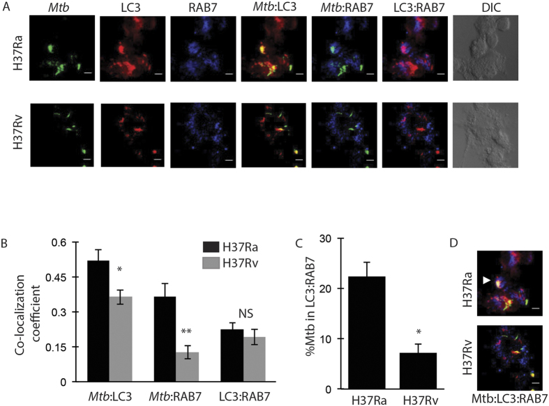
Here we report a novel regulatory mechanism for autophagy-mediated degradation of Mycobacterium tuberculosis (Mtb) and specific strategy exploited by the virulent Mtb to evade it. We show while both avirulent (H37Ra) and virulent (H37Rv) mycobacteria could readily localize to autophagosomes, their maturation into autolysosomes (flux) was significantly inhibited by the latter strain. The inhibition of autophagy flux by the virulent strain was highly selective, as it did not perturb the basal autophagy flux in the macrophages. Selective inhibition of flux of Mtb-containing autophagosomes required virulence regulators PhoP and ESAT-6. We show that the maturation of Mtb-containing autophagosomes into autolysosomes required recruitment of the late endosome marker RAB7, forming the intermediate compartment amphisomes. Virulent Mtb selectively evaded their targeting to the amphisomes. Thus we report a crosstalk between autophagy and phagosome maturation pathway and highlight the adaptability of Mtb, manifested by selective regulation of autophagy flux.


Figures

Figure 1
Figure 1. Virulent Mycobacterium tuberculosis selectively inhibits autophagy flux.
PMA differentiated (20 ng/ml, 24 hours) THP-1 macrophages were infected with PKH67 (green) labelled H37Ra or H37Rv at MOI of 1:10 (methods). Autophagosomes were visualized by LC3 immuno-staining at 6, 12, 24, 48 and 72 hours post infection with and without BafA1 treatment (100 nM, 3 hr). (A) Representative images showing co-localization of both H37Ra and H37Rv (green) to autophagosomes (LC3, red) at 48 hours post infection. Panels (B,C) show Co-localization Coefficient M1 of Mtb and LC3 for H37Ra and H37Rv infection, respectively with and without BafA1 treatment (Mean ± Standard Error Measurements, *p < 0.05). Panel (D) shows representative images of Mtb-infected mouse BMDMs with LC3 (red) at 48 hours post infection with and without BafA1. Panel E shows the corresponding quantification of the dataset (mean ± SEM, **p < 0.01). Panel (F) shows LC3 immunoblots from the cell lysates of H37Ra or H37Rv infected macrophages as well as uninfected control macrophages at 6 and 48 hours post infection. Cells were treated with BafA1 (100 nM, 3 hr) to assess autophagy flux. Panel (G) shows representative images of Mtb in autolysosomes (LC3:LysoTracker; (blue:red)) at 48 hours post infection. The plot for the same at 6, 12, 24, 48 and 72 hours post infection is depicted in Panel (F) (Mean ± Standard Error Measurements, *p < 0.05).
Figure 2
Figure 2. Mycobacterium tuberculosis PhoP and ESAT-6 help H37Rv inhibit autophagy flux.
H37Ra was complemented with wild type phoP (called as H37Ra:PhoP strain). Panel A shows M1 co-localization coefficient of H37Ra:PhoP with LC3 in the presence or absence of BafA1 treatment, 48 hours post infection. Panel (B) shows relative survival percentage in THP-1 macrophages with respect to H37Rv, H37Ra or H37Ra:PhoP at 48 hours post-infection (mean ± SD, *p-value < 0.05). M1 co-localization coefficient of ∆ESAT-6 mutant of H37Rv with LC3 compartment in BafA1 treated and untreated condition is shown in 2C (mean ± SEM, **p < 0.001). In 2D, relative survival was estimated in H37Rv and ∆ESAT-6 infected THP-1 macrophages, 48 hours post-infection. The ∆ESAT-6 strain infected cells were treated with 3-MA (5 mM, 12 hours) to observe their rescue upon autophagy inhibition (mean ± SD, *p-value < 0.05, **p-value < 0.001).
Figure 3
Figure 3. Virulent Mtb inhibits amphisome formation.
Co-localization of H37Ra and H37Rv (green) to LC3 (red) and RAB7 (blue) were examined at 48 hours post-infection. Representative images are shown in panel (A). Panel B shows the plot of Co-localization Coefficient of Mtb to LC3 (Mtb:LC3), Mtb to RAB7 (Mtb:RAB7) and LC3 to RAB7 (LC3:RAB7) at 48 hours post infection (Mean ± SEM, *p < 0.05, **p < 0.001). Panel C shows percentage of Mtb in LC3-RAB7 amphisomes at 48 hours time point (Mean ± SEM, *p-value < 0.05). Panel (D) shows the representative images of H37Ra and H37Rv in amphisomes (white arrow). Scale bar represents 5 μm.
Figure 4
Figure 4. Exclusion of RAB7 from Mtb-containing autophagosome allows H37Rv to escape lysosome targeting.
THP-1 macrophages infected with H37Ra or H37Rv were treated with specific siRNA against RAB7 (50 nM) till 48 hours post infection. Panel (A) shows co-localization coefficient M1 of H37Ra and H37Rv to autophagosomes in BafA1 treated and untreated conditions, with RAB7 siRNA treatment (RAB7) relative to control (SCR, scrambled) (mean ± SEM, *p < 0.05). Panel (B) shows co-localization coefficient M1 of H37Ra and H37Rv to LysoTracker (lysosomes) in the presence or absence of RAB7 siRNA treatment (mean ± SEM, *p < 0.05). Plot in panel C shows Mtb CFU with RAB7 siRNA treatment as compared to scrambled control.
Figure 5
Figure 5. Revisiting Mtb phagosome maturation pathway.
Mtb in the phagosomes could follow many pathways. Mtb, especially the virulent strains, is known to escape from the phagosome. Both phagosome bound and cytosolic Mtb may get trapped in the autophagosomes. Phagosome may also directly recruit LC3 to form a LAP vesicle. Further maturation of phagosomes, LAP or autophagosomes depends on recruitment of RAB7, which facilitates eventual fusion with the lysosome for degradation. Virulent strains of Mtb inhibit RAB7 recruitment on the phagosomes. This study shows, similar inhibition of RAB7 recruitment on autophagosomes and possibly on LAP by the virulent strain.

References

    1. Deretic V. & Levine B. Autophagy, immunity, and microbial adaptations. Cell host & microbe 5, 527–549 (2009). - PMC - PubMed
    1. Kroemer G., Marino G. & Levine B. Autophagy and the integrated stress response. Mol Cell 40, 280–293 (2010). - PMC - PubMed
    1. Mostowy S. & Cossart P. Bacterial autophagy: restriction or promotion of bacterial replication? Trends Cell Biol 22, 283–291 (2012). - PubMed
    1. Levine B. Eating oneself and uninvited guests: autophagy-related pathways in cellular defense. Cell 120, 159–162 (2005). - PubMed
    1. Baxt L. A., Garza-Mayers A. C. & Goldberg M. B. Bacterial subversion of host innate immune pathways. Science 340, 697–701 (2013). - PubMed

Publication types

Substances

LinkOut - more resources