Clinical Trial
. 2016 Aug;100(8):1052-7.
doi: 10.1136/bjophthalmol-2015-307575. Epub 2015 Nov 5.

Predicting vision gains with anti-VEGF therapy in neovascular age-related macular degeneration patients by using low-luminance vision

Affiliations
Clinical Trial

Predicting vision gains with anti-VEGF therapy in neovascular age-related macular degeneration patients by using low-luminance vision

Ronald E P Frenkel et al. Br J Ophthalmol. 2016 Aug.

Abstract

Background/aims: To evaluate baseline low-luminance visual acuity (LLVA) as a predictor of visual acuity improvement in patients with neovascular (wet) age-related macular degeneration (wAMD) receiving antivascular endothelial growth factor A (anti-VEGF) therapy.

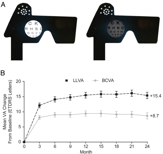
Methods: In the HARBOR trial, 1084 treatment-naïve patients ≥50 years of age with subfoveal wAMD received intravitreal ranibizumab 0.5 or 2.0 mg monthly or as needed. To measure LLVA, patients read a normally illuminated ETDRS (Early Treatment Diabetic Retinopathy Study) chart with a neutral density filter placed in front of the study eye. Patients were assigned into quartiles based on the magnitude of the difference between best-corrected visual acuity under optimal luminance (BCVA) and LLVA (BCVA-LLVA gap). The association between mean change in BCVA from baseline and BCVA-LLVA gap at baseline was analysed using a general linear model.

Results: A smaller baseline BCVA-LLVA gap predicted significantly higher BCVA gains over 24 months (p<0.0001 at each month; Pearson correlation), even after controlling for baseline BCVA or stratifying by treatment arm. Patients in the smallest baseline BCVA-LLVA gap quartile gained an average of +13.4 letters compared with +2.4 letters for patients in the widest baseline BCVA-LLVA gap quartile. At months 12 and 24, the smallest baseline BCVA-LLVA gap quartile had the highest proportion of ≥15-≥30-letter gain, and the widest baseline BCVA-LLVA gap quartile had the highest proportion of ≥15-/≥30-letter loss (p<0.0001; Fisher's exact test).

Conclusions: The baseline BCVA-LLVA gap is a significant predictor of visual acuity response to anti-VEGF treatment in patients with wAMD.

Trial registration number: NCT00891735; Post-results.

Keywords: Degeneration; Drugs; Macula; Neovascularisation; Vision.


Figures

Figure 1
Figure 1
(A) Assessing vision under standard and low-luminance conditions. (B) Mean VA change from baseline over 24 months. All treatment groups pooled. Error bars represent 95% CIs. BCVA, best-corrected visual acuity; ETDRS, Early Treatment Diabetic Retinopathy Study; LLVA, low-luminance visual acuity; VA, visual acuity.
Figure 2
Figure 2
Mean standard vision change from baseline over 24 months by baseline vision gap quartiles. Negative correlation between baseline BCVA–LLVA gap and BCVA change from baseline: p<0.0001 at each month based on Pearson correlation from linear regression. The n values for baseline gap quartiles 1, 2, 3 and 4 are 307, 263, 246 and 268, respectively. All treatment groups pooled. Error bars represent 95% CIs. BCVA, best-corrected visual acuity; ETDRS, Early Treatment Diabetic Retinopathy Study; LLVA, low-luminance visual acuity.
Figure 3
Figure 3
(A) Mean standard vision change over 24 months by baseline vision gap quartiles after controlling for baseline standard vision. (B) Mean standard vision change over 24 months by baseline vision gap quartiles after stratifying by treatment group. Association between baseline BCVA–LLVA gap and BCVA gain, controlling for baseline BCVA: p<0.0001 at each month based on a basic multivariate linear model with the gap magnitude considered a continuous measure. Association between baseline BCVA–LLVA gap and BCVA gain, stratifying by treatment group: p<0.05 at each month for each treatment group based on ANOVA. The n values for baseline quartiles of worst to best BCVA are 287, 269, 271 and 257, respectively. All treatment groups pooled. Baseline n values are 272, 270, 271 and 271 for ranibizumab 0.5 mg monthly, 2.0 mg monthly, 0.5 mg PRN and 2.0 mg PRN, respectively. ANOVA, analysis of variance; BCVA, best-corrected visual acuity; ETDRS, Early Treatment Diabetic Retinopathy Study; LLVA, low-luminance visual acuity; PRN, as needed.
Figure 4
Figure 4
Standard vision gain and loss at months 12 and 24 by baseline vision gap quartiles. (A) Patients who gained ≥15 ETDRS letters from baseline; (B) patients who lost ≥15 ETDRS letters from baseline; (C) patients who gained ≥30 ETDRS letters from baseline; (D) patients who lost ≥30 ETDRS letters from baseline. *p<0.0001; †p=0.0010; based on Fisher's exact test. BCVA, best-corrected visual acuity; ETDRS, Early Treatment Diabetic Retinopathy Study.

References

    1. Ho AC, Busbee BG, Regillo CD, et al. Twenty-four-month efficacy and safety of 0.5 mg or 2.0 mg ranibizumab in patients with subfoveal neovascular age-related macular degeneration. Ophthalmology 2014;121:2181–92. 10.1016/j.ophtha.2014.05.009 - DOI - PubMed
    1. Boyer DS, Antoszyk AN, Awh CC, et al. Subgroup analysis of the MARINA study of ranibizumab in neovascular age-related macular degeneration. Ophthalmology 2007;114:246–52. 10.1016/j.ophtha.2006.10.045 - DOI - PubMed
    1. Kaiser PK, Brown DM, Zhang K, et al. Ranibizumab for predominantly classic neovascular age-related macular degeneration: subgroup analysis of first-year ANCHOR results. Am J Ophthalmol 2007;144:850–7. 10.1016/j.ajo.2007.08.012 - DOI - PubMed
    1. Finger RP, Wickremasinghe SS, Baird PN, et al. Predictors of anti-VEGF treatment response in neovascular age-related macular degeneration. Surv Ophthalmol 2014;59:1–18. 10.1016/j.survophthal.2013.03.009 - DOI - PubMed
    1. Sunness JS, Rubin GS, Broman A, et al. Low luminance visual dysfunction as a predictor of subsequent visual acuity loss from geographic atrophy in age-related macular degeneration. Ophthalmology 2008;115:1480–8, 8 e1–2 10.1016/j.ophtha.2008.03.009 - DOI - PMC - PubMed

Publication types

MeSH terms

Associated data