. 2015 Nov 6:5:16188.
doi: 10.1038/srep16188.

Mechanisms underlying the activity-dependent regulation of locomotor network performance by the Na+ pump

Affiliations

Mechanisms underlying the activity-dependent regulation of locomotor network performance by the Na+ pump

Hong-Yan Zhang et al. Sci Rep. .

Erratum in

Abstract

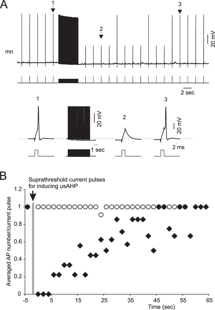
Activity-dependent modification of neural network output usually results from changes in neurotransmitter release and/or membrane conductance. In Xenopus frog tadpoles, spinal locomotor network output is adapted by an ultraslow afterhyperpolarization (usAHP) mediated by an increase in Na(+) pump current. Here we systematically explore how the interval between two swimming episodes affects the second episode, which is shorter and slower than the first episode. We find the firing reliability of spinal rhythmic neurons to be lower in the second episode, except for excitatory descending interneurons (dINs). The sodium/proton antiporter, monensin, which potentiates Na(+) pump function, induced similar effects to short inter-swim intervals. A usAHP induced by supra-threshold pulses reduced neuronal firing reliability during swimming. It also increased the threshold current for spiking and introduced a delay to the first spike in a train, without reducing subsequent firing frequency. This delay was abolished by ouabain or zero K(+) saline, which eliminate the usAHP. We present evidence for an A-type K(+) current in spinal CPG neurons which is inactivated by depolarization and de-inactivated by hyperpolarization, and accounts for the prolonged delay. We conclude that the usAHP attenuates neuronal responses to excitatory network inputs by both membrane hyperpolarization and enhanced de-inactivation of an A-current.


Figures

Figure 1
Figure 1. Swimming frequency and episode duration are affected by inter-swim interval.
(A) Pairs of fictive swimming episodes induced by the same skin stimuli with inter-swim intervals set at 5, 15 and 30 seconds. (B) Swimming bursts at the beginning of the first (Ep1) and second (Ep2) episodes shown in (A) respectively. Note that the cycle periods of Ep2 are longer compared to Ep1. (C) Average episode duration of Ep2 was significantly shorter than Ep1 at the 5 and 15 second intervals (*P < 0.05, n = 5). (D) Time series measurements of swimming frequencies of Ep1 (black) and Ep2 (grey) for the three defined intervals. Insert bar graphs: average normalised swimming frequency of Ep1 and Ep2. Ep2 swimming frequency is lower than Ep1 (*P < 0.05, n = 5).
Figure 2
Figure 2. Short inter-swim interval has neuron type-specific effects on the firing reliability of CPG neurons.
(A) Two swimming episodes with a short interval were induced by skin stimulation. A dIN was recorded simultaneously with ventral root (vr) activity. (B) MN firing during two swimming episodes with a short interval. Note that the firing reliability is lower during the second episode. (C) Mean dIN action potential (AP) number per swim cycle. The firing reliability of dINs is unaffected. (D) Mean non-dIN AP number per swim cycle. The firing reliability of non-dIN CPG neurons in the second episode was significantly reduced. (*P < 0.05, n = 17; paired t-test).
Figure 3
Figure 3. The usAHP reduces CPG neuron firing reliability.
(A) Control firing of a non-dIN neuron and simultaneous ventral root activity; note the different time scales. (B) same neuron as in (A); a swimming episode was evoked immediately after a train of pulses had been used to induce repetitive firing and a usAHP. (C) Firing reliability of non-dINs in episodes initiated during a usAHP was lower compared to the control episodes (***P < 0.001, n = 11; paired t-test). (D) The reduction of neuron firing reliability has been plotted against the membrane potential change (potential before the suprathreshold pulse train – potential before swimming). Each trial from neurons displaying a usAHP (circle) and not displaying a usAHP (cross) are shown.
Figure 4
Figure 4. Current threshold for generating action potentials is increased during the usAHP.
(A) Response of a MN to brief threshold current pulses (2 ms) before and during the usAHP. The suprathreshold current pulse train (30 ms pulse with 20 ms interval for 5 seconds) is used to trigger repetitive firing and a usAHP. (B) Pooled data of neuron responses to brief threshold current. Black diamonds: 9 non-dINs displayed a usAHP following suprathreshold current pulses (arrowed); open circles: 12 non-dINs without usAHP.
Figure 5
Figure 5. The effects of the usAHP on first spike delay, firing frequency and spike number.
(A) 30 ms suprathreshold depolarizing current pulses were applied every 2 seconds before (1) and after (3) a train of 30 ms depolarizing pulses (2) that induces continuous firing on top of the depolarization. A usAHP (5.5 mV) appears following the 5 second pulse train (as in Fig. 4). The first spike delay (arrow) is longer in pulse (3) than (1). (B) Changes in mean first spike delay, firing frequency and AP number per pulse. The grey line represents the train of pulses in (A). Filled circle: neurons displaying a usAHP; open circle: neurons not displaying a usAHP. Bar graphs represent pooled data of different groups. Open bar: control period (before the grey line); closed bar: the first 5 seconds following the long pulse train (2 in A). Paired t-test: **P < 0.01; ***P < 0.001; n = 13. (C) Action potentials induced by a series of suprathreshold pulses. Left panel: control period; right panel: in the presence of ouabain; black trace: action potential induced by the first pulse; dashed trace: action potentials induced by the 5th or 6th pulse. Note the differences in resting membrane potential and first spike delay in the left panel (arrow), but not in the right panel. (D) Pooled data of control (left) and block (right) groups. Open bar: the first pulse in both groups; closed bar: the 5th or 6th pulse. The spike delays in control are different, but not after blocking the Na+ pump with ouabain or zero K+ saline (paired t-test: **P < 0.01, n = 6).
Figure 6
Figure 6. Identifying a voltage-dependent transient outward current.
(A) In current clamp mode, a small negative membrane potential change (arrow) was occasionally observed during the first spike delay. Left panel shows neuron responses to depolarizing current pulses of two different amplitudes. Black: lower amplitude; red: higher amplitude; the negative membrane potential change appears. Middle panel shows neuron responses to the same depolarizing current pulse during control (black trace) and usAHP periods induced by suprathreshold pulse train (red trace). Right panel shows neuron responses to the same depolarization during control (black trace) and in the presence of TTX (red trace). (B) K+ currents recorded in voltage clamp mode. Red trace shows a test with pre- clamping potential of −30 mV and blue trace with −80 mV pre-clamping potential. Inset black trace is the difference between the two current traces at the beginning of the voltage step. (C) 4-AP at 2 mM preferentially blocks transient K+ currents. Red current trace is control, blue is in 4-AP and green is wash. Black trace is the difference in currents between control and 4-AP. (B,C) are recorded in the presence of 0.4 μM TTX and 200 μM Cd2+.
Figure 7
Figure 7. Increasing intracellular Na+ concentration using the Na+ ionophore monensin shortens swimming episode duration and decreases swimming frequency.
(A) Bath applying 10 μM monensin irreversibly reduced episode duration. (B) Swimming bursts at the beginning of each trace in (A). (C) Averaged swimming episode durations (***P < 0.001, n = 6). (D) Time series measurements showing swimming frequencies of an episode in control and in the presence of monensin. Inset bar graph shows the average normalized swim frequency (*P < 0.05, n = 6).
Figure 8
Figure 8. Monensin reduces the firing reliability of non-dINs.
(A) The firing reliability of non-dINs was reduced by 10 μM monensin. (B) dIN firing reliability was not affected. The episode duration was shortened in (A,B) by adding monensin in the bath. (C) Local application of monensin 20 μM directly to the recorded cell, firing reliability of all 5 non-dINs was reduced, whereas the swimming episode duration was almost not affected. (D) Averaged normalized firing reliability and episode duration before and after applying monensin in the bath. (E) Averaged normalized firing reliability and episode duration in control and during local application of monensin. Black bar: control; grey bar: monensin (*P < 0.05; ***P < 0.001, n = 5).
Figure 9
Figure 9
(A) Schematic diagram showing the organization of motor circuit in the spinal cord of Xenopus tadpoles at the time of hatching. dINs provide the excitatory drive for swimming and cINs couple the left and right sides. (B) Schematic diagram illustrating how the Na+ pump and A-type K+ current are involved in the short term memory of motor network output. At rest, most A-type K+ channels are inactivated. Weak activity (1) does not increase Na+ pump current sufficiently to hyperpolarize the membrane potential so when the membrane potential is subsequently depolarized above threshold (2) most A-type K+ channels cannot be activated, and thus the first spike delay is unaffected. Stronger activity (2) can potentiate Na+ pump function and induce a larger pump current which hyperpolarizes the membrane potential (usAHP). This hyperpolarization removes the inactivation of A-type K+ channels. Now, when depolarized above threshold (3), the A-type current is large enough to impede membrane repolarization, prolonging first spike delay, and reducing the total number of spikes.

References

    1. de Lores Arnaiz G. R. & Ordieres M. G. Brain Na+, K+-ATPase activity in aging and disease. Int. J. Biomed. Sci. 10, 85–102 (2014). - PMC - PubMed
    1. Parker D., Hill R. & Grillner S. Electrogenic pump and a Ca2+- dependent K+ conductance contribute to a posttetanic hyperpolarization in lamprey sensory neurons. J. Neurophysiol. 76, 540–553 (1996). - PubMed
    1. Ballerini L., Bracci E. & Nistri A. Pharmacological block of the electrogenic sodium pump disrupts rhythmic bursting induced by strychnine and bicuculline in the neonatal rat spinal cord. J. Neurophysiol. 77, 17–23 (1997). - PubMed
    1. Hess D., Nanou E. & El Manira A. Characterization of Na+-activated K+ currents in larval lamprey spinal cord neurons. J. Neurophysiol. 97, 3484–3493 (2007). - PubMed
    1. Scuri R. et al. Inhibition of Na+/K+ ATPase potentiates synaptic transmission in tactile sensory neurons of the leech. Eur. J Neurosci. 25, 159–167 (2007). - PubMed

Publication types

Substances

LinkOut - more resources