. 2015 Dec 1;408(1):99-108.
doi: 10.1016/j.ydbio.2015.10.008. Epub 2015 Nov 2.

En1 directs superior olivary complex neuron positioning, survival, and expression of FoxP1

Affiliations

En1 directs superior olivary complex neuron positioning, survival, and expression of FoxP1

Stefanie C Altieri et al. Dev Biol. .

Abstract

Little is known about the genetic pathways and transcription factors that control development and maturation of central auditory neurons. En1, a gene expressed by a subset of developing and mature superior olivary complex (SOC) cells, encodes a homeodomain transcription factor important for neuronal development in the midbrain, cerebellum, hindbrain and spinal cord. Using genetic fate-mapping techniques, we show that all En1-lineal cells in the SOC are neurons and that these neurons are glycinergic, cholinergic and GABAergic in neurotransmitter phenotype. En1 deletion does not interfere with specification or neural fate of these cells, but does cause aberrant positioning and subsequent death of all En1-lineal SOC neurons by early postnatal ages. En1-null cells also fail to express the transcription factor FoxP1, suggesting that FoxP1 lies downstream of En1. Our data define important roles for En1 in the development and maturation of a diverse group of brainstem auditory neurons.

Keywords: Auditory; Brainstem; Deafness; Hearing; Nucleogenesis.


Figures

Figure 1
Figure 1
SOC nuclear morphology. Cartoon of a coronal section through the brainstem shows the six nuclei that comprise the superior olivary complex (SOC). These include the lateral (LSO) and medial (MSO) superior olives, superior paraolivary nucleus (SPN) and lateral (LNTB) medial (MNTB) and ventral (VNTB) nuclei of the trapezoid body. Also indicated are the lateral (LOCB) and medial (MOCB) olivocochlear bundle neurons that reside within the SOC.
Figure 2
Figure 2
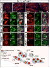
En1-lineal SOC cells are glycinergic, cholinergic and GABAergic neurons. (A-A‴) Coronal brainstem section from adult En1Cre; ROSAtdTomato mouse showing endogenous tdTomato signal with DAPI nuclear labeling. En1-lineal neurons are found in the LSO (A), MNTB (A′), VNTB (A″) and LNTB (A‴). Endogenous tdTomato signal (B) and immunostaining for MAP2 (B′) in the MNTB reveals that all tdTomato+ cells are also MAP2+ (B″). Immunostaining of En1Cre; ROSAtdTomato mouse brainstem sections for glycine shows that all tdTomato+ cells in the LSO, MNTB, and VNTB are glycinergic neurons (C-E″). Immunostaining for ChAT shows that En1-lineal VNTB neurons are also cholinergic (F-F″). Within the LNTB, all tdTomato+ neurons are GABA+ (G-G″). Glycinergic and GABAergic SPN neurons receive inputs from tdTomato+ cells (red perineuronal signal in H, I), but these neurons and ChAT+ neurons that contribute to the LOCB and MOCB are tdTomato- (J-K″). In panels B″-J″, the merged images contain DAPI staining in blue. (L) Schematic summarizing results. Scale bar: 40μm (A), 13μm (A′-A‴), 3.7μm (B-K″).
Figure 3
Figure 3
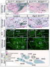
En1-lineal neurons are absent from the SOC of adult mice following En1 deletion. Color-coded dotted lines delineate SOC subdivisions in this and subsequent Figures. In animals with constitutive or conditional En1 deletion subdivisions are shown based on where they should be compared to control. Xgal+ cells are present in the SOC of adult En1Cre/+; ROSALacZ (A) but not En1Cre/flox; ROSALacZ mice (B). Comparison of in situ hybridization for the glycinergic neuron marker Glyt2 counterstained with Cresyl violet in adult control (C) and Egr2; En1CKO mice (D) indicates a loss of glycinergic neurons in the LSO, VNTB and MNTB of Egr2; En1CKO mice. (E-H) ChAT immunostaining demonstrates that LOCB and MOCB neurons are present in adult Egr2; En1CKO mice, but that cholinergic (Ach) neurons within the confines of the VNTB are absent. Immunostaining for GABA revealed GABAergic neurons present in the LNTB of control animals (I) were missing in Egr2; En1CKO mice (J). GABAergic boutons are present on cell bodies of both genotypes. Boxes show areas of small insets in (I, J). (K) Schematic summarizing results. Scale bar: 375μm (A-D), 150μm (E-H), 40μm (I, J), 15μm (insets).
Figure 4
Figure 4
En1-null SOC cells migrate aberrantly during development. Dotted vertical lines mark the brainstem midline. At E12.5, Xgal+ cells are found in similar locations in the presumptive SOC (pSOC) of the two genotypes (A, B). At E15.5, the distribution of Xgal+ cells in the medial pSOC is more diffuse in En1Cre/flox; ROSALacZ (D) compared to En1Cre/+; ROSALacZ mice (C). At P0, Xgal+ cells in the SOC are found in the adult distribution of En1Cre/+; ROSALacZ mice (E). In contrast, reduced numbers of Xgal+ cells are present in the SOC of En1Cre/flox; ROSALacZ mice (F), and these cells form an ectopic medial cell group. Compared to P0 control mice (G), Cresyl violet staining of Egr2; En1CKO (H) and En1-null (I) mice shows presence of an ectopic cell group close to the brainstem midline. Scale bar: 250μm (C-I), 150μm (A, B).
Figure 5
Figure 5
Ectopic cells derive from the En1-lineage and die in En1-null mice. Coronal brain sections through the SOC of P0 En1Cre/+; ROSAtdTomato (A) and En1Cre/Cre; ROSAtdTomato (B) mice showing endogenous tdTomato fluorescence. Dotted vertical lines mark the brainstem midline and boxed areas the regions of (A′-A′‴) and (B′-B′‴). Activated caspase-3 immunostaining (A″, B″) and DAPI staining (A‴, B‴) show the presence of apoptotic cells and pyknotic nuclei in En1Cre/Cre; ROSAtdTomato but not En1Cre/+; ROSAtdTomato mice. Yellow, purple and white arrowheads (B′-B‴) denote caspase-3+/tdTomato+ cells, pyknotic tdTomato+ cells, and pyknotic tdTomato- cells, respectively. Scale bar: 120μm (A, B), 25μm (A′-B‴).
Figure 6
Figure 6
En1 deletion does not affect neural cell fate in the SOC. Single-plane confocal microscope images of coronal brainstem sections through the presumptive SOC from E14.5 En1Cre/+; ROSAtdTomato and En1Cre/Cre; ROSAtdTomato mice (A-B″), MNTB of P0 En1Cre/+; ROSAtdTomato mice (C-C″, E-E″, G-G″) and ectopic cell group of P0 En1Cre/Cre; ROSAtdTomato mice (D-D″, F-F″, H-H″) showing endogenous tdTomato signal (A-H), immunostaining for the neural marker TUJ1 (A′-D′), the astrocyte marker ALDH1L1 (E′, F′), the oligodendrocyte marker Olig2 (G′, H′) and merged images (A″-H″). TUJ1 and tdTomato are colocalized in En1Cre/+; ROSAtdTomato and En1Cre/Cre; ROSAtdTomato mice, while no signal overlap is seen with the glial cell markers. Scale bar: 12μm.
Figure 7
Figure 7
FoxP1 expression, but not Sox2 or MafB expression, depends on En1 in the SOC. Boxed areas are shown in insets. Yellow arrowheads indicate double labeled neurons (A-D″). Coronal sections demonstrate endogenous tdTomato (A-D) and FoxP1 immunostaining (A′-D′) in the presumptive SOC of E14.5 En1Cre/+; ROSAtdTomato (A-A″) and En1Cre/Cre; ROSAtdTomato (B-B″) mice. FoxP1+ cells are found throughout the presumptive SOC of En1Cre/+; ROSAtdTomato mice but are limited to the nascent SPN of En1Cre/Cre; ROSAtdTomato mice. tdTomato+/FoxP1+ cells are absent from the presumptive SOC of En1Cre/Cre; ROSAtdTomato mice. The MNTB marker Sox2 labels tdTomato+ cells in both En1Cre/+; ROSAtdTomato (C-C″) and En1Cre/Cre; ROSAtdTomato mice (D-D″). The LSO and MSO neuron marker MafB is present in the presumptive SOC at E14.5 in En1Cre/+; ROSAtdTomato (E-E″) and En1Cre/Cre; ROSAtdTomato (F-F″) but is not co-expressed in tdTomato+ neurons in either genotype. (G) Summary graphic placing En1 into genetic hierarchies that regulate SOC neuron development. Scale bar: 120μm (A-F″), 12μm (insets).

Similar articles

Cited by

References

    1. Atlas, A.B. n.d. http://mouse.brain-map.org.
    1. Bacon C, Schneider M, Le Magueresse C, Froehlich H, Sticht C, Gluch C, Monyer H, Rappold GA. Brain-specific Foxp1 deletion impairs neuronal development and causes autistic-like behaviour. Mol Psychiatry. 2014 doi: 10.1038/mp.2014.116. - DOI - PMC - PubMed
    1. Benson CG, Cant NB. The ventral nucleus of the lateral lemniscus of the gerbil (Meriones unguiculatus): organization of connections with the cochlear nucleus and the inferior colliculus. J Comp Neurol. 2008;510:673–690. doi: 10.1002/cne.21820. - DOI - PMC - PubMed
    1. Bilovocky NA, Romito-DiGiacomo RR, Murcia CL, Maricich SM, Herrup K. Factors in the genetic background suppress the engrailed-1 cerebellar phenotype. Journal of Neuroscience. 2003;23:5105–5112. - PMC - PubMed
    1. Bledsoe SC, Snead CR, Helfert RH, Prasad V, Wenthold RJ, Altschuler RA. Immunocytochemical and lesion studies support the hypothesis that the projection from the medial nucleus of the trapezoid body to the lateral superior olive is glycinergic. Brain Research. 1990;517:189–194. - PubMed

Publication types