. 2016 Jan 1;409(1):218-233.
doi: 10.1016/j.ydbio.2015.10.022. Epub 2015 Nov 2.

Multiple mouse models of primary lymphedema exhibit distinct defects in lymphovenous valve development

Affiliations

Multiple mouse models of primary lymphedema exhibit distinct defects in lymphovenous valve development

Xin Geng et al. Dev Biol. .

Abstract

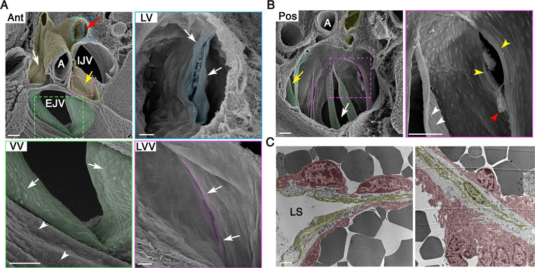
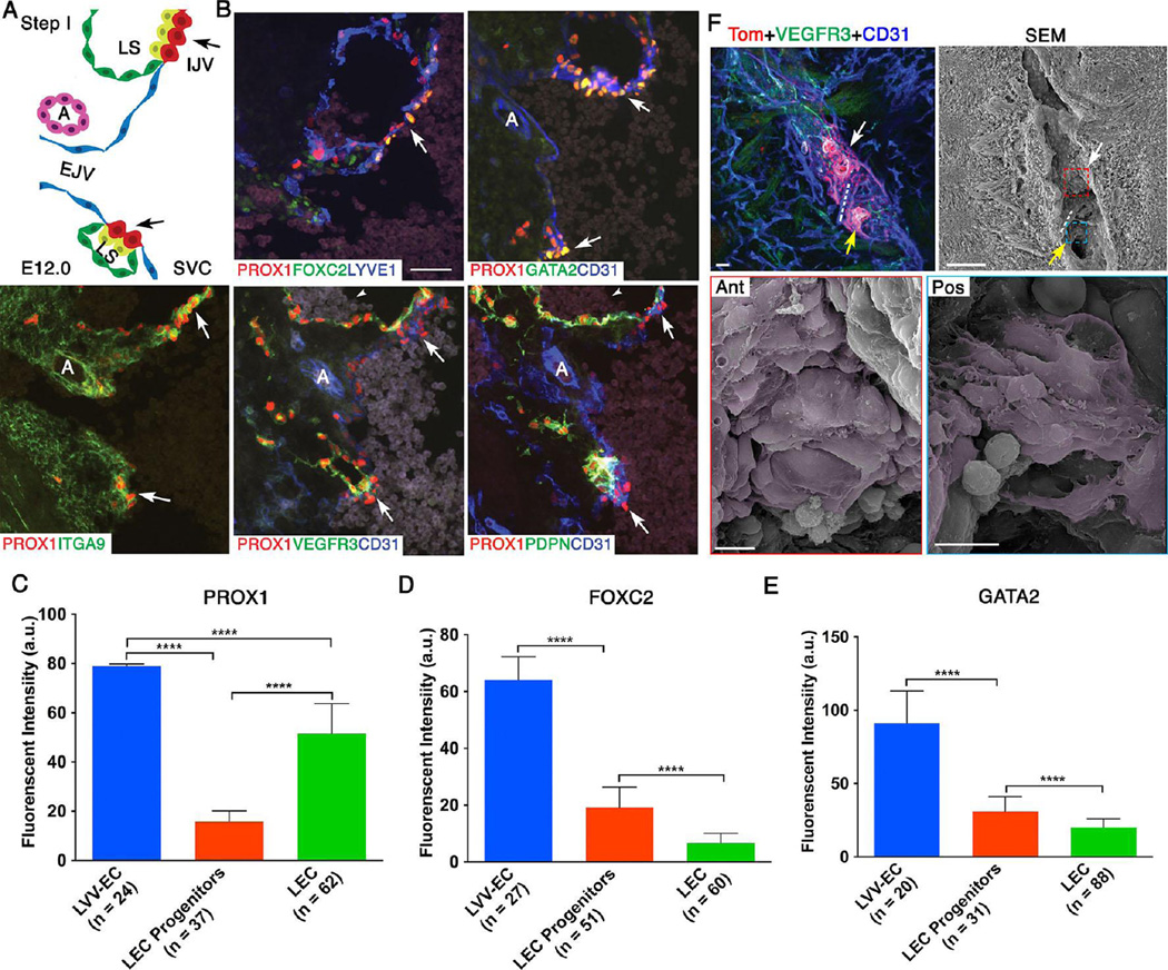
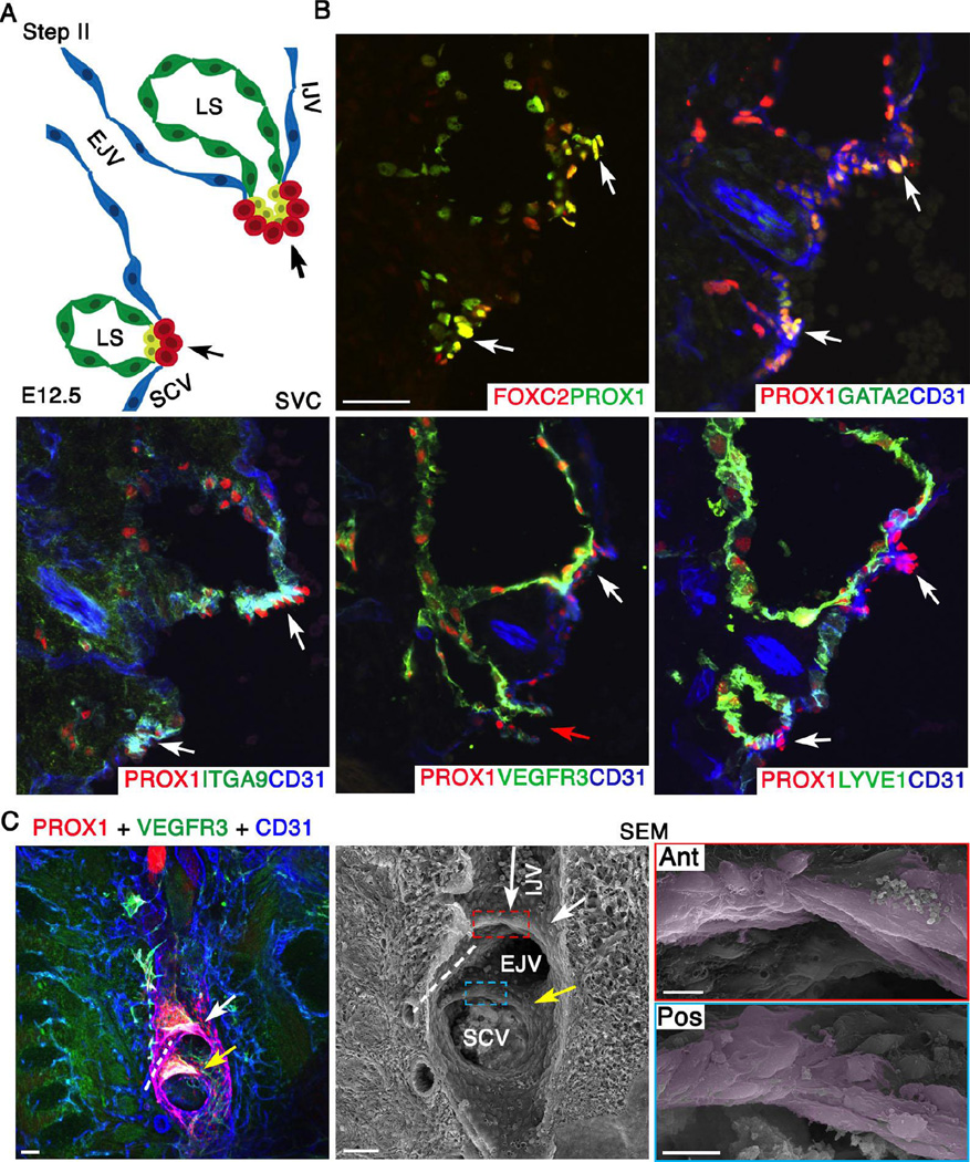
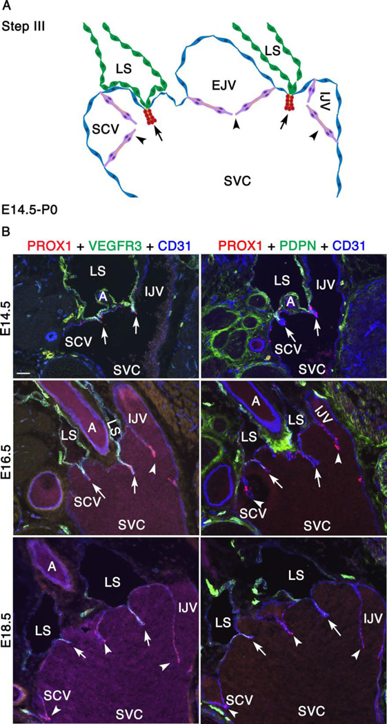
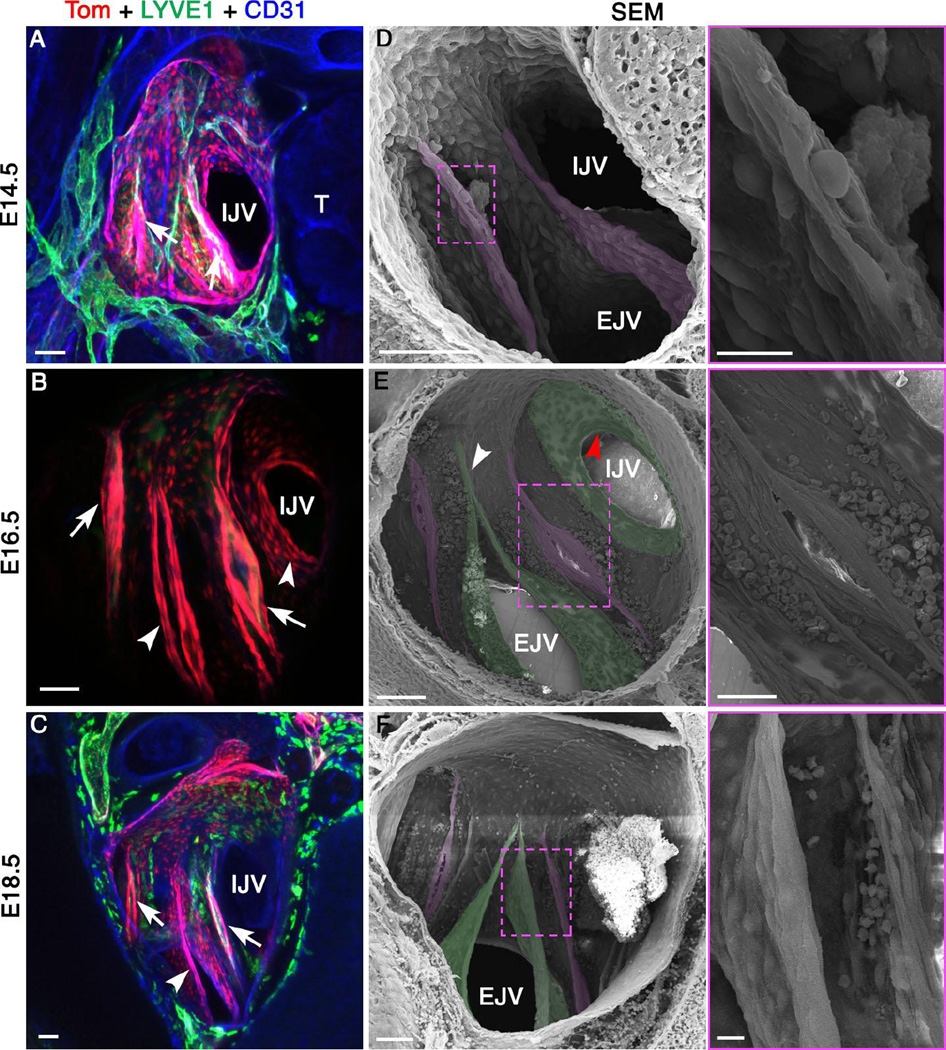
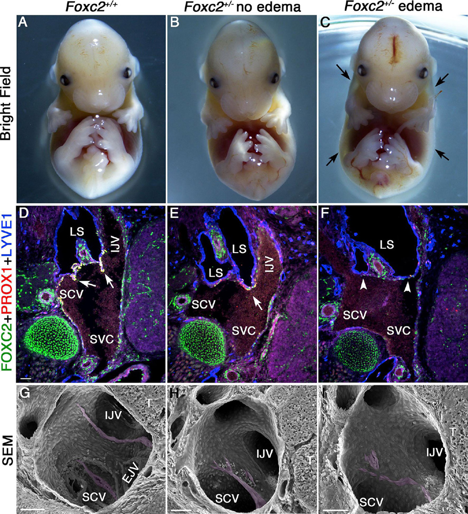
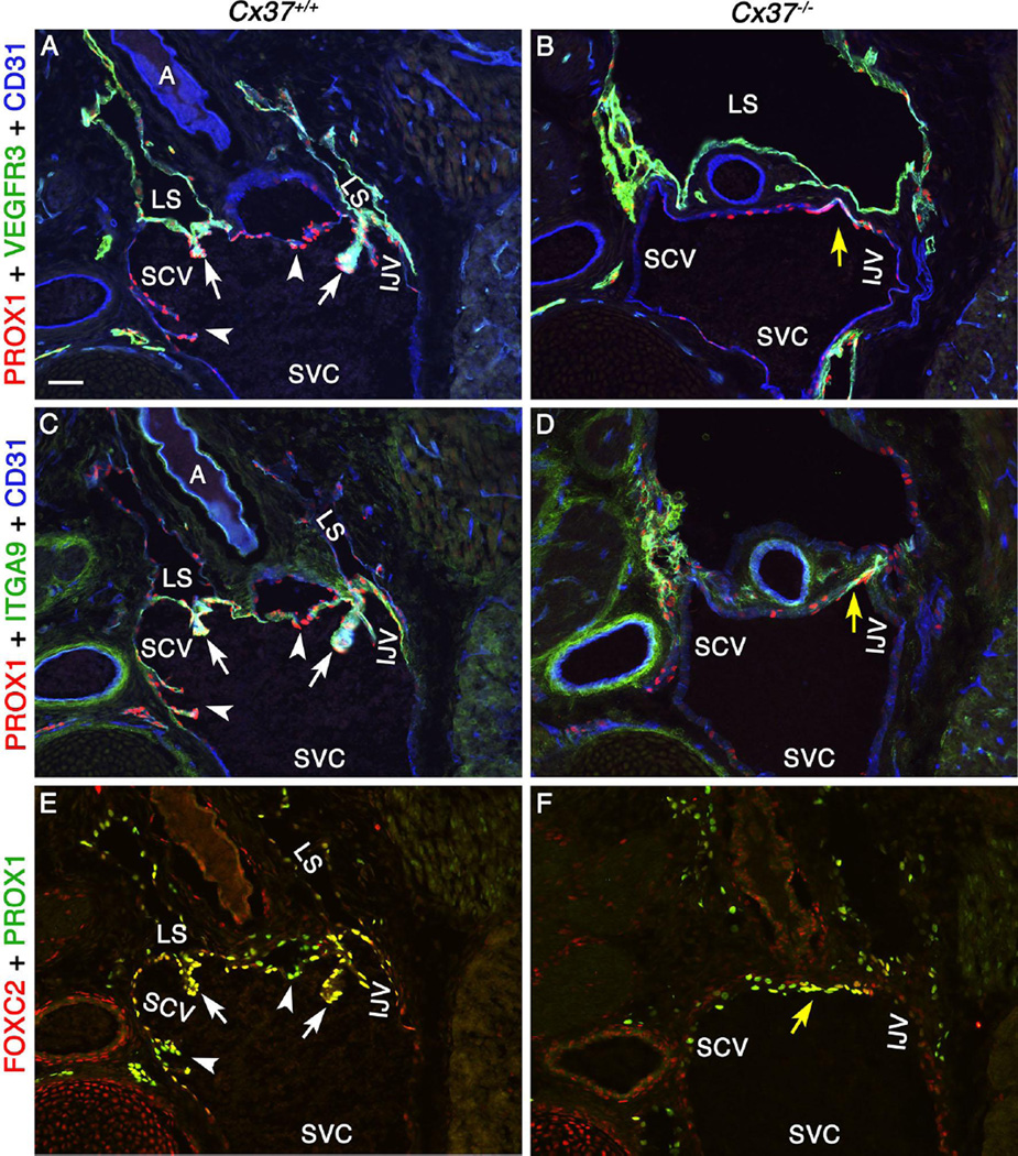
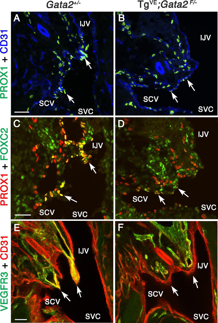
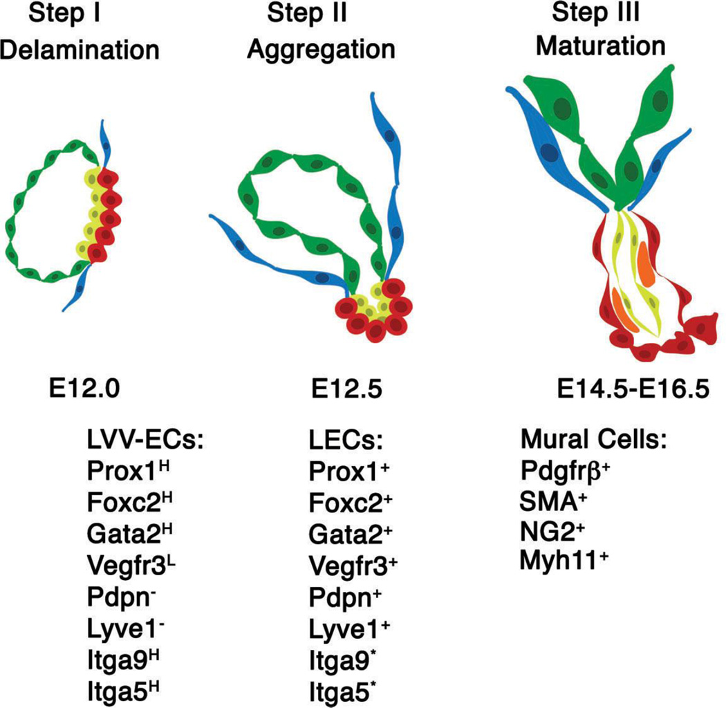
Lymph is returned to the blood circulation exclusively via four lymphovenous valves (LVVs). Despite their vital importance, the architecture and development of LVVs is poorly understood. We analyzed the formation of LVVs at the molecular and ultrastructural levels during mouse embryogenesis and identified three critical steps. First, LVV-forming endothelial cells (LVV-ECs) differentiate from PROX1(+) progenitors and delaminate from the luminal side of the veins. Second, LVV-ECs aggregate, align perpendicular to the direction of lymph flow and establish lympho-venous connections. Finally, LVVs mature with the recruitment of mural cells. LVV morphogenesis is disrupted in four different mouse models of primary lymphedema and the severity of LVV defects correlate with that of lymphedema. In summary, we have provided the first and the most comprehensive analysis of LVV development. Furthermore, our work suggests that aberrant LVVs contribute to lymphedema.


Figures

Figure 1
Figure 1. Organization of cells in the lymphovenous valve (LVV) complex of newborn mice
(A) SEM in the anterior-to-posterior (Ant) orientation shows lymph sac (pseudo colored in yellow), upstream sides of LVVs (white and yellow arrows), VV within EJV (pseudo colored in green) and the downstream side of a LV (pseudo colored in blue). At higher magnification, LVVs appear to have a smaller opening compared to VV. And, cells at the entrance of LVVs are elongated perpendicular to the direction lymph flow, which is away from the reader. Cells within EJV are seen aligned parallel to the direction of blood flow (VV, arrowheads). Instead, cells within VV are cuboidal (VV, arrows). (B) SEM in the posterior-to-anterior (Pos) orientation shows the downstream side of LVVs (magenta) and VVs (green). White arrow indicates EJV and yellow arrow indicates SCV. At higher magnification cells in both LVVs and VVs (yellow and white arrowheads respectively) appear elongated. Cells on the downstream side of LVs are also elongated (A, LV, arrows). Fibrin clots at the entrance of LVVs are also seen (red arrowhead). (C) TEM shows thin and elongated lymphatic endothelial cells in the upstream end of LVVs (yellow). LVV-forming endothelial cells on the veins are in magenta. Statistics: n= 4 for A, B. n= 2 for C. Abbreviations: LS, lymph sac; A, artery; IJV, internal jugular vein; EJV, external jugular vein; SCV, subclavian vein; SVC, superior vena cava. Scale bars: 50 µm for A (Ant, VV) and B; 10 µm for A (LVV, LV) and 1 µm for C.
Figure 2
Figure 2. At E12.0 LVV-forming endothelial cells (LVV-ECs) delaminate from the veins in the luminal orientation
(A) Organization of veins and lymph sacs (LS) in the frontal orientation at this first step of LVV development. Lymphatic endothelial cells (LECs) and LVV-ECs that form LVVs are in yellow and red respectively. The remaining LECs that form LS and venous endothelial cells are in green and blue respectively. EJV is perpendicular to IJV and SVC. (B) Immunohistochemistry for the indicated markers in the region from panel A. LVV-ECs are indicated by arrows. PROX1 is expressed at higher level in LVV-ECs compared to LECs. FOXC2 and GATA2 are expressed almost exclusively in LVV-ECs. In contrast, PDPN is restricted to LECs. VEGFR3 is higher in LVV forming LECs compared to LVV-ECs and the rest of LECs. ITGA9 is strongly expressed in the LVV forming LECs and LVV-ECs. Arrowheads point to the blood cells seen within the lymph sacs. (C-E) After performing immunohistochemistry on sections as described above, the fluorescent signals produced by antibodies were measured in arbitrary units (a.u.) using ImageJ software. PROX1 (C), FOXC2 (D) and GATA2 (E) are expressed at significantly higher levels in LVV-ECs compared to LEC progenitors and LECs. (F) 800 µm sagittal section of an E12.0 ProxTom embryo was immunostained and imaged by confocal microscopy. Two loose clusters of Tomhigh LVV-ECs are seen within the vein. Dotted line represents the artery located between the LVV-ECs clusters. SEM of the same section revealed delaminating LVV-ECs that overlap each other (pseudo colored in magenta) in both anterior (white arrow) and posterior (yellow arrow) clusters. Statistics: n= 3 for B; n= 6 for F. For panels C-E, the indicated numbers of cells from a single embryo were analyzed. This data is representative of three-independent experiments. **** p<0.0001. Abbreviations: LS, lymph sac; IJV, internal jugular vein; EJV, external jugular vein; SCV, subclavian vein; SVC, superior vena cava. Scale bars: 50 µm for B and the top two panels of C; 10 µm for the bottom two panels of C.
Figure 3
Figure 3. At E12.5 LVV-EC clusters invaginate into the veins and elongate perpendicular to blood flow
(A) Schematic of the LVV complex in frontal orientation at E12.5. This is the second step of LVV development. Cells are color coded as in Figure 2. Note that the EJV has rotated clockwise towards IJV by 45°. (B) Immunohistochemistry revealed that the LVV-ECs (white arrows) have invaginated into the veins. And, a connection between LS and veins is seen (red arrow). LYVE1 is excluded from LVV-ECs at E12.5. Expression pattern of other markers is identical to that at E12.0. (C) Confocal imaging followed by SEM of an 800 µm sagittal section from a E12.5 ProxTom embryo revealed two compact Tomhigh LVV-EC clusters. Cells in both anterior (white arrow) and posterior (yellow arrow) clusters have elongated perpendicular to the direction of blood flow. The long arrow indicates the direction of flow in IJV. The flow from EJV and SCV are towards the reader. Dotted line indicates the artery between the two LVVs. Statistics: n= 3 for B. n= 8 for C. Abbreviations: LS, lymph sac; IJV, internal jugular vein; EJV, external jugular vein; SCV, subclavian vein; SVC, superior vena cava. Scale bars: 50 µm for B and the first two panels of C; 10 µm for the last two panels of C.
Figure 4
Figure 4. Between E14.5 and P0, VVs are formed and LEC markers are gradually downregulated in lymph sacs
(A) This is the third and final step of LVV development between E14.5 and P0. The schematic depicts the overall arrangement of the LVV complex in frontal orientation. The only obvious change during this interval is the appearance of VVs at E16.5. VVs are in magenta. Rest of the cells is color coded as in Figure 2. (B) Immunohistochemistry revealed that the LEC markers VEGFR3 and podoplanin (PDPN) are gradually downregulated in LSs. However, they remain strongly expressed in the LECs that form LVVs (arrows). Statistics: n= 3 for each developmental stage. Abbreviations: LS, lymph sac; IJV, internal jugular vein; EJV, external jugular vein; SCV, subclavian vein; SVC, superior vena cava. Scale bars: 50 µm for B and 1 µm for C.
Figure 5
Figure 5. Mural cells are progressively recruited to lymph sacs, LVVs and VVs
Mural cells are progressively recruited to lymph sacs, LVVs and VVs. LVV-forming region of E14.5, E16.5 and E18.5 embryos were analyzed using mural cell markers. PDGFRβ (A-C) is a pan-mural cell marker. SMA (D-F) and MYH11 (G-I) are vascular smooth muscle cell markers. NG2 is a pericyte marker (J-L). At E14.5, PDGFRβ+ cells are is seen surrounding the lymph sacs (A, red arrowheads) and within LVVs (A, white arrows). Other mural cell markers are not seen in lymph sacs or LVVs at this stage (D, G, J). At subsequent developmental time points, downregulation of LEC marker LYVE1 in lymph sacs coincides with the expression of mature mural cell markers (red arrowheads). Scattered mural cells are also seen within LVVs (white arrows) and VVs (white arrowheads). Yellow arrows and yellow arrowheads point to the arterial and venous perivascular cells respectively. And, red arrows point to the muscles. Statistics: n= 3 for each developmental stage. Abbreviations: LS, lymph sac; IJV, internal jugular vein; EJV, external jugular vein; SCV, subclavian vein; SVC, superior vena cava. Scale bars: 50 µm
Figure 6
Figure 6. LVV-ECs undergo limited change while VVs rapidly develop between E14.5 and E18.5
(A-C) Confocal imaging of LVV-complex from ProxTom embryos in the Pos orientation reveals LVVs (arrows). VVs are seen at E16.5 and E18.5 (arrowheads). (D-F) SEM of the same samples reveals LVVs (pseudo colored in magenta) and VVs (pseudo colored in green). Higher magnification picture of the boxed areas are shown in the adjacent panels. The overall structure of LVVs remains stable in this time window. LVV-ECs appear to become more elongated with time. Diameter of venous junction increases during this time period. (D) VVs are not seen at E14.5. (E) At E16.5 VV at the entrance of IJV appears as a circular shelf (stage 2 according to the scheme of Bazigou et al., 2011). A commissure is clearly seen in the VV located in EJV (arrowhead) suggesting developmental stage 3. (F) At E18.5 VV of EJV is dome shaped and fully matured (stage 4). Statistics: n= 10 for A, D; n= 12 for B, E; n= 3 for C, F. Abbreviations: T, thymus; IJV, internal jugular vein; EJV, external jugular vein. Scale bars: 10 µm for E, G and I. 50 µm for the rest.
Figure 7
Figure 7. Variable penetrance of edema phenotype in Foxc2+/− embryos correlates with defective LVVs
Approximately half of the Foxc2-heterozygoes embryos generated in C57BL/6 background are grossly indistinguishable from wild type embryos (A, B). The other half develops edema (C, arrows) by E14.5. Analysis of frontal sections from these embryos revealed one LVV in the normal-looking Foxc2+/− embryos (E, arrow). The edematous embryos lack LVVs (F, arrowheads). (G-I) SEM of the LVV-complex from control embryos revealed two LVVs (G, magenta) in control and one LVV (H, magenta) in non-edematous Foxc2-heterozygote embryos. In contrast, no LVVs are seen in edematous Foxc2-heterozygoes (I). Instead, a few elongated cells with the characteristics of LVV-ECs are seen (magenta). Statistics: n= 3 for A-G; n = 4 for H; n = 2 for I. Abbreviations: SVC, superior vena cava; SCV, subclavian vein; LS, lymph sac; IJV, internal jugular vein. Scale bars: 50 µm
Figure 8
Figure 8. LVV-EC invagination is defective in Cx37−/− embryos
(A-F) E16.5 Cx37+/+ and Cx37−/− embryos were sectioned and analyzed after immunostaining for the indicated markers. LVVs (arrows) and VVs (arrowheads) are seen in Cx37+/+ embryos. LVV-ECs are observed in the valve-forming region of Cx37−/− embryos (yellow arrows) but they do not invaginate into veins to form clear LVVs or VVs. Statistics: n= 2 for A, C and E; n = 4 for B, D and F. Abbreviations: A, artery; LS, lymph sac; IJV, internal jugular vein; EJV, external jugular vein; SCV, subclavian vein; SVC, superior vena cava. Scale bars: 50 µm
Figure 9
Figure 9. GATA2 is necessary for the proper differentiation of LVV-ECs
(A-F) E13.5 Gata2+/− or TgVE;Gata2f/− (in which Gata2 is conditionally deleted from all endothelial cells using CreERT2 after tamoxifen injection at E10.0) embryos were analyzed using the indicated markers. A-D are 12 µm frontal cryosections. PROX1 (A, C arrows) and FOXC2 (C, arrows) are strongly expressed in LVV-ECs of Gata2+/− embryos. In contrast, PROX1 (B, D arrows) and FOXC2 (D, arrows) are weakly expressed in the LVV-forming region of TgVE;Gata2f/− embryos. E and F are projections of 100 µm thick frontal cryosections imaged by confocal microscopy. LVVs (E, arrows) seen in Gata2+/− are absent in TgVE;Gata2f/− embryos (F, arrows). Statistics: n= 3. Abbreviations: LS, lymph sac; IJV, internal jugular vein; SCV, subclavian vein; SVC, superior vena cava. Scale bars: 50 µm.
Figure 10
Figure 10. Summary of the morphological, cellular and molecular mechanisms controlling LVV morphogenesis
LVVs morphogenesis occurs in a stepwise manner. Step I: LVV-ECs (red) are specified from venous endothelial cells (blue). Step II: LVV-ECs and LECs (yellow) invaginate into the vein. Step III: Mural cells are recruited in between LECs and LVV-ECs. The expression pattern of various markers is indicated below the respective schematics. Abbreviations: LVV-EC, LVV-forming endothelial cell; LECs, lymphatic endothelial cell; H-higher expression; L-lower expression; *-modest expression is observed in all LECs. However, expression appears to be enriched in the LECs that are juxtaposed to LVV-ECs.

Similar articles

Cited by

References

    1. Adams RH, Alitalo K. Molecular regulation of angiogenesis and lymphangiogenesis. Nature reviews Molecular cell biology. 2007;8:464–478. - PubMed
    1. Armstrong EJ, Bischoff J. Heart valve development: endothelial cell signaling and differentiation. Circulation research. 2004;95:459–470. - PMC - PubMed
    1. Armulik A, Genove G, Betsholtz C. Pericytes: developmental, physiological, and pathological perspectives, problems, and promises. Developmental cell. 2011;21:193–215. - PubMed
    1. Bazigou E, Lyons OT, Smith A, Venn GE, Cope C, Brown NA, Makinen T. Genes regulating lymphangiogenesis control venous valve formation and maintenance in mice. The Journal of clinical investigation. 2011;121:2984–2992. - PMC - PubMed
    1. Bazigou E, Xie S, Chen C, Weston A, Miura N, Sorokin L, Adams R, Muro AF, Sheppard D, Makinen T. Integrin-alpha9 is required for fibronectin matrix assembly during lymphatic valve morphogenesis. Developmental cell. 2009;17:175–186. - PMC - PubMed

Publication types