. 2016 Jan 1;409(1):55-71.
doi: 10.1016/j.ydbio.2015.10.033. Epub 2015 Nov 2.

Primary cilia are critical for Sonic hedgehog-mediated dopaminergic neurogenesis in the embryonic midbrain

Affiliations

Primary cilia are critical for Sonic hedgehog-mediated dopaminergic neurogenesis in the embryonic midbrain

Mary Gazea et al. Dev Biol. .

Abstract

Midbrain dopaminergic (mDA) neurons modulate various motor and cognitive functions, and their dysfunction or degeneration has been implicated in several psychiatric diseases. Both Sonic Hedgehog (Shh) and Wnt signaling pathways have been shown to be essential for normal development of mDA neurons. Primary cilia are critical for the development of a number of structures in the brain by serving as a hub for essential developmental signaling cascades, but their role in the generation of mDA neurons has not been examined. We analyzed mutant mouse lines deficient in the intraflagellar transport protein IFT88, which is critical for primary cilia function. Conditional inactivation of Ift88 in the midbrain after E9.0 results in progressive loss of primary cilia, a decreased size of the mDA progenitor domain, and a reduction in mDA neurons. We identified Shh signaling as the primary cause of these defects, since conditional inactivation of the Shh signaling pathway after E9.0, through genetic ablation of Gli2 and Gli3 in the midbrain, results in a phenotype basically identical to the one seen in Ift88 conditional mutants. Moreover, the expansion of the mDA progenitor domain observed when Shh signaling is constitutively activated does not occur in absence of Ift88. In contrast, clusters of Shh-responding progenitors are maintained in the ventral midbrain of the hypomorphic Ift88 mouse mutant, cobblestone. Despite the residual Shh signaling, the integrity of the mDA progenitor domain is severely disturbed, and consequently very few mDA neurons are generated in cobblestone mutants. Our results identify for the first time a crucial role of primary cilia in the induction of mDA progenitors, define a narrow time window in which Shh-mediated signaling is dependent upon normal primary cilia function for this purpose, and suggest that later Wnt signaling-dependent events act independently of primary cilia.

Keywords: Dopaminergic neurons; Ift88; Intraflagellar transport; Midbrain; Primary cilia; Shh.


Conflict of interest statement

The authors declare no competing financial interests or other conflicts of interest.

Figures
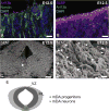
Fig. 1
Fig. 1
Primary cilia are present on radial glia at the ventricular zone of the embryonic ventral midbrain. Immunofluorescent staining (A and B) and scanning electron microscopy (SEM) (C and D) of cilia projecting into the ventral mesencephalic ventricle of wild-type E12.5 embryos. A and B, Primary cilia were detected using an antibody against Arl13b (A, magenta, B, green). Neural progenitors were labeled with antibodies against nestin (A, green) or BLBP (B, magenta). Blue, DAPI-labeled nuclei. C and D: arrow in C indicates the ventral ventricular surface of the midbrain shown in D; arrowheads in D indicate primary cilia. Scale bars: A and B, 10 µm; C, 500 µm; D, 1 µm. (E) Schematic representation of the location of mDA progenitors and neurons in the E12.5 midbrain.
Fig. 2
Fig. 2
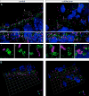
Super-resolution microscopy of primary cilia in the ventral midbrain of E11.5 wild-type and cobblestone embryos does not show major structural differences in the primary cilia or basal bodies. (A) Primary cilia were detected using an antibody against Arl13b (magenta). Basal bodies were detected using an antibody against γ-tubulin (green). Blue, Hoechst-labeled nuclei. Note that the number of visible nuclei does not reflect the total number of cells that make contact with the ventricular surface and extend primary cilia into the ventricle. The top image depicts a 30 µm (x) by 20 µm (y) maximum-intensity z-projection of a 7.25 µm-thick image stack. Bottom image is a maximum intensity y-projection of the corresponding xz slices of the recorded volumes. Higher magnifications of the boxed areas are shown in the bottom. Scale bar: 5 µm. (B) 3-dimensional reconstruction of the corresponding 30 × 20 × 7.25 µm3 (xyz) image stacks. The squares are 3 µm in length.
Fig. 3
Fig. 3
Disorganization of the mDA progenitor domain in cobblestone mutant mice. (A–F′, J–K′) RNA in situ hybridization for Shh (A–B′), Gli1 (C–D′), Gli3 (E–F′), and Cyclin D1 (J–K′) showing clusters of progenitor cells. (G) Schematic overview of the relevant gene expression domains in the wild-type ventral midbrain. (H–I′) Hoechst-labeled nuclei within the ventral midbrain show disorganization of the mDA progenitor domain in cobblestone mutant mice. (B′,D′–F′,H′–K′) are higher magnifications of the boxed areas in (B,D–F,H–K). Ve: Ventricle; PS: Pial surface. Scale bars: A–D,E′,F′,H,I,J′,K′, 100 µm; B′,D′,H′,I′, 20 µm; E,F,J,K, 200 µm.
Fig. 4
Fig. 4
Expression profiles of progenitor domains are intermingled in cobblestone mutants. (A and B) RNA in situ hybridization for Lmx1a on coronal midbrain sections of wild-type and cobblestone mutant embryos at E11.5. (A′ and B′) higher magnification of boxed areas in A,B, respectively. Inset in B’: Immunofluorescence for FoxA2 (magenta) and Nkx6-1 (green) on adjacent sections (B″). (C–D‴), Immunofluorescence for neural progenitor markers FoxA2 (magenta) and Nkx6-1 (green). C′–D‴, higher magnification of boxed areas in C, D. (E–H) RNA in situ hybridization for Axin2 (E,F) and Wnt1 (G,H) on coronal midbrain sections of wild-type and cobblestone mutant embryos at E9.5 (E,F) and E11.5 (G,H). (I and J) RNA in situ hybridization for Corin (green) combined with immunofluoresence for Lmx1a (magenta) (I,J) and RNA in situ hybridization for Arx (K,L) on coronal midbrain sections of wild-type and cobblestone mutant embryos at E10.5. Note that the image of the Corin RNA in situ hybridization was false colored and image color was inverted to visualize overlap with Lmx1a. Corin and Arx expression could be detected in the ventral midbrain in one of three E10.5 embryos, while Lmx1a expression was absent in all analyzed embryos. (M) Schematic overview of expression domains of FoxA2, Nkx6-1 and Lmx1a in the wild-type ventral midbrain. Scale bars: A, B, 200 µm; A′, B′, C, I–L, 40 µm, E–H, 100 µm.
Fig. 5
Fig. 5
Number of mDA neurons is severely reduced in cobblestone mutants. (A–D) RNA in situ hybridization for TH to visualize mDA neurons on sagittal sections of control and cobblestone mutant embryos at E12.5. (E–L) Immunofluorescent staining for TH on coronal midbrain sections in wild-type and cobblestone mutant embryos at E12.5. Co-staining with Lmx1a (G, H) and FoxA2 (I–L). (M, N) Immunofluorescent staining for TH and Nurr1 (M) or TH and Pitx3 (N) shows that mDA neurons surround rosette-like structures in the cobblestone mutant. (O) Immunofluorescent staining for primary cilia marker Arl13b and axoneme protein γ-tubulin shows that the primary cilia extend into the center of the rosette like structures. Blue, Hoechst-labeled nuclei. P, Schematic representation of FoxA2 and Lmx1a expression domains and localization of mDA neurons in the wild-type. Scale bars: A–F, I, J, 200 µm; G,H, K–N, 20 µm; O, 10 µm.
Fig. 6
Fig. 6
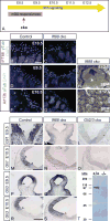
Conditional inactivation of Ift88 leads to a loss of primary cilia and impaired Shh signaling. (A) Schematic representation of the timeline of Shh signaling, mDA progenitor responsiveness to Shh signaling and timepoint of conditional gene inactivation (cko). (B–G) Immunofluorescent staining for Ift88 (B, C, magenta) or ciliary marker Arl13b (D–G, magenta) and γ-tubulin (B–G, green) at the ventricular surface of the ventral midbrain of control and Ift88 cko embryos at E9.5 or E10.5. Blue, Hoechst-labeled nuclei. (H,I) SEM of the ventral midbrain at E12.5 in Ift88 cko embryos. Arrowhead in H indicates the area imaged in I. In Ift88 cko embryos, primary cilia start to disappear at E9.5 and are lost completely at E10.5. (J–S) RNA in situ hybridization for Gli1 (J–O) and Gli3 (P–S) on control, Ift88 cko and Gli2/3 cko embryos at E9.5 (J–L,P,Q) and E10.5 (M–O, R,S). Black arrowheads indicate the domain in the control, red arrowheads indicate the changes in the expression domain in the Ift88 cko embryos. Similarly to Gli2/3 cko, Ift88 cko mutant embryos show a loss in Shh-responsiveness (Gli1 expression). (T) Western blot of protein lysates of E12.5 brain from wild-type (+/+) and cbbs/cbbs (−/−) embryos, using an anti-C-terminal Ift88 antibody.Scale bars: B–G, 10 µm; H, 500 µm; I, 1 µm; J–S, 200 µm.
Fig. 7
Fig. 7
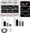
The size of the mDA progenitor domain is reduced in Ift88 cko and Gli2/3 cko mutant embryos. (A–L) RNA in situ hybridization for Shh (A–C), Wnt1 (J–L) and immunofluorescence staining for FoxA2 (D–F) and Lmx1a (G–I) on ventral midbrain sections of wild-type, Ift88 cko and Gli2/3 cko embryos at E10.5. The VZ is outlined. Arrowheads in C,E point to striped expression patterns of Shh and FoxA2 in the mutants. (M–O) Immunofluorescent staining for FoxA2 and Nkx6-1. The progenitor cells co-expressing FoxA2 and Nkx6-1 are absent in Ift88 cko and Gli2/3 cko embryos. (P,Q) Quantification of the size of the FoxA2- (P) and Lmx1a-positive (Q) domains in E10.5 control, Ift88 cko and Gli2/3 cko embryos. Values are means ± SD. The perimeter of the Lmx1a or Foxa2 positive domain was measured and normalized for the perimeter of the ventricle. ANOVA with Tukey’s multiple comparison test. n ≥ 3. Foxa2: F(2,13) = 12.63, Lmx1a: F(2,8) = 8.67, * p < 0.05, **p < 0.01, ***p < 0.001. (R) Schematic representation of expression domains of the markers given in A–O. Scale bars: 100 µm
Fig. 8
Fig. 8
mDA neurons are reduced in Ift88 cko and Gli2/3 cko mutant embryos. (A–F′) Immunofluorescent staining for TH and FoxA2 on control and Ift88 cko ventral midbrains at E13.5. (E′,F′), higher magnification of boxed areas in E,F. Yellow arrow indicates the loss of mDA neurons in the midline. (G–I′) Immunofluorescent staining for TH and FoxA2 on wild-type, Ift88 cko and Gli2/3 cko ventral midbrains at E18.5. (G’-I’) Higher magnification of boxed areas in G–I. Yellow arrow indicates the loss of mDA neurons in the midline. (J–M) Immunofluorescent staining for DAT and Pitx3 (J, K) and for Nurr1 (L, M) on E18.5 control and Ift88 cko ventral midbrain coronal sections. (N–Q) Immunofluorescent staining for TH and RNA in situ hybridization for Vmat2 and Aadc. Note that the image of the Vmat2 and Aadc RNA in situ hybridization signal was false colored and image color was inverted to visualize overlap with TH. (R, S) Quantification of TH-positive cells in control versus Ift88 cko (R) and control versus Gli2/3 cko brains at E18.5 (S). The number of TH-positive cells is significantly reduced in Ift88 cko and Gli2/3 cko embryos as compared to wild-type. Values are given as mean percentage from wild-type TH-positive cells ± SD. Student's t-test: Gli2/3 cko: n ≥ 3, t(6) = 2.60, Ift88 cko: n ≥ 3, t(7) = 4.99, *p < 0.05, **p < 0.01. TH-positive neurons were quantified in 14 µm frozen sections for Ift88 cko embryos and control littermates and in 7 µm paraffin sections for Gli2/3 cko embryos and control littermates. Thus, the results are represented independently. Scale bars: A–F, 100 µm; G–I, 200 µm; G′–J′, N–Q 40 µm; E′,F′, J–M, 20 µm.
Fig. 9
Fig. 9
Primary cilia are required for Shh pathway activation downstream of Smo. (A–K, M) RNA in situ hybridization for Gli1 (A,B), Shh (C, D), FoxA2 (E, F, K), Lmx1a (G, H, M) and Wnt1 (I, J) on SmoM2/Ift88 cko and SmoM2 ca mutant embryos at E10.5 (A–J) and E12.5 (K–P). (L, N–P) Immunofluorescent staining for FoxA2 (L), Lmx1a (N), and TH (O,P) on SmoM2/Ift88 cko and SmoM2 ca embryos at E12.5. (Q, R) Quantification of the size of the Lmx1a- (Q) and Foxa2-positive (R) domains in E10.5 Ift88 cko and SmoM2/Ift88 cko embryos. Values are means ± SD. The perimeter of the Lmx1a or Foxa2 positive domain was measured and normalized for the perimeter of the ventricle. Values are means ± SD. Student’s t-test. Foxa2 domain: n ≥ 3, t(5) = 0.90, Lmx1a domain: n ≥ 3, t(4) = 1.60. Constitutive activation of the Shh pathway results in the ventralization of the midbrain and an abnormal distribution of mDA progenitors and neurons. Conditional inactivation of Ift88 on the SmoM2 background (SmoM2/Ift88 cko) results in a phenotype similar to Ift88 cko embryos indicating that primary cilia are necessary for Shh pathway activation downstream of Smo. Compare with Figs. 7 and 8 for controls and Ift88 cko brains. Scale bars: A–J, 200 µm; K–P, 100 µm.
Movie 1
Movie 1
A 3-dimensional reconstruction of an image stack of Fig. 2A recorded by 3D SIM in super-resolution is shown rotating through 360 degrees. A video clip is available online.Supplementary material related to this article can be found online at http://dx.doi.org/10.1016/j.ydbio.2015.10.033.
Movie 2
Movie 2
A 3-dimensional reconstruction of an image stack of Fig. 2B recorded by 3D SIM in super-resolution is shown rotating through 360 degrees. A video clip is available online.Supplementary material related to this article can be found online at http://dx.doi.org/10.1016/j.ydbio.2015.10.033.

Similar articles

Cited by

References

    1. Abdelhamed ZA, Wheway G, Szymanska K, Natarajan S, Toomes C, Inglehearn C, Johnson CA. Variable expressivity of ciliopathy neurological phenotypes that encompass Meckel-Gruber syndrome and Joubert syndrome is caused by complex de-regulated ciliogenesis, Shh and Wnt signalling defects. Hum. Mol. Genet. 2013;22:1358–1372. http://dx.doi.org/10.1093/hmg/dds546. - DOI - PMC - PubMed
    1. Albin RL, Young AB, Penney JB. The functional-anatomy of basal ganglia disorders. Trends Neurosci. 1989;12:366–375. - PubMed
    1. Andersson E, Tryggvason U, Deng Q, Friling S, Alekseenko Z, Robert B, Perlmann T, Ericson J. Identification of intrinsic determinants of midbrain dopamine neurons. Cell. 2006;124:393–405. http://dx.doi.org/10.1016/j.cell.2005.10.037. - DOI - PubMed
    1. Andersson ER, Saltó C, Villaescusa JC, Cajanek L, Yang S, Bryjova L, Nagy II, Vainio SJ, Ramirez C, Bryja V. Wnt5a cooperates with canonical Wnts to generate midbrain dopaminergic neurons in vivo and in stem cells. Proc. Natl. Acad. Sci. USA. 2013;110:E602–E610. http://dx.doi.org/10.1073/pnas.1208524110/-/DCSupplemental. - DOI - PMC - PubMed
    1. Baala L, Romano S, Khaddour R, Saunier S, Smith UM, Audollent S, Ozilou C, Faivre L, Laurent N, Foliguet B, Munnich A, Lyonnet S, Salomon R, EnchaRazavi F, Gubler M-C, Boddaert N, de Lonlay P, Johnson CA, Vekemans M, Antignac C, Attié-Bitach T. The Meckel-Gruber syndrome gene, MKS3, is mutated in Joubert syndrome. Am. J. Hum. Genet. 2006;80:186–194. http://dx.doi.org/10.1086/510499. - DOI - PMC - PubMed

Publication types

MeSH terms

LinkOut - more resources