. 2016 Apr;137(4):1206-1215.e6.
doi: 10.1016/j.jaci.2015.09.013. Epub 2015 Nov 2.

Expansion of inflammatory innate lymphoid cells in patients with common variable immune deficiency

Affiliations

Expansion of inflammatory innate lymphoid cells in patients with common variable immune deficiency

Montserrat Cols et al. J Allergy Clin Immunol. 2016 Apr.

Abstract

Background: Common variable immunodeficiency (CVID) is an antibody deficiency treated with immunoglobulin; however, patients can have noninfectious inflammatory conditions that lead to heightened morbidity and mortality.

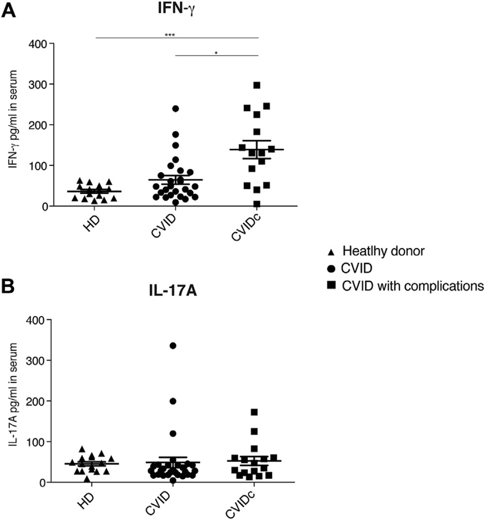
Objectives: Modular analyses of RNA transcripts in whole blood previously identified an upregulation of many interferon-responsive genes. In this study we sought the cell populations leading to this signature.

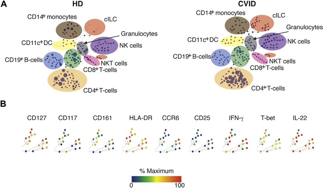
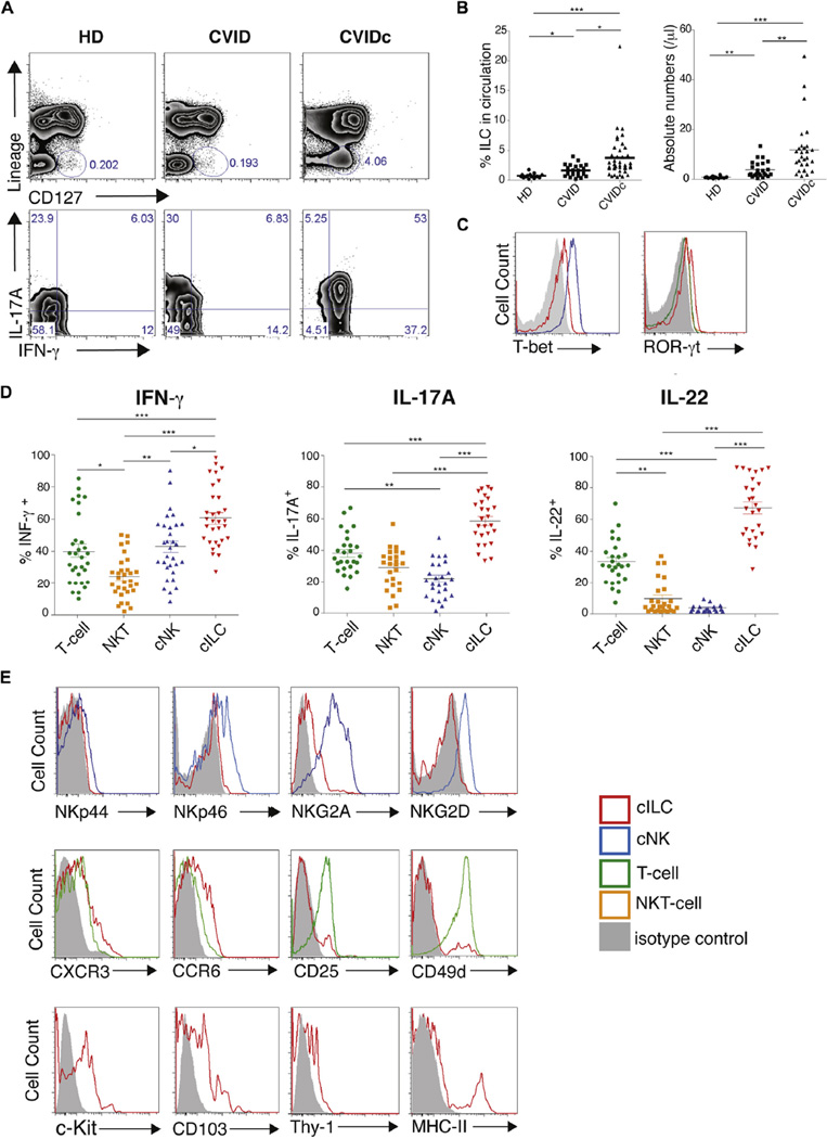
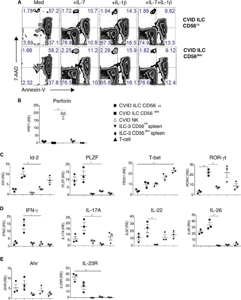
Methods: Lymphoid cells were measured in peripheral blood of 55 patients with CVID (31 with and 24 without inflammatory/autoimmune complications) by using mass cytometry and flow cytometry. Surface markers, cytokines, and transcriptional characteristics of sorted innate lymphoid cells (ILCs) were defined by using quantitative PCR. Gastrointestinal and lung biopsy specimens of subjects with inflammatory disease were stained to seek ILCs in tissues.

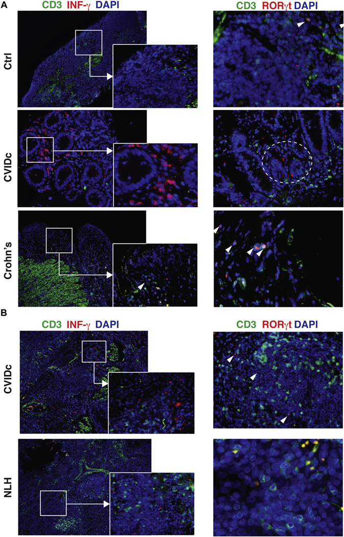
Results: The linage-negative, CD127(+), CD161(+) lymphoid population containing T-box transcription factor, retinoic acid-related orphan receptor (ROR) γt, IFN-γ, IL-17A, and IL-22, all hallmarks of type 3 innate lymphoid cells, were expanded in the blood of patients with CVID with inflammatory conditions (mean, 3.7% of PBMCs). ILCs contained detectable amounts of the transcription factors inhibitor of DNA binding 2, T-box transcription factor, and RORγt and increased mRNA transcripts for IL-23 receptor (IL-23R) and IL-26, demonstrating inflammatory potential. In gastrointestinal and lung biopsy tissues of patients with CVID, numerous IFN-γ(+)RORγt(+)CD3(-) cells were identified, suggesting a role in these mucosal inflammatory states.

Conclusions: An expansion of this highly inflammatory ILC population is a characteristic of patients with CVID with inflammatory disease; ILCs and the interferon signature are markers for the uncontrolled inflammatory state in these patients.

Keywords: Common variable immunodeficiency; inflammatory complications; innate lymphoid cells; mucosal disease.


Conflict of interest statement

Disclosure of potential conflict of interest: The rest of the authors declare that they have no relevant conflicts of interest.

Figures

FIG 1
FIG 1
CyTOF analyses. A, CyTOF was used to compare cell lineages in the blood of 4 patients with CVIDc compared with control subjects (HD). B, Lin cells positive for CD127 and CD161 were observed in samples from patients with CVID. Other markers tested are shown here. ILC populations are highlighted. Node color is scaled to the median intensity of marker expression. One representative experiment is displayed.
FIG 2
FIG 2
Immunophenotyping of circulating ILC3s. A, Fluorescence-activated cell sorting analyses show LinIFN-γ+ and IL-17A+CD127+ cells in peripheral blood in patients with CVIDc. B, ILC3s were significantly increased in these patients with CVID. C, Intracellular T-bet and RORγt of ILC3 populations (red), NK cells (blue), NK T cells (green), or T cells (orange). D, Percentages of IFN-γ+, IL17-A+, and IL-22+ immune cells in these patients with CVID. E, Fluorescence-activated cell sorting comparison of ILC3s from patients with CVID with NK cells (NKp44, NKp46, and NKG2D). Middle row, CXCR3, CCR6, CD25, and CD49d levels for ILC3s from patients with CVID to T cells. Bottom row, ILC3s from patients with CVID are c-Kit+, CD103+, and Thy-1+. Representative plots from 22 healthy control subjects, 31 patients with CVID with complications, and 24 patients with CVID without complications are shown in Fig 2, A, C, and E. For Fig 2, B and D: *P < .05, **P < .01, and ***P < .001 (1-way ANOVA). HD, Healthy donors.
FIG 3
FIG 3
CD56 expression and transcriptional features. A, Viability of sorted CD56hi and CD56lo ILC3s from patients with CVIDc cultured with medium alone or IL-7, IL-1β, or both × 5 days, as assessed by using fluorescence-activated cell sorting. B, RT-PCR for perforin (PRF1) mRNA expression of sorted cILC3s or NK cells from spleens of patients with CVID or control subjects. C, Quantitative RT-PCR analysis of mRNA for Id-2 (ID2), PLZF, T-bet (TBX21), and RORγt (RORC) in sorted ILC3s, NK cells, or CD3+ T cells. D, Quantitative RT-PCR mRNA encoding for IFN-γ, IL-17A, IL-22, and IL-26. Results were normalized and expressed as above (Fig 3, C). E, Quantitative RT-PCR mRNA for aryl hydrocarbon receptor and the cytokine receptor IL-23R. All RT-PCR results were normalized to β-actin mRNA expressed as relative expression (RE) to PBMCs. Data are from 3 experiments. For Fig 3, B–E: *P < .05, 1-way ANOVA. Error bars = SEMs.
FIG 4
FIG 4
ILC3s are detected in mucosal tissues. A, Ileal biopsy specimens of a patient with CVID with enteropathy were compared with tissue from a nonimmunodeficient donor (top) and a patient with Crohn disease (bottom). Tissue was stained for CD3+ T cells (green), IFN-γ (left panels), and RORc (right panels; red) and counterstained with 4′,6-diamidino-2-phenylindole dihydrochloride (DAPI) for nuclei (blue). Solid squares, Area of magnification; dashed circles, IFN-γ+ and RORc+ CD3 cells. CD3+RORc+ double-positive T cells (arrowheads) in noninflamed control ileum (upper rightmost panel) and T cells in patients with Crohn disease. Magnification ×10; inset magnification ×40. Magnification ×20 and ×40 for Crohn disease. B, Lung biopsy specimen of a patient with CVID with lymphoid hyperplasia (CVIDc). The control subject was a nonimmunodeficient patient with nodular lymphoid hyperplasia. Tissue was stained as in Fig 4, A. Magnification ×10; inset magnification ×40. Right panel magnification ×20 and ×40 for nodular lymphoid hyperplasia. Data are from one 4 experiments.
FIG 5
FIG 5
Cytokine profile of patients with CVID. IFN-γ (A) and IL-17A (B) levels in serum, as measured by using ELISA. *P < .05 and ***P < .001, 1-way ANOVA, followed by the Dunn multiple comparison post hoc test). Data for 15 healthy subjects (HD), 26 patients with CVID without inflammatory complications, and 16 patients with CVID with such complications were pooled for Fig 5, B. Bars denote means with SEMs.

References

    1. Al-Herz W, Bousfiha A, Casanova JL, Chatila T, Conley ME, Cunningham-Rundles C, et al. Primary immunodeficiency diseases: an update on the classification from the international union of immunological societies expert committee for primary immunodeficiency. Front Immunol. 2014;5:162. - PMC - PubMed
    1. Warnatz K, Voll RE. Pathogenesis of autoimmunity in common variable immunodeficiency. Front Immunol. 2012;3:210. - PMC - PubMed
    1. Quinti I, Soresina A, Spadaro G, Martino S, Donnanno S, Agostini C, et al. Long-term follow-up and outcome of a large cohort of patients with common variable immunodeficiency. J Clin Immunol. 2007;27:308–316. - PubMed
    1. Wehr C, Kivioja T, Schmitt C, Ferry B, Witte T, Eren E, et al. The EUROclass trial: defining subgroups in common variable immunodeficiency. Blood. 2008;111:77–85. - PubMed
    1. Resnick ES, Moshier EL, Godbold JH, Cunningham-Rundles C. Morbidity and mortality in common variable immune deficiency over 4 decades. Blood. 2012;119:1650–1657. - PMC - PubMed

Publication types

MeSH terms