. 2015 Nov 6:5:16204.
doi: 10.1038/srep16204.

Pachychoroid neovasculopathy and age-related macular degeneration

Affiliations

Pachychoroid neovasculopathy and age-related macular degeneration

Masahiro Miyake et al. Sci Rep. .

Abstract

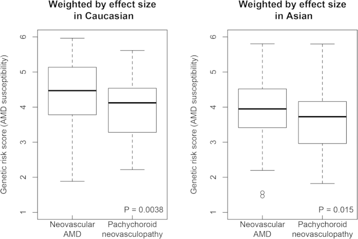
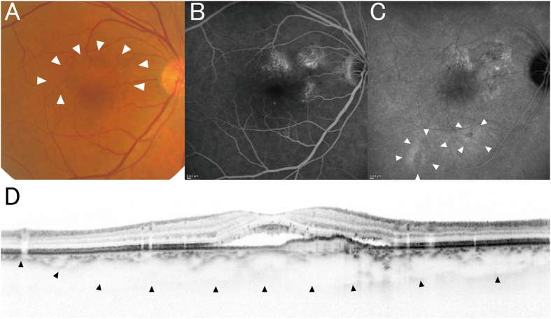
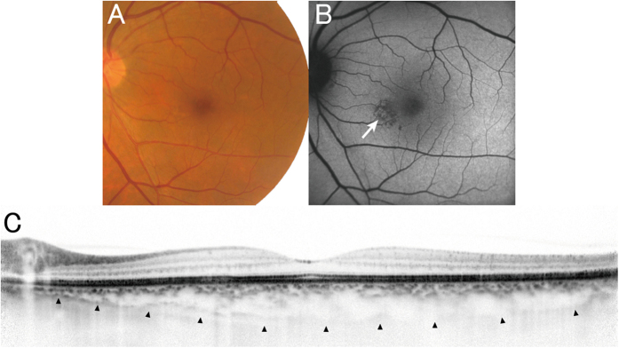
Pachychoroid neovasculopathy is a recently proposed clinical entity of choroidal neovascularization (CNV). As it often masquerades as neovascular age-related macular degeneration (AMD), it is currently controversial whether pachychoroid neovasculopathy should be distinguished from neovascular AMD. This is because its characteristics have yet to be well described. To estimate the relative prevalence of pachychoroid neovasculopathy in comparison with neovascular AMD and to investigate the phenotypic/genetic differences of the two diseases, we evaluated 200 consecutive Japanese patients who agreed to participate in the genetic study and diagnosed with pachychoroid neovasculopathy or neovascular AMD. Pachychoroid neovasculopathy was observed in 39 individuals (19.5%), which corresponds to one fourth of neovascular AMD. Patients with pachychoroid neovasculopathy were significantly younger (p = 5.1 × 10(-5)) and showed a greater subfoveal choroidal thickness (p = 3.4 × 10(-14)). Their genetic susceptibility to AMD was significantly lower than that of neovascular AMD; ARMS2 rs10490924 (p = 0.029), CFH rs800292 (p = 0.013) and genetic risk score calculated from 11 AMD susceptibility genes (p = 3.8 × 10(-3)). Current results implicate that the etiologies of the two conditions must be different. Thus, it will be necessary to distinguish these two conditions in future studies.


Figures

Figure 1
Figure 1. Distribution of genetic risk score calculated from 11 SNPs of 11 AMD susceptibility genes.
We constructed a multi-locus genetic risk score by summing up the number of risk alleles of each single nucleotide polymorphism (SNP), weighted by its reported effect size (log odds ratio, OR). We evaluated both genetic risk score using effect size in Caucasian (A) and that using effect size in Asian (B). The effect sizes applied in this analysis are summarized in Supplementary Table 2. Pachychoroid neovasculopathy patients had significantly lower genetic risk scores for AMD than did neovascular AMD patients.
Figure 2
Figure 2. Kaplan-Meier curve for retreatment-free period after loading dose of ranibizumab.
We selected study participants who had received a loading dose of ranibizumab (3 × one-monthly injections, 0.5 mg), been treated as needed afterwards, and been followed up for more than 12 months. The duration from the third ranibizumab injection to the administration of additional treatment (event), or to the final visit by June 2014, or loss to follow up) was reported on a daily basis. A log-rank test revealed that the curves for pachychoroid neovasculopathy and neovascular AMD differed significantly from one another (p = 0.0095). Pachychoroid neovasculopathy showed longer retreatment-free period.
Figure 3
Figure 3
(A) Choroidal vascular hyperpermeability. Choroidal vascular hyperpermeability is evidenced by focal areas of hyperfluorescence, which appear during the middle phase of indocyanine green angiography (IA) and expand over time, eventually forming a ring shape (arrowheads). The center of the initially hyperfluorescent area becomes hypofluorescent during the late phase. (B) Retinal pigment epithelium abnormality. Retinal pigment epithelium (RPE) abnormality is seen as patchy areas of granular hypoautofluorescence with occasional discrete hyperautofluorescent specks scattered throughout the fundus autofluorescence (FAF) images (arrowheads). However, this was not considered indicative of RPE abnormality when this finding was adjacent to a choroidal neovascularization (CNV; arrows) lesion.
Figure 4
Figure 4. A case of pachychoroid neovasculopathy.
A 50-year-old man visited the macular service at Kyoto University Hospital with a chief complaint of central scotoma in his right eye. (A,D) The color fundus photograph from the patient’s first visit shows serous retinal detachment without drusen. Fluorescein angiography (FA) shows late leakage (ink blot pattern; white arrow). Indocyanine green angiography (IA) image shows no choroidal neovascularization (CNV). Optical coherence tomography (OCT; vertical scan thorough the center of the fovea) shows serous retinal detachment and an almost flat retinal pigment epithelium (RPE) band. All these findings were compatible with a diagnosis of central serous chorioretinopathy (CSC). (E) Four months after the patient’s first visit: OCT shows persistent serous retinal detachment. (B,F) Ten months after the patient’s first visit: CNV is not apparent in either FA or IA. However, there are several protrusions in the OCT image. (G–I) 2.5 yr (G), 4.5 yr (H), and 6.5 yr (I) after the patient’s first visit. After spontaneous resolution of subretinal fluid, it again increased. The RPE band gradually elevated, suggesting the development of CNV (red arrows). (C,J) Seven years after the patient’s first visit, CNV is apparent in both FA/IA (white arrowheads) and OCT (red arrow) images. Choroid is thick all over the macula. Color fundus photography contains no drusen.
Figure 5
Figure 5. A case of pachychoroid neovasculopathy.
A 68-year-old male was visually impaired in the right eye (best-corrected visual acuity = 30/20). (A) Color fundus photograph shows serous retinal detachment (arrowheads) without drusen. (B) FA suggests occult CNV. (C) Late-phase IA shows choroidal vascular hyperpermeability (arrowheads). (D) Enhanced depth imaging OCT (vertical scan thorough the center of the fovea) reveals type 1 CNV and subretinal fluid. Choroid is thick throughout the macula, and subfoveal choroidal thickness was measured as 353 μm. He had GT genotype at ARMS2 A69S (rs10490924) and AG genotype at CFH I62V (rs800292).
Figure 6
Figure 6. Pachychoroid pigment epitheliopathy observed in the fellow eye of the patient in Fig. 5.
(A) Color fundus photography shows no drusen. (B) FAF shows RPE abnormality associated with choroidal vascular dilation (arrow), but without hyperautofluorescent lesions, which would suggest the previous presence of subretinal fluid. (C) Enhanced depth imaging OCT (horizontal scan thorough the center of the fovea) shows thick choroid throughout the macula, and subfoveal choroidal thickness was 335 μm. All these findings are compatible with a diagnosis of pachychoroid pigment epitheliopathy.

References

    1. Kawasaki R. et al. Prevalence and risk factors for age-related macular degeneration in an adult Japanese population: the Funagata study. Ophthalmology 115, 1376–81, 1381 e1-2 (2008). - PubMed
    1. Kawasaki R. et al. The prevalence of age-related macular degeneration in Asians: a systematic review and meta-analysis. Ophthalmology 117, 921–7 (2010). - PubMed
    1. Varma R., Fraser-Bell S., Tan S., Klein R. & Azen S. P. Prevalence of age-related macular degeneration in Latinos: the Los Angeles Latino eye study. Ophthalmology 111, 1288–97 (2004). - PubMed
    1. Edwards A. O. et al. Complement factor H polymorphism and age-related macular degeneration. Science 308, 421–4 (2005). - PubMed
    1. Haines J. L. et al. Complement factor H variant increases the risk of age-related macular degeneration. Science 308, 419–21 (2005). - PubMed

Publication types