Review
. 2015 Dec;8(12):1495-515.
doi: 10.1242/dmm.022103. Epub 2015 Nov 5.

Non-muscle myosin II in disease: mechanisms and therapeutic opportunities

Affiliations
Review

Non-muscle myosin II in disease: mechanisms and therapeutic opportunities

Karen A Newell-Litwa et al. Dis Model Mech. 2015 Dec.

Abstract

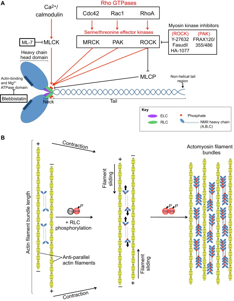
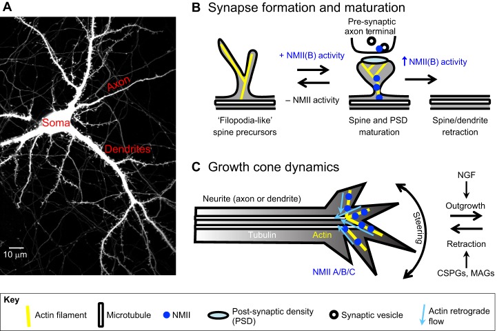
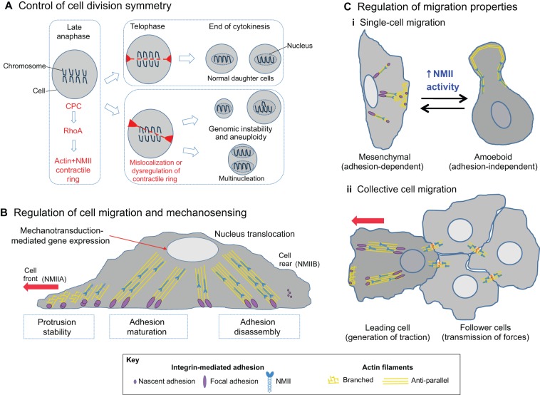
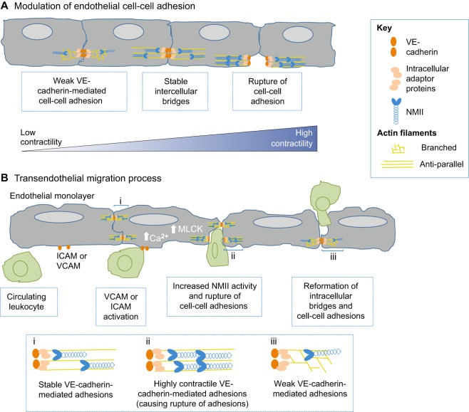
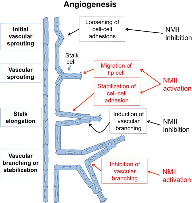
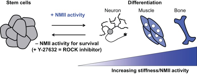
The actin motor protein non-muscle myosin II (NMII) acts as a master regulator of cell morphology, with a role in several essential cellular processes, including cell migration and post-synaptic dendritic spine plasticity in neurons. NMII also generates forces that alter biochemical signaling, by driving changes in interactions between actin-associated proteins that can ultimately regulate gene transcription. In addition to its roles in normal cellular physiology, NMII has recently emerged as a critical regulator of diverse, genetically complex diseases, including neuronal disorders, cancers and vascular disease. In the context of these disorders, NMII regulatory pathways can be directly mutated or indirectly altered by disease-causing mutations. NMII regulatory pathway genes are also increasingly found in disease-associated copy-number variants, particularly in neuronal disorders such as autism and schizophrenia. Furthermore, manipulation of NMII-mediated contractility regulates stem cell pluripotency and differentiation, thus highlighting the key role of NMII-based pharmaceuticals in the clinical success of stem cell therapies. In this Review, we discuss the emerging role of NMII activity and its regulation by kinases and microRNAs in the pathogenesis and prognosis of a diverse range of diseases, including neuronal disorders, cancer and vascular disease. We also address promising clinical applications and limitations of NMII-based inhibitors in the treatment of these diseases and the development of stem-cell-based therapies.

Keywords: Migration; Myosin; NMII; Stem cell; Synapse.


Conflict of interest statement

Competing interests

The authors declare no competing or financial interests.

Figures

Fig. 1.
Fig. 1.
The structure of NMII and its regulation by serine/threonine kinases. (A) NMII consists of a heavy chain, which includes a globular head domain that binds both actin and ATP; a neck region, which binds both the essential and regulatory light chains (ELC and RLC, respectively); and a tail region, which homodimerizes in a helical fashion, as well as a non-helical tail region that directs the subcellular localization of the NMII isoform. Serine/threonine kinases regulate NMII activity by phosphorylation of the myosin RLC on residues Thr18 and Ser19. These kinases function downstream of small Rho GTPases, as well as downstream of Ca2+/calmodulin signaling pathways. This figure also indicates pharmacological inhibitors of myosin regulatory kinases that can be used to modulate NMII activity. (B) NMII filaments associate with each other in an anti-parallel fashion, allowing them to crosslink and slide actin filaments past each other. RLC Ser19 phosphorylation increases NMII ATPase activity, leading to contraction of actin filament bundles, and phosphorylation of both Ser19 and Thr18 increases NMII ATPase activity, driving the association of multiple actin filaments into actomyosin filament bundles, often referred to as stress fibers. MLCK, myosin light chain kinase; MRCK, myotonic dystrophy kinase-related Cdc42-binding kinase; PAK, p21-associated kinase; ROCK, RhoA-associated kinase; MLCP, myosin light chain phosphatase.
Fig. 2.
Fig. 2.
NMII regulates neuronal plasticity. (A) Confocal image of a GFP-expressing primary rat hippocampal neuron, highlighting the cell body, or soma, and processes, including post-synaptic dendrites, which form spines, and pre-synaptic axons, which form axon terminals containing synaptic vesicles. (B) NMII drives dynamic changes in neuronal morphology, including changes in dendritic spine formation and maturation, driven primarily by the isoform NMIIB. At the post-synaptic spine, NMII drives changes in actin organization that regulate spine and post-synaptic density (PSD) morphology and size, whereas, on the pre-synaptic side, NMII participates in synaptic vesicle recycling. The absence or inhibition of NMIIB activity results in dynamic ‘filopodia-like’ spine precursors and prevents spine maturation. In contrast, NMIIB activity drives spine and PSD maturation, although further NMIIB activity might result in spine and even dendrite retraction. (C) At the growth cone, all three NMII isoforms are present, and regulate substrate attachment and actin retrograde flow underlying neurite outgrowth. NGF, nerve growth factor; CSPGs, chondroitin sulfate proteoglycans; MAGs, myelin-associated glycoproteins.
Fig. 3.
Fig. 3.
NMII drives cancer cell progression. (A) NMII in cell division. At the end of anaphase during mitosis, the chromosomal passenger complex (CPC) induces RhoA-mediated formation of an actin-NMII contractile ring (red), resulting in cytokinesis and division into two daughter cells (upper box; telophase). However, mislocalization of the contractile ring (lower box) can result in genetic abnormalities that are linked to cancer, such as aneuploidy or multinucleation. (B) NMII in cell migration and metastasis. During adhesion-dependent cell migration (see Box 1), cells polymerize actin at the cell front (unaligned yellow lines), while integrin-based adhesions (purple circles) mediate attachment to the extracellular matrix. NMIIA (blue) generates forces that alter actin organization at the cell front and initiate adhesion maturation (purple ellipses indicate adhesion elongation). NMIIB (blue) is involved in the formation of stress fibers (aligned yellow lines), nucleus translocation and in the detachment of adhesions at the cell rear. These mechanical forces induced by NMII can also influence other biochemical pathways (mechanotransduction) that are able to modify cell behavior. (C) NMII participates in various modes of cell migration. (i) Invasion of a single tumor cell is either mesenchymal (adhesion-dependent) or amoeboid (adhesion-independent). For mesenchymal-like, single-cell migration, myosin regulates the migration process as described in B; for amoeboid-like single-cell migration, increased NMII-mediated tension affects the cortical actin network (Ruprecht et al., 2015), allowing for adhesion-independent migration through porous matrices. (ii) During collective cell migration, the leading cell generates NMII-mediated traction forces that are propagated to the follower cells through cell–cell adhesions (see Fig. 4 for details).
Fig. 4.
Fig. 4.
NMII regulates vascular biology and disease. (A) NMII remodels endothelial adherens junctions. VE-cadherin (orange ovals) mediates intercellular links between neighboring cells. Intracellularly, catenins (beige ovals) link VE-cadherin with the actin cytoskeleton (yellow lines). Myosin (blue) generates forces that stabilize intercellular bridges. However, enhanced NMII-mediated contractility can lead to the rupture of cell-cell adhesions. (B) NMII facilitates transendothelial migration. During rolling, leukocytes (green) interact with ICAM or VCAM receptors at the membrane of endothelial cells, resulting in an influx of Ca2+. At the adherens junctions at the cell border, increased NMII activity mediated by MLCK results in the rupture of cell–cell adhesions. After leukocytes squeeze and pass by the border of the two endothelial cells (paracellular), there is a neo-formation of VE-cadherin. ICAM, intercellular adhesion molecule; VCAM, vascular cell adhesion molecule; MLCK, myosin light chain kinase.
Fig. 5.
Fig. 5.
Differential NMII activity defines distinct stages of angiogenesis. After the detection of stimuli (hypoxia or growth factors), NMII inhibition results in the loosening of endothelial adherens junctions, favoring the detachment of migrating endothelial cells (tip cell). The tip cell migrates into the connective tissue, in a mechanism that relies on NMII activation (see Fig. 3B). During collective cell migration (stalk elongation), NMII activation stabilizes the junctions of the follower endothelial cells, resulting in stalk elongation. During stalk elongation, the localized activation or inhibition of NMII results in the inhibition or induction of vascular branching, respectively.
Fig. 6.
Fig. 6.
NMII determines stem cell fate. Pluripotent stem cells are cultured in the presence of the ROCK inhibitor Y-27632 to prevent apoptosis (Chen et al., 2010; Watanabe et al., 2007). NMII activity directs the differentiation of stem cells to specific tissue lineages: decreased NMII activity leads to neuronal fates, whereas increased NMII activity promotes the formation of stiffer tissues, such as muscle and bone (Engler et al., 2006; Seo et al., 2014; Wang et al., 2013b).

References

    1. Abraham S., Yeo M., Montero-Balaguer M., Paterson H., Dejana E., Marshall C. J. and Mavria G. (2009). VE-cadherin-mediated cell-cell interaction suppresses sprouting via signaling to MLC2 phosphorylation. Curr. Biol. 19, 668-674. 10.1016/j.cub.2009.02.057 - DOI - PubMed
    1. Adyshev D. M., Moldobaeva N., Mapes B., Elangovan V. and Garcia J. G. N. (2013). MicroRNA regulation of nonmuscle myosin light chain kinase expression in human lung endothelium. Am. J. Respir. Cell Mol. Biol. 49, 58-66. 10.1165/rcmb.2012-0397OC - DOI - PMC - PubMed
    1. Alon U., Barkai N., Notterman D. A., Gish K., Ybarra S., Mack D. and Levine A. J. (1999). Broad patterns of gene expression revealed by clustering analysis of tumor and normal colon tissues probed by oligonucleotide arrays. Proc. Natl. Acad. Sci. USA 96, 6745-6750. 10.1073/pnas.96.12.6745 - DOI - PMC - PubMed
    1. Amano M., Ito M., Kimura K., Fukata Y., Chihara K., Nakano T., Matsuura Y. and Kaibuchi K. (1996). Phosphorylation and activation of myosin by Rho-associated kinase (Rho-kinase). J. Biol. Chem. 271, 20246-20249. 10.1074/jbc.271.34.20246 - DOI - PubMed
    1. Arozarena I., Sanchez-Laorden B., Packer L., Hidalgo-Carcedo C., Hayward R., Viros A., Sahai E. and Marais R. (2011). Oncogenic BRAF induces melanoma cell invasion by downregulating the cGMP-specific phosphodiesterase PDE5A. Cancer Cell 19, 45-57. 10.1016/j.ccr.2010.10.029 - DOI - PubMed

Publication types