Review
. 2016 Feb;73(4):757-74.
doi: 10.1007/s00018-015-2086-9. Epub 2015 Nov 5.

Epigenesis and plasticity of mouse trophoblast stem cells

Affiliations
Review

Epigenesis and plasticity of mouse trophoblast stem cells

Julie Prudhomme et al. Cell Mol Life Sci. 2016 Feb.

Abstract

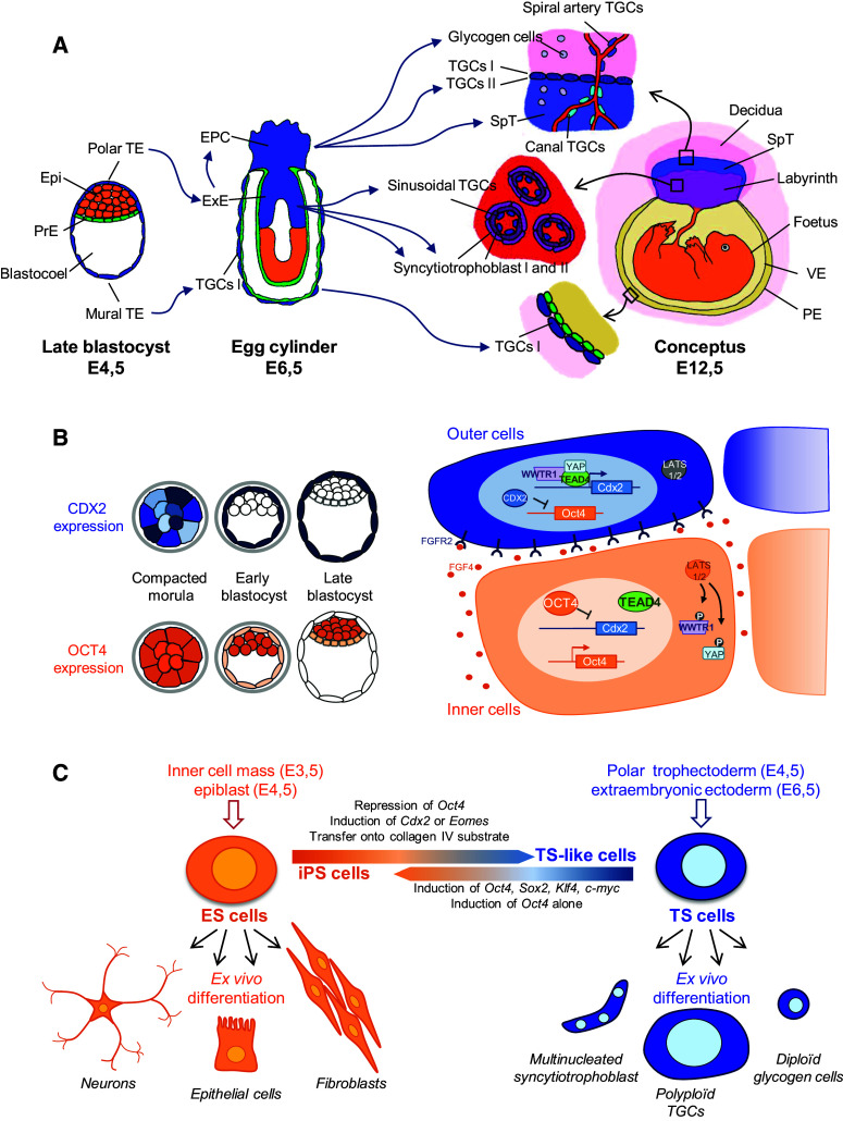
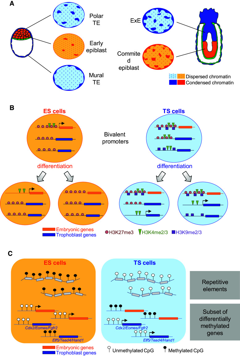
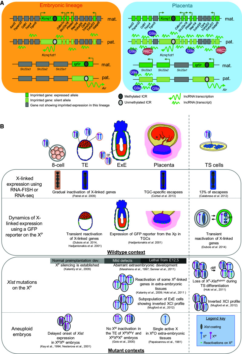
The critical role of the placenta in supporting a healthy pregnancy is mostly ensured by the extraembryonic trophoblast lineage that acts as the interface between the maternal and the foetal compartments. The diverse trophoblast cell subtypes that form the placenta originate from a single layer of stem cells that emerge from the embryo when the earliest cell fate decisions are occurring. Recent studies show that these trophoblast stem cells exhibit extensive plasticity as they are capable of differentiating down multiple pathways and are easily converted into embryonic stem cells in vitro. In this review, we discuss current knowledge of the mechanisms and control of the epigenesis of mouse trophoblast stem cells through a comparison with the corresponding mechanisms in pluripotent embryonic stem cells. To illustrate some of the more striking manifestations of the epigenetic plasticity of mouse trophoblast stem cells, we discuss them within the context of two paradigms of epigenetic regulation of gene expression: the imprinted gene expression of specific loci and the process of X-chromosome inactivation.

Keywords: Epigenetic plasticity; Mouse extraembryonic development; Multipotency; Placenta; Trophoblast stem cells.


Figures

Fig. 1
Fig. 1
Phenotypic plasticity of the mouse trophoblast lineage. a The development of the trophoblast lineage highlights the diversity of cell phenotypes among specialised subtypes. TE trophectoderm, Epi epiblast, PrE primitive endoderm, EPC ectoplacental cone, ExE extraembryonic ectoderm, TGC trophoblast giant cell, SpT spongiotrophoblast, VE visceral endoderm, PE parietal endoderm. b Progressive restriction of CDX2 and OCT4 expression, respectively, in outer and inner cells at the morula–blastocyst transition (left panel) involves a cascade of regulation mediated by the Hippo pathway, FGF signalling and OCT4/CDX2 mutual repression (right panel). c ES and TS cells derived from the blastocyst lineages and maintained in culture can be differentiated or transconverted into the other lineage
Fig. 2
Fig. 2
Characteristics of the epigenome of mouse TS and ES cells. a Global chromatin architecture of the embryonic and of the trophoblast lineages in vivo. Pluripotent cells of the early epiblast are characterised by a widely dispersed chromatin whereas committed epiblast cells and cells of the extraembryonic ectoderm (ExE) display large domains of condensed chromatin. Cells of the polar trophectoderm (TE) exhibit an intermediate chromatin architecture, which is markedly distinct from the chromatin structure of committed cells of the mural TE. b Characteristics of bivalent promoters in ES and TS cells. Bivalent promoters in ES cells carry both the repressive H3K27me3 histone mark and the permissive H3K4me2/3 histone marks. Upon differentiation, these marks resolve into H3K27me3 at repressed trophoblast-specific gene promoters and into H3K4me2/3 at the promoter of genes specific of each differentiated lineage. In the trophoblast lineage an additional repressive histone mark, H3K9me2/3, comes into play. It participates in the initial repression of developmental genes at the TS cell state and, upon TS cell differentiation, in the repression of specific genes in each trophoblast subtype. c DNA methylation profiles in ES and in TS cells. Repetitive elements are hypomethylated in TS cells and hypermethylated in ES cells. The majority of gene promoters are undermethylated in ES cells specifically. However, differential methylation is observed at a number of lineage-specific genes
Fig. 3
Fig. 3
Trophoblast-specific features of imprinted gene expression and X-chromosome inactivation in the mouse. a Distinct regulations of the Kcnq1 and of the Igf2r imprinted gene clusters in the placenta compared to the embryonic lineages. At both clusters, differential methylations of the imprinted control regions (ICR) control the mono-allelic expression of a lncRNA which, in turn, regulates the allelic expression of protein coding genes. The association of the lcnRNAs Kncnq1ot1 and Air extends to additional placenta-specific imprinted genes of the Kcnq1 and of the Igf2r loci, respectively in the placenta compared to embryonic lineages. This association is accompanied by extraembryonic-specific recruitment of histone modifiers G9a and PRC2 (polycomb repressive complex 2). b Main features characterising the plasticity of X-chromosome inactivation in the mouse trophoblast lineage and in mouse TS cells. The upper panel summarises observations made in wildtype mouse embryos and the lower panel summarises observations made in various mutant contexts. References to the ad hoc studies are indicated. X P paternal X chromosome, X M maternal X-chromosome, TE trophectoderm, ExE extraembryonic ectoderm, TGC trophoblast giant cells, XCI X-chromosome inactivation

Similar articles

Cited by

References

    1. Rossant J, Tam PP. Blastocyst lineage formation, early embryonic asymmetries and axis patterning in the mouse. Development. 2009;136(5):701–713. doi: 10.1242/dev.017178. - DOI - PubMed
    1. Saiz N, Plusa B. Early cell fate decisions in the mouse embryo. Reproduction. 2013;145(3):R65–R80. doi: 10.1530/REP-12-0381. - DOI - PubMed
    1. Cross JC, et al. Trophoblast functions, angiogenesis and remodeling of the maternal vasculature in the placenta. Mol Cell Endocrinol. 2002;187(1–2):207–212. doi: 10.1016/S0303-7207(01)00703-1. - DOI - PubMed
    1. Georgiades P, Ferguson-Smith AC, Burton GJ. Comparative developmental anatomy of the murine and human definitive placentae. Placenta. 2002;23(1):3–19. doi: 10.1053/plac.2001.0738. - DOI - PubMed
    1. John R, Hemberger M. A placenta for life. Reprod Biomed Online. 2012;25(1):5–11. doi: 10.1016/j.rbmo.2012.03.018. - DOI - PubMed

Publication types

LinkOut - more resources