. 2015 Nov 6:5:16255.
doi: 10.1038/srep16255.

Highly efficient radiosensitization of human glioblastoma and lung cancer cells by a G-quadruplex DNA binding compound

Affiliations

Highly efficient radiosensitization of human glioblastoma and lung cancer cells by a G-quadruplex DNA binding compound

Patrick Merle et al. Sci Rep. .

Abstract

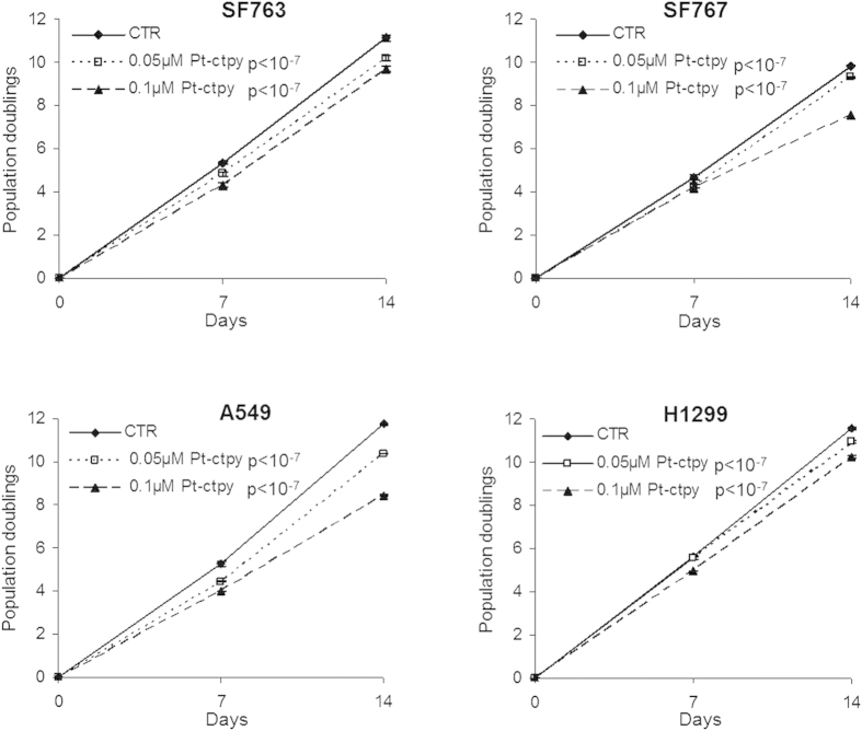
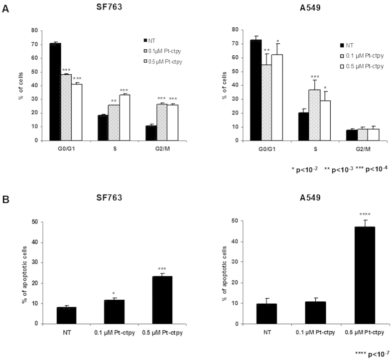
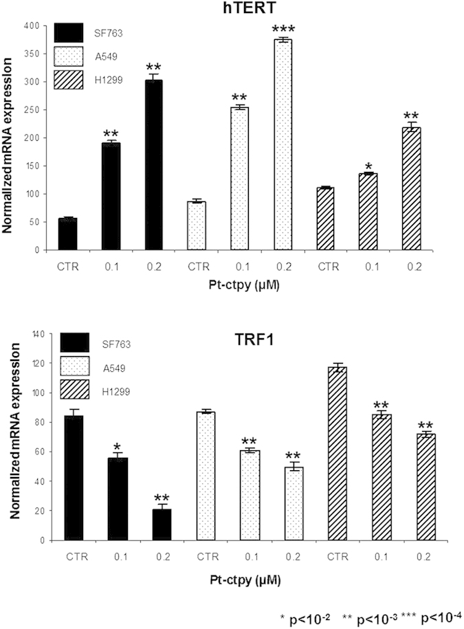
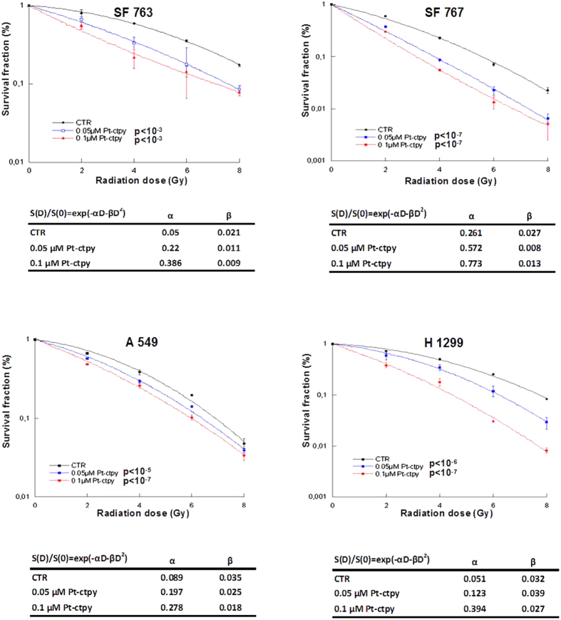
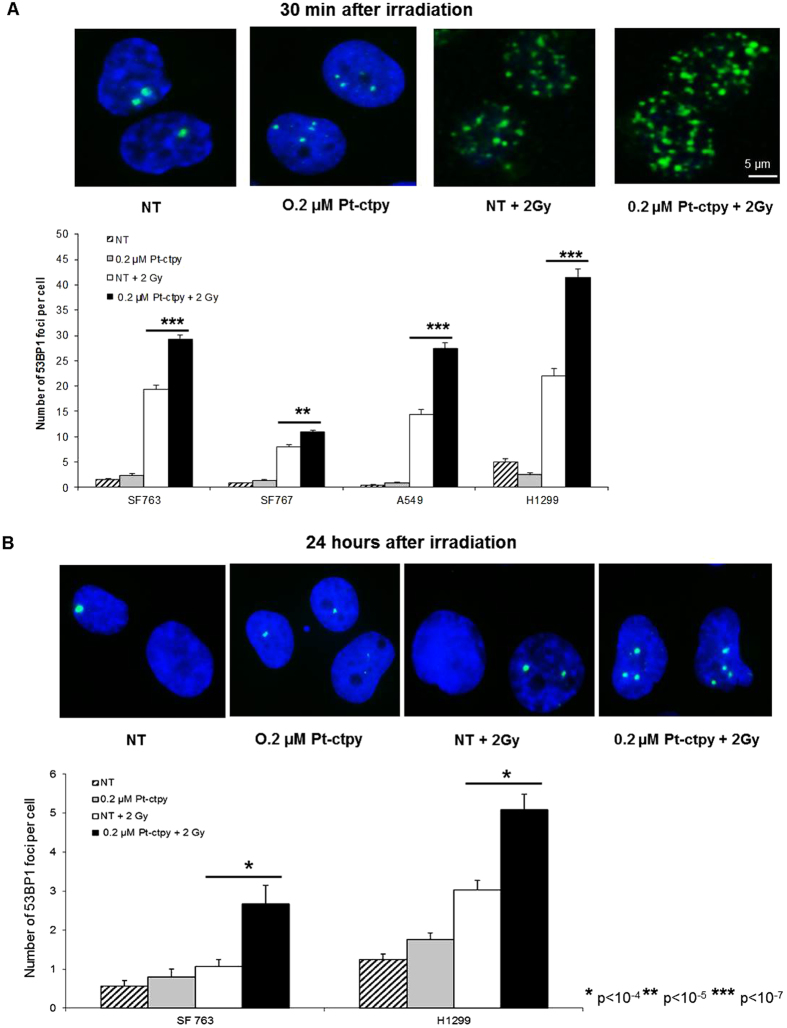
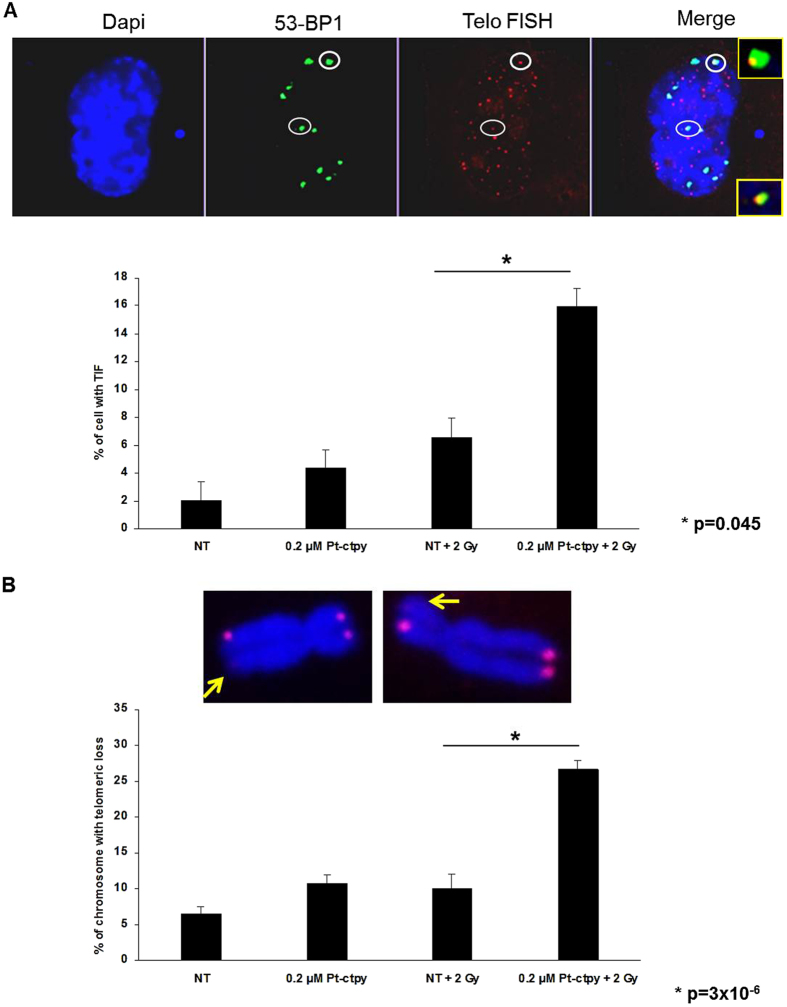
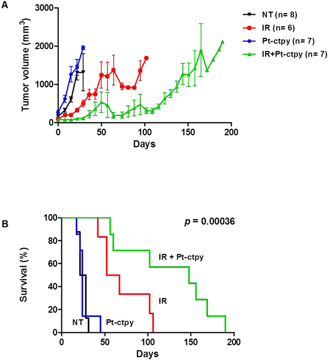
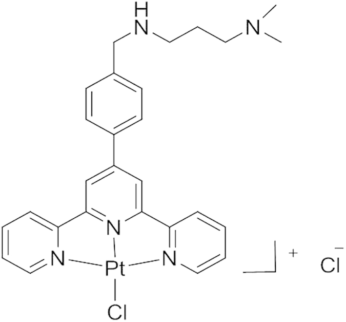
Telomeres are nucleoprotein structures at the end of chromosomes which stabilize and protect them from nucleotidic degradation and end-to-end fusions. The G-rich telomeric single-stranded DNA overhang can adopt a four-stranded G-quadruplex DNA structure (G4). Stabilization of the G4 structure by binding of small molecule ligands enhances radiosensitivity of tumor cells, and this combined treatment represents a novel anticancer approach. We studied the effect of the platinum-derived G4-ligand, Pt-ctpy, in association with radiation on human glioblastoma (SF763 and SF767) and non-small cell lung cancer (A549 and H1299) cells in vitro and in vivo. Treatments with submicromolar concentrations of Pt-ctpy inhibited tumor proliferation in vitro with cell cycle alterations and induction of apoptosis. Non-toxic concentrations of the ligand were then combined with ionizing radiation. Pt-ctpy radiosensitized all cell lines with dose-enhancement factors between 1.32 and 1.77. The combined treatment led to increased DNA breaks. Furthermore, a significant radiosensitizing effect of Pt-ctpy in mice xenografted with glioblastoma SF763 cells was shown by delayed tumor growth and improved survival. Pt-ctpy can act in synergy with radiation for efficient killing of cancer cells at concentrations at which it has no obvious toxicity per se, opening perspectives for future therapeutic applications.


Figures

Figure 1
Figure 1. Effects of Pt-ctpy on proliferation of GBM (SF763, SF767) and NSCLC cells lines (A549, H1299).
Cells were treated continuously with Pt-ctpy (0.05 and 0.1 μM) for 14 days and compared with non-treated cells (NT). Population doublings of the cell lines are shown. Mean ± SE values of 3 independent experiments, each performed in triplicate, are presented. Pt-ctpy induced a significant dose-dependent proliferation inhibition in both cell lines (ANOVA).
Figure 2
Figure 2. Effects of Pt-ctpy on cell cycle and apoptosis.
(A) Cell cycle analysis: distribution of cells in the different cell cycle phases. SF763 and A549 cells exposed to Pt-ctpy showed significant modifications in this distribution. (B) Flow cytometry assessment of sub-G0/G1 fraction, corresponding to apoptotic cells. Tumor cells treated with Pt-ctpy exhibited a significant increase in Sub-G0/G1 population (t-test).
Figure 3
Figure 3. Pt-ctpy effects on hTERT and TRF1 mRNA expression levels.
Quantitative real-time RT-PCR results for the expression of hTERT and telomere-related genes TRF1. In both cell lines, the treatment with Pt-ctpy led to an increase in the expression of hTERT (A), whereas the expression of TRF1 (B) was decreased after Pt-ctpy treatment (t-test).
Figure 4
Figure 4. Pt-ctpy radiosensitizes GBM and NSCLC cells.
After continuous treatment with 0.05 μM and 0.1 μM Pt-ctpy for one week and/or X-ray irradiation with doses ranging from 2 to 8 Gy, survival of GBM and NSCLC were determined using colony formation assay. Mean ± SE values of 3 independent experiments each performed in triplicate are shown. Survival (S) data after a radiation dose (D) were fit according to the linear-quadratic model. The linear parameter α and the quadratic parameter β are given for each experimental condition. Radiation-induced killing of Pt-ctpy treated cells was significantly enhanced in a concentration-dependent manner (ANOVA).
Figure 5
Figure 5. Induced and residual DNA damage in GBM or NSCLC cells treated with radiation and Pt-ctpy.
Results of 53BP1 immunofluorescence analysis of GBM (SF763, SF767) and NSCLC (A549, H1299) cells pre-treated by Pt-ctpy during 24 hours and irradiated with 2 Gy. Cells were fixed 0.5 h (A) or 24 h (B) after irradiation. (A) Representative images of SF763 cell nuclei fixed 0.5 h following irradiation stained with 4′,6-diamidino-2-phenylindole (DAPI, blue) and 53BP1 (green) exposed to Pt-ctpy and/or irradiation. The increase of 53BP1 foci number per cell was significant in GBM or NSCLC cells exposed to 0.2 μM Pt-ctpy and radiation compared with non-treated (NT) cells and cells treated with radiation alone (H-test). (B) Representative images of SF763 cell nuclei fixed 24 h after irradiation stained with 4′,6-diamidino-2-phenylindole (DAPI, blue) and 53BP1 (green) after exposure to Pt-ctpy and/or irradiation. The number of residual DSB 24 h after irradiation was significantly increased after combined treatment (H-Test).
Figure 6
Figure 6. Telomere damage persists in NSCLC cells treated with radiation and Pt-ctpy.
Results of 53BP1 immunofluorescence analysis, telomere FISH and telomeric loss of NSCLC (H1299) cells treated with Pt-ctpy for 24 h and then irradiated with 2 Gy. Cells were fixed 24 h after irradiation. (A) Representative images of H1299 cell nuclei stained with 4′,6-diamidino-2-phenylindole (DAPI, blue), 53BP1 (green), telomere FISH (red) exposed to Pt-ctpy and/or irradiation. The increase in the number of TIF was significant in NSCLC cells exposed to 0.2 μM Pt-ctpy and radiation versus non treated (NT) and radiation (t-test). (B) Representative images of H1299 metaphase chromosomes stained with 4′,6-diamidino-2-phenylindole (DAPI, blue) and telomere FISH (red) from cells exposed to Pt-ctpy and/or irradiation. After combined treatment, we observed a significant increase in the number of complete telomere loss (t-test).
Figure 7
Figure 7. Antitumor efficacy of Pt-ctpy in combination with radiation on SF763 xenografts.
GBM human xenografts with SF763 cells were done in the leg of nude mice. Pt-ctpy treatment was given intra- and peritumoraly at 2 mg/kg/day. In the combination experiments, Pt-ctpy was administered every 24 h for 5 days prior to single irradiation of 15 Gy and then 5 days after irradiation. (A) Representation of tumor growth kinetics in untreated group (NT) and in groups treated with radiation alone, Pt-ctpy alone or radiation + Pt-ctpy group. (B) Survival curves in these groups are significantly different.
Figure 8
Figure 8. Chemical structure of Pt-ctpy.

References

    1. Stupp R. et al. Radiotherapy plus concomitant and adjuvant temozolomide for glioblastoma. N. Engl. J. Med. 352, 987–996 (2005). - PubMed
    1. Auperin A. et al. Meta-analysis of concomitant versus sequential radiochemotherapy in locally advanced non-small-cell lung cancer. J. Clin. Oncol. 28, 2181–2190 (2010). - PubMed
    1. Begg A. C., Stewart F. A. & Vens C. Strategies to improve radiotherapy with targeted drugs. Nat. Rev. Cancer 11, 239–253 (2011). - PubMed
    1. Wong K. K. et al. Telomere dysfunction impairs DNA repair and enhances sensitivity to ionizing radiation. Nat. Genet. 26, 85–88 (2000). - PubMed
    1. Oganesian L. & Karlseder J. Telomeric armor: the layers of end protection. J. Cell Sci. 122, 4013–4025 (2009). - PMC - PubMed

Publication types

MeSH terms