. 2016 Jan;209(2):576-89.
doi: 10.1111/nph.13739. Epub 2015 Nov 6.

Giant peroxisomes in a moss (Physcomitrella patens) peroxisomal biogenesis factor 11 mutant

Affiliations

Giant peroxisomes in a moss (Physcomitrella patens) peroxisomal biogenesis factor 11 mutant

Yasuko Kamisugi et al. New Phytol. 2016 Jan.

Abstract

Peroxisomal biogenesis factor 11 (PEX11) proteins are found in yeasts, mammals and plants, and play a role in peroxisome morphology and regulation of peroxisome division. The moss Physcomitrella patens has six PEX11 isoforms which fall into two subfamilies, similar to those found in monocots and dicots. We carried out targeted gene disruption of the Phypa_PEX11-1 gene and compared the morphological and cellular phenotypes of the wild-type and mutant strains. The mutant grew more slowly and the development of gametophores was retarded. Mutant chloronemal filaments contained large cellular structures which excluded all other cellular organelles. Expression of fluorescent reporter proteins revealed that the mutant strain had greatly enlarged peroxisomes up to 10 μm in diameter. Expression of a vacuolar membrane marker confirmed that the enlarged structures were not vacuoles, or peroxisomes sequestered within vacuoles as a result of pexophagy. Phypa_PEX11 targeted to peroxisome membranes could rescue the knock out phenotype and interacted with Fission1 on the peroxisome membrane. Moss PEX11 functions in peroxisome division similar to PEX11 in other organisms but the mutant phenotype is more extreme and environmentally determined, making P. patens a powerful system in which to address mechanisms of peroxisome proliferation and division.

Keywords: Physcomitrella patens; gene disruption; organelle size; peroxisomal biogenesis factor 11 (PEX11); peroxisome.


Figures

Figure 1
Figure 1
Targeted knockout of the Physcomitrella patens Phypa_PEX11‐1 gene (PEX11: peroxisomal biogenesis factor 11). (a) Structure of the Phypa_PEX11‐1 gene and design of the targeting construct. Exons are denoted by boxes (grey, untranslated region (UTR); black, protein‐coding region). The gene targeting construct comprised 505‐bp and 880‐bp 5′‐ and 3′‐targeting sequences amplified from the Phypa_PEX11‐1 gene, and interrupted by replacing the sequences between the SphI and SalI sites with the nptII selection cassette. Dashed lines indicate the relationship of PCR primer pairs p2 and p3, p22 and p5, p6 and p4 and p22 to the sequence, and the fragments amplified from targeted transgenic strains using these primer combinations are indicated. (b) Identification of targeted strains by PCR. Targeting by the 5′ end of the targeting construct is indicated by the amplification of a 677‐bp fragment with primers p22 and p5 (top panel); targeting by the 3′ end of the construct by amplification of a 1374‐bp fragment using primers p4 and p6 (middle panel). Strains in which targeting at both ends has occurred are indicated by a black dot above the cognate tracks. The bottom panel identifies strains in which targeted replacement of the native locus with a single copy of the targeting construct has occurred, by amplification of a 3777‐bp fragment with primers p22 and p4. Track ‘c’ shows the amplification of the expected fragment in a control amplification of the disrupted, cloned gene from plasmid DNA. ‘wt’ indicates amplification of the native gene (2.9 kb) in a wild‐type (WT) strain. Lines indicated by upward arrowheads have undergone single‐copy targeted gene replacement. Lines indicated by downward arrowheads contain a multicopy replacement of the endogenous gene (too large for amplification by external primers) and lines indicated by open circles contain multicopy replacements of the endogenous locus but also a wild‐type copy of the native gene: these lines are polyploids formed by protoplast fusion during PEG‐mediated transformation. (c) Identification of strains lacking adventitious transgene insertion by Southern blot hybridization using the selection cassette sequence as a probe. Lines 2‐13 and 3‐8 are lines in which multiple copies of the transforming DNA have inserted at nontargeted sites within the genome. Line 2‐12 appears to contain a single off‐target insertion in addition to a correctly targeted locus and lines 3‐1, 3‐18 and 3‐27 contain only a single copy of the targeting cassette correctly targeted to the Phypa_PEX11‐1 locus. (d) Western blot of wild‐type and three targeted mutants. Upper panel, probed with anti‐PEX11 antibody; lower panel, the same samples probed with anti‐ATPβ as a loading control. Samples 1 and 2 derive from targeted replacement lines. Sample 3 derives from a line containing a targeted insertion at the PEX11‐1 locus, but retains a wild‐type copy of the gene. The intense band at c. 17 kDa in the upper panel is an unrelated cross‐reactive protein.
Figure 2
Figure 2
Growth of wild‐type (WT) and pex11‐1 mutants. (a) Appearance of wild‐type (upper 7) Physcomitrella patens plants and Phypa_pex11‐1‐KO (lower 7) plants grown on BCD agar medium for 31 d following inoculation with protonemal explants. A dark‐green central zone (principally chloronemata) is surrounded by a pale‐green diffuse network of caulonemal filaments, from which gametophores can be seen developing in profusion in the wild‐type strain. Gametophore differentiation is comparatively retarded in the pex11KO mutant. (b) Appearance of wild‐type (upper 12) plants and Phypa_pex11‐1‐KO (lower 12) plants grown on BCD containing 1 mM CaCl2 and 5 mM ammonium tartrate (BCDAT) for 31 d following inoculation with protonemal explants. Supplementation with ammonium tartrate favours chloronemal development. While the wild‐type plants have developed a large number of gametophores, the mutant plants remain entirely protonemal in character on BCDAT medium. (c) Growth rate of wild‐type (solid line) and mutant (dashed line) plants on BCD agar medium. Growth is measured by determining the surface area of each plant (mean ± SD). (d) Growth rate of wild‐type (solid line) and mutant (dashed line) plants on BCDAT agar medium. Growth is measured by determining the surface area of each plant (mean ± SD) (arbitrary units).
Figure 3
Figure 3
Reduced growth of the mutant is attributable to reduced cell elongation. (a) Regenerating protonemata of wild‐type Physcomitrella patens 4 d following fragmentation. The subapical cells measured in the determination of cell growth are indicated by the arrowed lines. While the branch to the left is chloronemal in nature, the main filament has already commenced differentiation into a caulonema, as evidenced by the oblique cross‐wall between the apical and subapical cells (arrowhead), the more sharply pointed apical dome, and the reduced number of chloroplasts. (b) Regenerating protonemata of the pex11 mutant, 4 d following fragmentation. The subapical cells measured in the determination of cell growth are indicated by the arrowed lines. Large intracellular globular structures are evident in these cells (arrowheads). (c) Subapical cell lengths of regenerating protonemata measured at 2–7 d following fragmentation. Between 25 and 35 cells were measured at each time‐point. Average subapical cell length (arbitrary units) were measured in the wild type varies from 240% greater (2 d) to 128% greater (7 d) than in the pex11 mutant. Error bars represent ± SD (= 12–24).
Figure 4
Figure 4
Physcomitrella patens pex11‐1‐KO mutants accumulate giant peroxisomes. (a–d) Bright‐field images of wild‐type (a, c) and Phypa_pex11‐1‐KO (b, d) protonemal cells visualized using standard (a, b) and Nomarski (c, d) optics. Giant peroxisomes are indicated by arrows in (b) and (d). (e, f) Confocal imaging of accumulation of the peroxisomal marker GFPSKL in wild‐type and mutant protonemata. (e) Wild type; (f) Phypa_pex11‐1‐KO mutant.
Figure 5
Figure 5
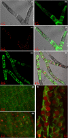
The giant organelles are peroxisomes not vacuoles. (a–d) Confocal images of vacuoles and peroxisomes in wild‐type protonemata of Physcomitrella patens transformed with the vacuolar reporter Arabidopsis homologue of S. cerevisiae VAM3 (AtVAM3)‐GFP and the peroxisome marker mRFPSRL. (a) Bright‐field image; (b) GFP fluorescence; (c) RFP fluorescence; (d) merged image. (e, f) Confocal images of vacuoles and peroxisomes in protonemata of the Phypa_pex11‐1‐KO mutant. (e) AtVAMGFP decorates the vacuolar membrane; (f) merged image for AtVAMGFP and mRFPSRL shows the peroxisomal marker filling large structures that distort the vacuolar membrane around them. (g, h) Epifluorescence microscopic images of vacuoles and peroxisomes in gametophore tissue of wild type (g) and pex11 mutant (h) transformed with the vacuolar reporter AtVAM3‐GFP and the peroxisome marker mRFPSRL: merged fluorescent and bright‐field images. (i) Confocal z stack image showing giant peroxisomes (red) and vacuolar membrane (green) in gametophore cells of the Phypa_pex11‐1‐KO mutant transformed with the vacuolar reporter AtVAM3‐GFP and the peroxisome marker mRFPSRL: merged GFP and RFP images.
Figure 6
Figure 6
Phypa_PEX11‐1 is located in the peroxisome membrane and overexpression causes tabulation. (a) Merged bright‐field and GFP image of Physcomitrella patens gametophore cells showing GFPPEX11 localization to the membranes of peroxisomes greatly reduced in size. Peroxisomes showing this particularly clearly are arrowed. (b–d) Tubulation can be seen in GFP (b) and merged (c, d) images of gametophore (c) and chloronemal (d) cells overexpressing the GFP‐Phypa_PEX11‐1 gene.
Figure 7
Figure 7
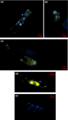
Physcomitrella patens PEX11‐1 interacts with fission factors. (a–c) Protonemal tissue of the Phypa_pex11‐1‐KO mutant expressing a CFPSKL transgene was co‐bombarded with a Phypa_PEX11‐ YFPN fusion construct and a PpFIS1A‐ YFPC fusion construct. YFP fluorescence detected by epifluorescence microscopy is clearly visible around the periphery of the CFP‐marked peroxisomes, indicating an interaction between the Pex11 and Fission 1A (Fis1A) proteins at the peroxisome membrane. (d) Control experiment in which the CFPSKL expressing strain was co‐bombarded with an unfused YFPN construct and a PpFIS1A‐ YFPC construct. (e) Control experiment in which the CFPSKL expressing strain was co‐bombarded with an unfused YFPC construct and a Phypa_PEX11‐1‐ YFPN fusion construct. Each panel in this figure shows the merged YFP and CFP fluorescence signal.
Figure 8
Figure 8
Recovery from low temperature. Protonemata of the Physcomitrella patens pex11‐1‐KO line expressing the mRFPSRL and AtVam3‐GFP reporters that had been archived at low temperature for over 6 months were homogenized and the homogenate fragments subcultured on cellophane‐overlaid agar medium for regeneration under standard growth conditions. Merged bright‐field, GFP and RFP epifluorescence images of protonemata regenerated for 1 (a), 2 (b) and 4 (c, d) d are shown. The cell walls dividing the filament are indicated by arrows in (c) and (d). The image in (c) contains two complete cells and a part of a third (the antepenultimate cell for the filament), while (d) contains four cells and part of a fifth. The cell wall between the apical and subapical cell in this filament is slightly oblique, indicating that the apical cell is commencing differentiation into a caulonemal initial. Note also the smaller peroxisomes in the older cells of each filament.

Similar articles

Cited by

References

    1. Abramoff M, Magalhaes P, Ram S. 2004. Processing with ImageJ. Biophotonics International 11: 36–42.
    1. Barton K, Mathur N, Mathur J. 2013. Simultaneous live‐imaging of peroxisomes and the ER in plant cells suggests contiguity but no luminal continuity between the two organelles. Frontiers in Physiology 4: 196. - PMC - PubMed
    1. Bonekamp NA, Grille S, Cardoso MJ, Almeida M, Aroso M, Gomes S, Magalhaes AC, Ribeiro D, Islinger M, Schrader M. 2013. Self‐interaction of human Pex11p beta during peroxisomal growth and division. PLoS ONE 8: e53424. - PMC - PubMed
    1. Chang J, Klute MJ, Tower RJ, Mast FD, Dacks JB, Rachubinski RA. 2015. An ancestral role in peroxisome assembly is retained by the divisional peroxin Pex11 in the yeast Yarrowia lipolytica . Journal of Cell Science 128: 1327–1340. - PubMed
    1. Desai M, Hu J. 2008. Light induces peroxisome proliferation in Arabidopsis seedlings through the photoreceptor phytochrome A, the transcription factor HY5 HOMOLOG, and the peroxisomal protein PEROXIN11b. Plant Physiology 146: 1117–1127. - PMC - PubMed

Publication types