

Screening of Transcription Factors Involved in Fetal Hemoglobin Regulation Using Phylogenetic Footprinting
- PMID: 26543346
- PMCID: PMC4624090
- DOI: 10.4137/EBO.S15364
Screening of Transcription Factors Involved in Fetal Hemoglobin Regulation Using Phylogenetic Footprinting
Abstract
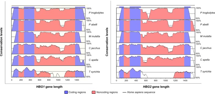
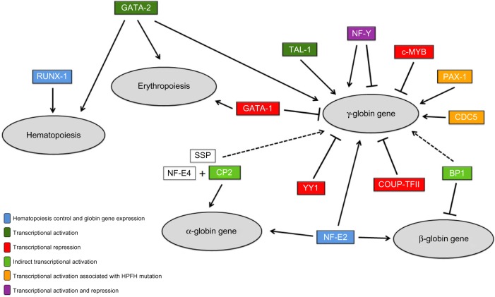
Fetal hemoglobin (Hb F) is an important genetic modulator of the beta-hemoglobinopathies. The regulation of Hb F levels is influenced by transcription factors. We used phylogenetic footprinting to screen transcription factors that have binding sites in HBG1 and HBG2 genes' noncoding regions in order to know the genetic determinants of the Hb F expression. Our analysis showed 354 conserved motifs in the noncoding regions of HBG1 gene and 231 motifs in the HBG2 gene between the analyzed species. Of these motifs, 13 showed relation to Hb F regulation: cell division cycle-5 (CDC5), myelo-blastosis viral oncogene homolog (c-MYB), transcription factor CP2 (TFCP2), GATA binding protein 1 (GATA-1), GATA binding protein 2 (GATA-2), nuclear factor erythroid 2 (NF-E2), nuclear transcription factor Y (NF-Y), runt-related transcription factor 1 (RUNX-1), T-cell acute lymphocytic leukemia 1 (TAL-1), YY1 transcription factor (YY1), beta protein 1 (BP1), chicken ovalbumin upstream promoter-transcription factor II (COUP-TFII), and paired box 1 (PAX-1). The last three motifs were conserved only in the noncoding regions of the HBG1 gene. The understanding of genetic elements involved in the maintenance of high Hb F levels may provide new efficient therapeutic strategies in the beta-hemoglobinopathies treatment, promoting reduction in clinical complications of these genetic disorders.
Keywords: Hb F; beta-hemoglobinopathies; phylogenetic footprinting; transcription factors.
Figures
Similar articles
-
Functional Analysis of an (A)γ-Globin Gene Promoter Variant (HBG1: g.-225_-222delAGCA) Underlines Its Role in Increasing Fetal Hemoglobin Levels Under Erythropoietic Stress.Hemoglobin. 2016;40(1):48-52. doi: 10.3109/03630269.2015.1107842. Epub 2015 Nov 16. Hemoglobin. 2016. PMID: 26575252
-
A new unstable variant of the fetal hemoglobin HBG2 gene: Hb F-Turritana [(G) γ64(E8)Gly→Asp, HBG2:c.194G>A] found in cis to the Hb F-Sardinia gene [(A) γ(E19)Ile→Thr, HBG1:c.227T>C].Eur J Haematol. 2014 Jun;92(6):510-3. doi: 10.1111/ejh.12277. Epub 2014 Feb 26. Eur J Haematol. 2014. PMID: 24483321
-
GATA-and SP1-binding sites are required for the full activity of the tissue-specific promoter of the tal-1 gene.Oncogene. 1994 Sep;9(9):2623-32. Oncogene. 1994. PMID: 8058326
-
[Regulation of the β-globin gene family expression, useful in the search for new therapeutic targets for hemoglobinopathies].Medicina (B Aires). 2016;76(6):383-389. Medicina (B Aires). 2016. PMID: 27959850 Review. Spanish.
-
Regulation of the MIR155 host gene in physiological and pathological processes.Gene. 2013 Dec 10;532(1):1-12. doi: 10.1016/j.gene.2012.12.009. Epub 2012 Dec 14. Gene. 2013. PMID: 23246696 Review.
Cited by
-
TAL1 overexpression in induced pluripotent stem cells promotes the formation of hematopoietic cell-forming complexes but inhibits enucleation in vitro.Front Cell Dev Biol. 2025 Apr 24;13:1474631. doi: 10.3389/fcell.2025.1474631. eCollection 2025. Front Cell Dev Biol. 2025. PMID: 40342930 Free PMC article.
-
Distinct miRNA Signatures and Networks Discern Fetal from Adult Erythroid Differentiation and Primary from Immortalized Erythroid Cells.Int J Mol Sci. 2021 Mar 31;22(7):3626. doi: 10.3390/ijms22073626. Int J Mol Sci. 2021. PMID: 33807258 Free PMC article.
-
Atypical β-S haplotypes: classification and genetic modulation in patients with sickle cell anemia.J Hum Genet. 2019 Mar;64(3):239-248. doi: 10.1038/s10038-018-0554-4. Epub 2019 Jan 9. J Hum Genet. 2019. PMID: 30622282
-
Investigating the role of FOX gene family in development and stress response in Labeo rohita: A multi-faceted analysis of phylogeny and genome characterization.PLoS One. 2025 Aug 21;20(8):e0323740. doi: 10.1371/journal.pone.0323740. eCollection 2025. PLoS One. 2025. PMID: 40839711 Free PMC article.
References
-
- Menzel S, Garner C, Gut I, et al. A QTL influencing F cell production maps to a gene encoding a zinc-finger protein on chromosome 2p15. Nat Genet. 2007;39(10):1197–9. - PubMed
-
- Galanello R, Cao A. Relationship between genotype and phenotype. Ann N Y Acad Sci. 1998;850:325–33. - PubMed
-
- Thein SL. Genetic modifiers of β-thalassemia. Haematologica. 2005;90:649–60. - PubMed
LinkOut - more resources
Full Text Sources
Research Materials
Miscellaneous
