

The Role of Ultrasound Imaging of Callus Formation in the Treatment of Long Bone Fractures in Children
- PMID: 26543512
- PMCID: PMC4614376
- DOI: 10.12659/PJR.894548
The Role of Ultrasound Imaging of Callus Formation in the Treatment of Long Bone Fractures in Children
Abstract
Background: In the process of diagnosis and treatment of fractures, an X-ray study is typically performed. In modern medicine very important is the development of new diagnostic methods without adverse effects on the body. One of such techniques is ultrasound imaging. It has a high value in imaging most areas of the body, including the musculoskeletal system. Reports on the use of ultrasound in the evaluation of the callus are rare and this could be a method equivalent to or even better than standard radiographs. The aim of the study was to analyze the correlation of ultrasound with radiographs in imaging of callus formation after fractures of long bones in children and to analyze the correlation of vascular resistance index (RI) and the degree of vascularization of the callus with a subjective radiological assessment of the bone union quality.
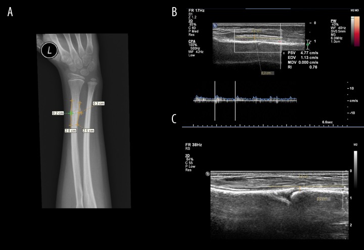
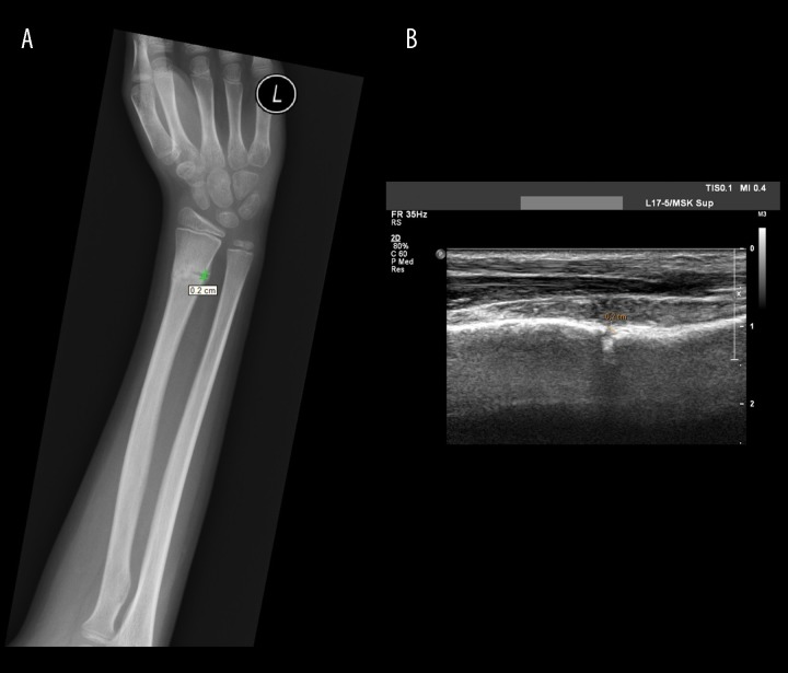
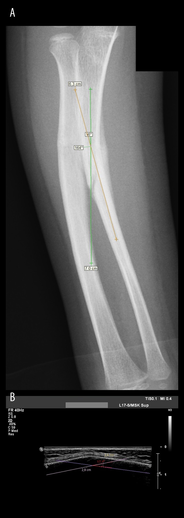
Material/methods: The prospective study was planned to qualify 50 children treated for long bones fractures of the arm, forearm, thigh and lower leg. Ultrasound diagnosis was carried out using a Philips iU22 camera equipped with a linear probe with 17-5-MHz resolution and MSK Superficial program. During ultrasound examination measurements of the callus were performed. Using the Power Doppler callus vascularity was visualized and vascular resistance index (RI) was measured. The same measurements were made within the corresponding area of the healthy limb. The results obtained by ultrasound were compared with radiograph measurements and with the subjective assessment of the callus quality.
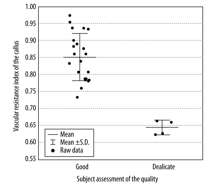
Results: Preliminary results were developed on a group of 24 patients, where 28 fractured bones and 28 corresponding healthy bones were examined. Fifteen boys and 9 girls participated in the study. The average age at injury was, respectively, 11 and 9 years. In both groups fractures without displacement were the most frequent. A similar frequency was observed in fractures requiring reposition and subperiosteal fractures. In contrast, fractures with a slight displacement of the fragments, were 3 times more common in girls. Statistical analysis of the measurements of length and width of the callus demonstrated that the differences between results obtained in the ultrasound in comparison with X-rays were not statistically significant. Moreover, preliminary results showed a significantly higher degree of vascularization of the callus than of the healthy periosteum.
Conclusions: Preliminary results indicate the high efficacy of ultrasound in the evaluation of callus formation after fractures of long bones in children and the possibility of its alternative use to X-ray examinations.
Keywords: Child Care; Fracture Healing; Fractures, Bone; Radiography; Ultrasonography.
Figures

References
-
- Grigoryan M, Lynch JA, Fierlinger AL, et al. Quantitative and qualitative assessment of closed fracture healing using computed tomography and conventional radiography. Academic Radiology. 2003;10(11):1267–73. - PubMed
-
- Webb J, Herling G, Gardner T, et al. Manual assessment of fracture stiffnes. Injury. 1996;27(5):319–20. - PubMed
-
- Wong LC, Chiu WK, Russ M, Liew S. Review of techniques for monitoring the healing fracture of bones for implementation in an internally fixated pelvis. Med Eng Physics. 2012;34(2):140–52. - PubMed
-
- U.S Department of Health and Human Services. 13th Report on Carcinogenes. 2014. [Accessed 2 October 2014]. http://ntp.niehs.nih.gov/pubhealth/roc/roc13/index.html.
LinkOut - more resources
Full Text Sources