

Dataset for the quantitative proteomics analysis of the primary hepatocellular carcinoma with single and multiple lesions
- PMID: 26543886
- PMCID: PMC4589833
- DOI: 10.1016/j.dib.2015.08.036
Dataset for the quantitative proteomics analysis of the primary hepatocellular carcinoma with single and multiple lesions
Abstract
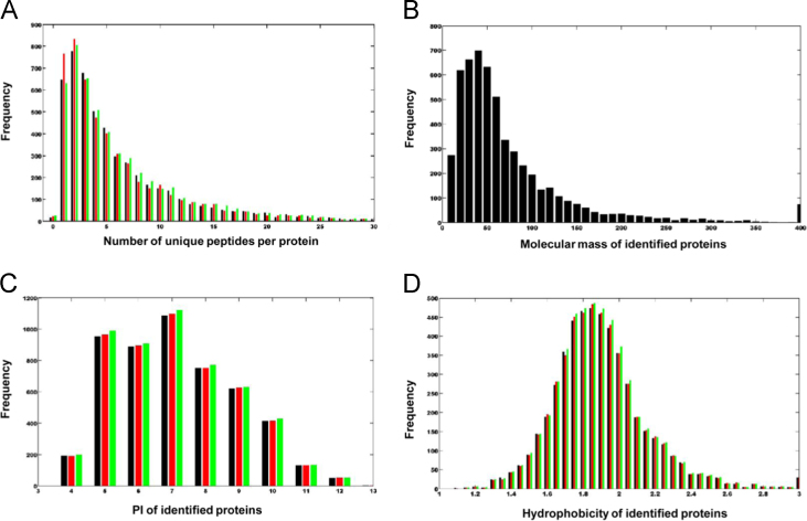
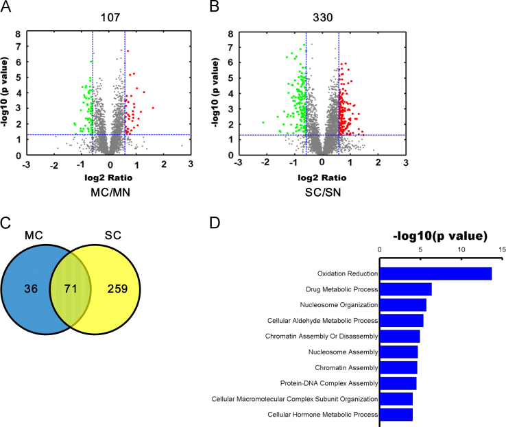
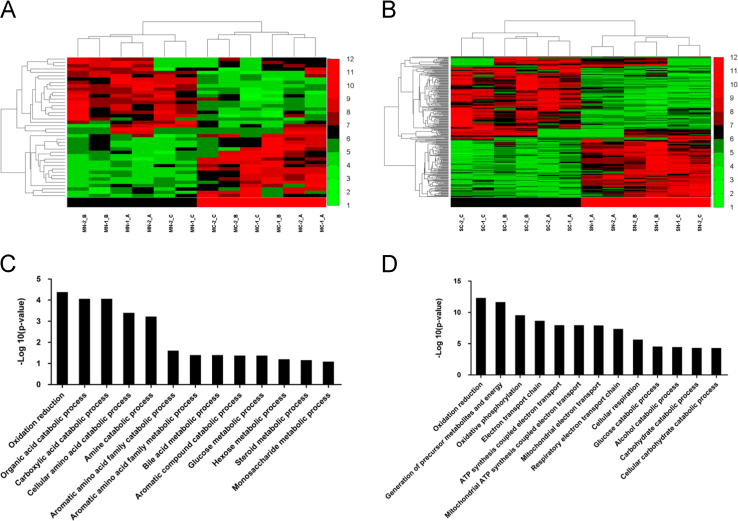
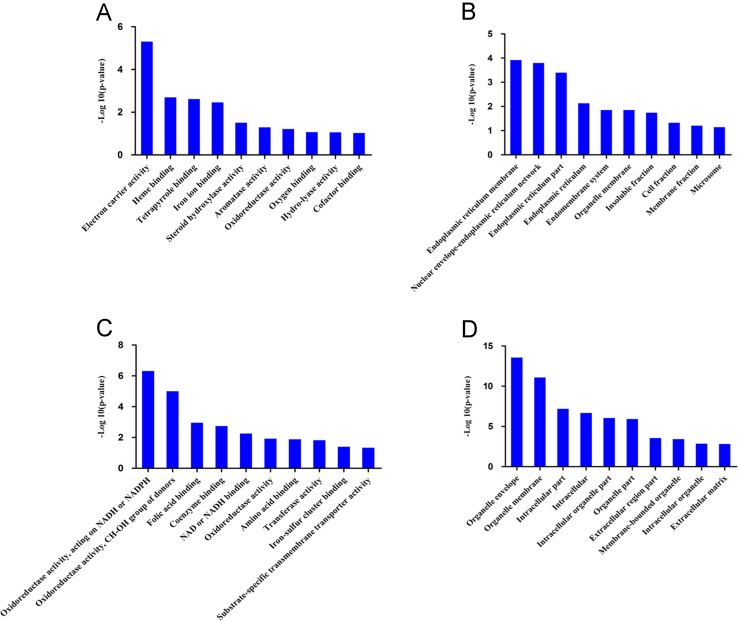
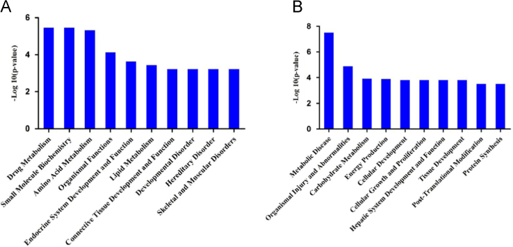
Hepatocellular Carcinoma (HCC) is one of the most common malignant tumor, which is causing the second leading cancer-related death worldwide. The tumor tissues and the adjacent noncancerous tissues obtained from HCC patients with single and multiple lesions were quantified using iTRAQ. A total of 5513 proteins (FDR of 1%) were identified which correspond to roughly 27% of the total liver proteome. And 107 and 330 proteins were dysregulated in HCC tissue with multiple lesions (MC group) and HCC tissue with a single lesion (SC group), compared with their noncancerous tissue (MN and SN group) respectively. Bioinformatics analysis (GO, KEGG and IPA) allowed these data to be organized into distinct categories. The data accompanying the manuscript on this approach (Xing et al., J. Proteomics (2015), http://dx.doi.org/10.1016/j.jprot.2015.08.007[1]) have been deposited to the iProX with identifier IPX00037601.
Figures
References
-
- Xiaohua Xing, Yao Huang, Sen Wang, Minhui Chi, Yongyi Zeng, Lihong Chen, Jinhua Zeng, Minjie Lin, Xiaolong Liu, Jingfeng Liu. Comparative analysis of the primary multiple and single hepatocellular carcinoma by iTRAQ based quantitative proteomics. J. Proteomics 128 (2015) 262–271. 〈http://dx.doi.org/10.1016/j.jprot.2015.08.007〉 (in press). - DOI - PubMed
-
- Gilar M., Olivova P., Daly A.E., Gebler J.C. Two-dimensional separation of peptides using RP–RP–HPLC system with different pH in first and second separation dimensions. J. Sep. Sci. 2005;28:1694–1703. - PubMed
LinkOut - more resources
Full Text Sources
Other Literature Sources
Molecular Biology Databases
