. 2015 Nov 6;11(11):e1005643.
doi: 10.1371/journal.pgen.1005643. eCollection 2015 Nov.

UFBP1, a Key Component of the Ufm1 Conjugation System, Is Essential for Ufmylation-Mediated Regulation of Erythroid Development

Affiliations

UFBP1, a Key Component of the Ufm1 Conjugation System, Is Essential for Ufmylation-Mediated Regulation of Erythroid Development

Yafei Cai et al. PLoS Genet. .

Abstract

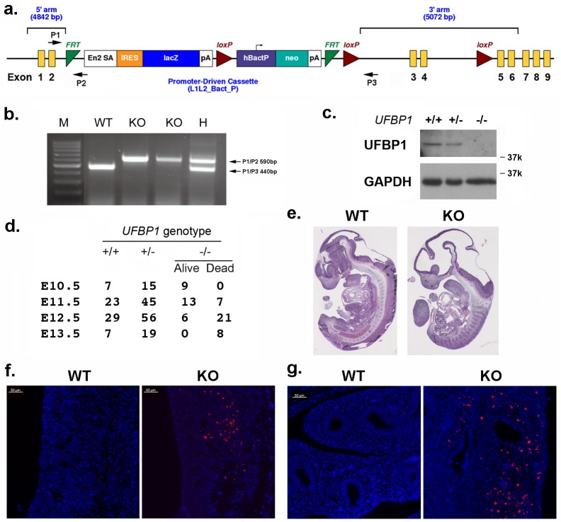
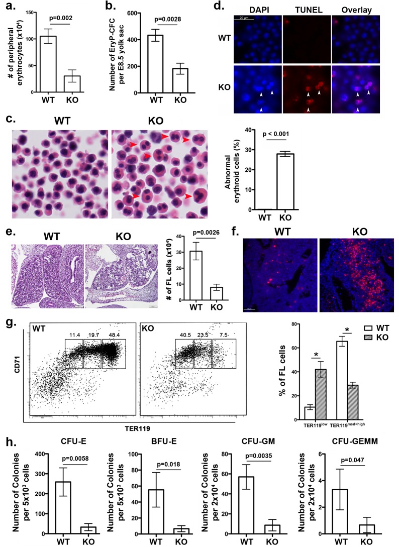
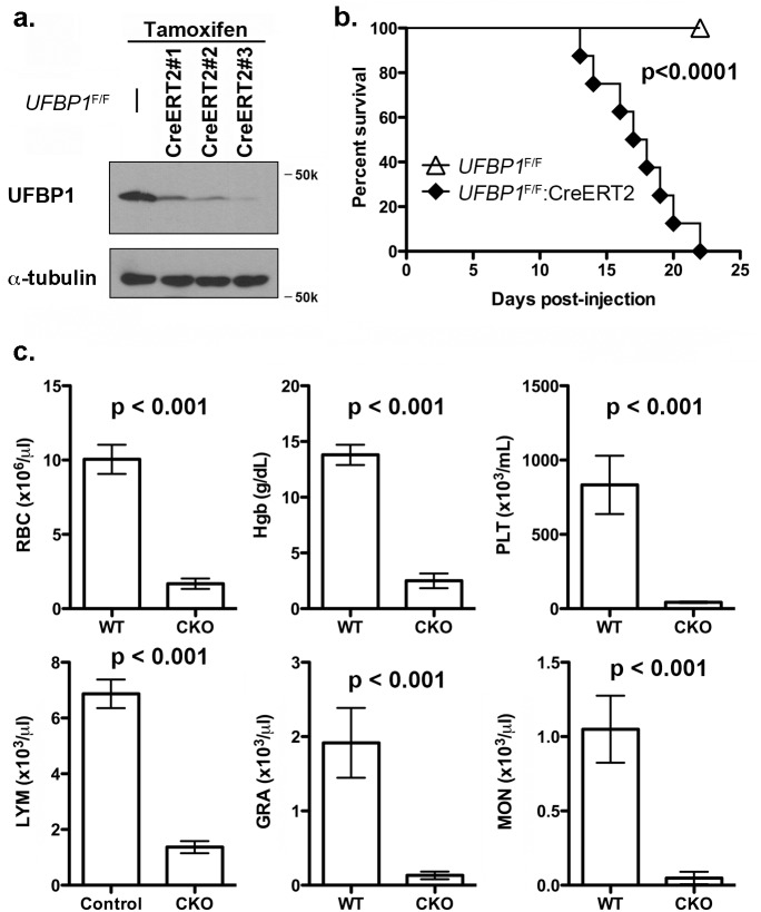
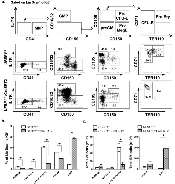
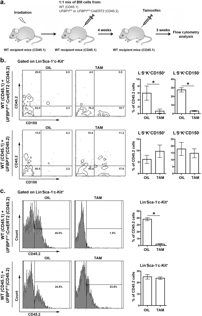
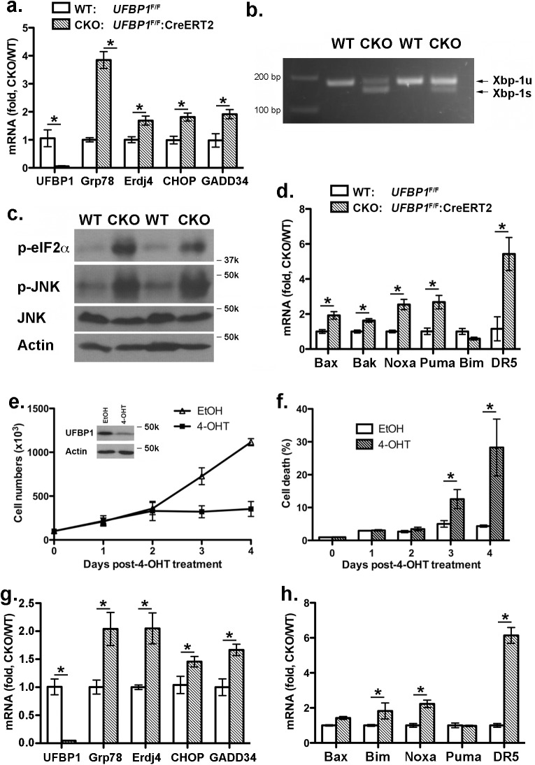
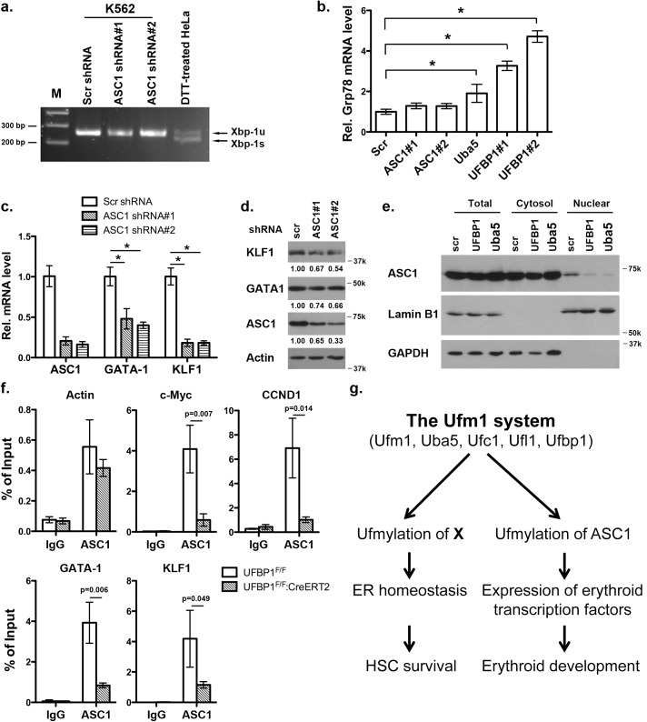
The Ufm1 conjugation system is an ubiquitin-like modification system that consists of Ufm1, Uba5 (E1), Ufc1 (E2), and less defined E3 ligase(s) and targets. The biological importance of this system is highlighted by its essential role in embryogenesis and erythroid development, but the underlying mechanism is poorly understood. UFBP1 (Ufm1 binding protein 1, also known as DDRGK1, Dashurin and C20orf116) is a putative Ufm1 target, yet its exact physiological function and impact of its ufmylation remain largely undefined. In this study, we report that UFBP1 is indispensable for embryonic development and hematopoiesis. While germ-line deletion of UFBP1 caused defective erythroid development and embryonic lethality, somatic ablation of UFBP1 impaired adult hematopoiesis, resulting in pancytopenia and animal death. At the cellular level, UFBP1 deficiency led to elevated ER (endoplasmic reticulum) stress and activation of unfolded protein response (UPR), and consequently cell death of hematopoietic stem/progenitor cells. In addition, loss of UFBP1 suppressed expression of erythroid transcription factors GATA-1 and KLF1 and blocked erythroid differentiation from CFU-Es (colony forming unit-erythroid) to proerythroblasts. Interestingly, depletion of Uba5, a Ufm1 E1 enzyme, also caused elevation of ER stress and under-expression of erythroid transcription factors in erythroleukemia K562 cells. By contrast, knockdown of ASC1, a newly identified Ufm1 target that functions as a transcriptional co-activator of hormone receptors, led to down-regulation of erythroid transcription factors, but did not elevate basal ER stress. Furthermore, we found that ASC1 was associated with the promoters of GATA-1 and Klf1 in a UFBP1-dependent manner. Taken together, our findings suggest that UFBP1, along with ASC1 and other ufmylation components, play pleiotropic roles in regulation of hematopoietic cell survival and differentiation via modulating ER homeostasis and erythroid lineage-specific gene expression. Modulating the activity of this novel ubiquitin-like system may represent a novel approach to treat blood-related diseases such as anemia.


Conflict of interest statement

The authors have declared that no competing interests exist.

Figures

Fig 1
Fig 1. UFBP1 is essential for embryonic development.
a. The targeting vector of UFBP1 allele. b. PCR-based genotyping of UFBP1 allele with the gene trap cassette. c. Immunoblotting of UFBP1 protein in wild-type (WT) and KO embryos. d. Embryonic lethality of UFBP1 null embryos. e. Hematoxylin & eosin (H & E) staining of representative WT and KO embryos (E11.5). f. TUNEL staining of the bottom of the fourth ventricle of embryonic brain in E11.5 embryos. g. TUNEL staining of lumen of duodenum in E11.5 embryos. Scale bar: 50 μm.
Fig 2
Fig 2. UFBP1 deficiency impairs embryonic erythroid development.
a. The number of erythrocytes in peripheral blood of WT and KO E11.5 embryos. Blood cells were collected from suspension solution during separation of yolk sac from embryos and manually counted. Data are presented as means ± SD (n = 3). b. The number of EryP-CFC in E8.5 yolk sac. Data are presented as means ± SD (n = 3). c. Abnormal multi-nucleated erythroid cells in E11.5 embryos. Multi-nucleated erythrocytes are marked by red arrowheads in H & E stained sections. Data are presented as means ± SD (n = 3). d. Representative photographs of TUNEL staining of circulating erythrocytes in E11.5 embryos. e. H & E staining and number of fetal liver cells in E11.5 embryos. The number of fetal liver cells was scored by counting total cells from dissected fetal livers. Data are presented as means ± SD (n = 3). f. Representative photographs of TUNEL staining of fetal liver sections (E11.5). g. Representative flow cytometry analysis of fetal liver cells from E11.5 embryos. Fetal liver cells were freshly isolated from E11.5 embryos and subjected to flow cytometry analysis of CD71 and TER119 markers. Percentages of each population of cells are presented as means ± SD (n = 3). h. CFU-Es, BFU-Es, CFU-GMs, and CFU-GEMMs in E11.5 fetal livers. Data are presented as means ± SD (n = 3).
Fig 3
Fig 3. Loss of UFBP1 in adult mice results in severe pancytopenia and animal death.
a. Confirmation of UFBP1 depletion in bone marrow cells of TAM-injected CKO mice. Floxed mice were IP injected with tamoxifen according to a standard protocol. BM cells were collected and subject to immunoblotting of UFBP1. b. Survival curve of UFBP1 deficient mice after TAM injection, p < 0.001 (n = 12 each group). c. The result of CBC counts. Data are presented as means ± SD. The blood was drawn from control (UFBP1 F/F) (n = 5) and UFBP1 deficient mice (UFBP1 F/F:CreERT2) (n = 5) when UFBP1 deficient mice became moribund and/or lost 20% of body weight after TAM injection (between 2–3 weeks post-TAM treatment). Blood samples were subjected to CBC analysis. The mice used in this experiment were ~ 8-week old male mice. RBC: red blood cell; Hgb: hemoglobin; PLT: platelet; LYM: lymphocyte; GRA: granulocyte; MON: monocyte.
Fig 4
Fig 4. UFBP1 deficiency impairs lineage development of erythroid progenitors.
a. Flow cytometry analysis of erythroid and myeloid lineages in control and UFBP1 deficient mice after TAM injection. BM cells were collected from TAM-injected control (n = 4) and floxed (n = 4) male mice when UFBP1 deficient mice exhibited significant weight loss and became moribund. The following markers were used for flow analysis: BV510-Sca-1, APC780-c-Kit, PE-Cy7-CD150, Alexa 700-CD16/32, PE-IL-7R, APC-CD41, BV650-CD105, FITC-CD71, BV421-TER119, and lineage markers including PerCP-Cy5.5-conjugated CD4, CD8, CD3, CD5, Gr-1, CD11b, CD19 and B220. b. The percentages of each lineage in L-S-K+ cells. *p <0.01, and **p <0.05 (n = 4). MkP: megakaryocyte progenitor; GMP: granulocyte macrophage progenitor; Pre GM: granulocyte macrophage precursor; Pre MegE: megakaryocyte erythroid precursor; Pre CFU-E: CFU-E precursor; CFU-E: erythroid colony-forming unit; Pro Ery: proerythroblast. c. The absolute cell numbers of each lineage in control and UFBP1-depleted BMs. UFBP1 depletion led to reduction of total BM cells (1 x 10⁸ cells in control versus 5 x 10⁷ in UFBP1 deficient mice). *p <0.01 (n = 4).
Fig 5
Fig 5. UFBP1 is essential for HSC function.
a. Experimental scheme for competitive repopulation assay. b. Contribution of UFBP1 deficient cells (CD45.2) to long-term HSCs (L-S+K+CD150+) and multipotent progenitors (L-S+K+CD150-). * p < 0.001 (n = 5). c. Contribution of UFBP1 deficient cells (CD45.2) to oligopotent progenitor cells (L-S-K+). * p < 0.001 (n = 5). Data are presented as means ± SD. The unfractionated BM cells from either UFBP1 F/F or UFBP1 F/F:CreERT2 (CD45.2) were mixed with wild-type (CD45.1) BM cells at a 1:1 ratio, and co-transplanted into lethally irradiated recipient CD45.1 mice. Four weeks after transplantation, the mice were treated with either oil or TAM. Three weeks after initiation of the treatment, the BM cells were isolated and subjected to flow analysis using indicated markers.
Fig 6
Fig 6. Loss of UFBP1 activates the UPR and cell death program.
a. Up-regulation of Grp78, ERdj4 and CHOP in UFBP1 deficient BM cells. Total RNAs were purified from BM cells and subject to quantitative RT-PCR analysis (normalized to β-actin gene). TAM-injected UFBP1 F/Fmice were used as WT control mice, while TAM-injected UFBP1 F/F:CreERT2 mice were designated as CKO mice. The ratio of CKO to WT was presented. * p < 0.01 (n = 3). Data are presented as means ± SD. b. Xbp-1 mRNA splicing in UFBP1 deficient BM cells. Total RNAs were isolated from BM cells of control and CKO mice, and subjected to Xbp-1 mRNA splicing assay. c. Elevation of phosphorylation of eIF2α and JNK in UFBP1 deficient cells. Total cell lysates of BM cells were subjected to immunoblotting of indicated antibodies. d. Up-regulation of cell death genes in UFBP1 deficient BM cells. Total RNAs were purified from BM cells and subject to quantitative RT-PCR analysis (normalized to β-actin gene). The ratio of CKO to WT was presented. * p < 0.01 (n = 3). e. Proliferation of wild-type and UFBP1 deficient LSK cells. LSK cells were sorted from BM of UFBP1 F/F:CreERT2 mice, and cultured in the absence or presence of 4-OHT (1 μM) for indicated period of time. Cell numbers were manually scored. The experiment was performed three times independently. f. Cell death of UFBP1 deficient HLSK cells. Cell death was elevated by DNA dye exclusion assay. g. Up-regulation of UPR genes in UFBP1 deficient LSK cells. Total RNAs were purified from LSK cells and subject to quantitative RT-PCR analysis (normalized to β-actin gene). The ratio of CKO to WT was presented. * p < 0.01 (n = 3). h. Up-regulation of cell death genes in UFBP1 deficient LSK cells. The ratio of CKO to WT was presented (normalized to β-actin gene). * p < 0.01 (n = 3).
Fig 7
Fig 7. Depletion of Uba5 activates the UPR and suppresses expression of erythroid transcription factors in K562 cells.
a. Elevation of phosphorylation of eIF2α in Uba5- and UFBP1-depleted K562 cells. b. Xbp-1 mRNA splicing in Uba5- and UFBP1-depleted K562 cells. mRNA from DTT (dithiothreitol)-treated HeLa cells was used as a positive control. c. Under-expression of GATA-1 and Klf1 in Uba5- and UFBP1-depleted K562 cells. Total RNAs were purified from K562 cells and subject to quantitative RT-PCR analysis (normalized to β-actin gene). The data are presented as the relative level to the K562 cells treated with scrambled shRNA. * p < 0.01 (n = 3). Total cell lysates were subject to immunoblotting of indicated antibodies.
Fig 8
Fig 8. Knockdown of ASC1 down-regulates expression of erythroid transcription factors but does not activate the UPR.
a. Xbp-1 mRNA splicing in ASC1-depleted K562 cells. mRNA from DTT (dithiothreitol)-treated HeLa cells was used as a positive control. b. Expression of Grp78 in K562 cells with knockdown of ASC1, Uba5 and UFBP1. The data are presented as the relative level to K562 cells treated with scrambled shRNA. * p < 0.01 (n = 3). c. Expression of GATA-1 and Klf1 in ASC1-knockdown K562 cells. The data are presented as the relative level to the cells with scrambled shRNA. * p < 0.01 (n = 3). d. GATA-1 and KLF1 protein level in ASC1 knockdown cells. e. Subcellular localization of ASC1 in UFBP1 and Uba5 knockdown K562 cells. Subcellular fractionation of K562 cells was performed according to [6]. Lamin B1 was used as the nuclear marker, while GAPDH was used as the cytosolic marker. f. ChIP analysis of ASC1 association to the promoters of GATA-1 and Klf1 genes in BM cells. BM and spleen cells were isolated from phenylhydrazine-treated control and UFBP1 CKO mice, and subjected to ChIP assays. c-Myc and CCND1 promoters were used as positive ASC1 targets while actin promoter was the negative control. Data are presented as means ± SD (n = 3). g. A working model for the role of ufmylation in regulation of hematopoiesis.

Similar articles

Cited by

References

    1. Komatsu M, Chiba T, Tatsumi K, Iemura S, Tanida I, Okazaki N, et al. A novel protein-conjugating system for Ufm1, a ubiquitin-fold modifier. The EMBO journal 2004, 23(9): 1977–1986. - PMC - PubMed
    1. Tatsumi K, Sou YS, Tada N, Nakamura E, Iemura S, Natsume T, et al. A novel type of E3 ligase for the Ufm1 conjugation system. The Journal of biological chemistry 2010, 285(8): 5417–5427. 10.1074/jbc.M109.036814 - DOI - PMC - PubMed
    1. Daniel J, Liebau E. The ufm1 cascade. Cells 2014, 3(2): 627–638. 10.3390/cells3020627 - DOI - PMC - PubMed
    1. Tatsumi K, Yamamoto-Mukai H, Shimizu R, Waguri S, Sou YS, Sakamoto A, et al. The Ufm1-activating enzyme Uba5 is indispensable for erythroid differentiation in mice. Nat Commun 2011, 2: 181 10.1038/ncomms1182 - DOI - PMC - PubMed
    1. Neziri D, Ilhan A, Maj M, Majdic O, Baumgartner-Parzer S, Cohen G, et al. Cloning and molecular characterization of Dashurin encoded by C20orf116, a PCI-domain containing protein. Biochim Biophys Acta 2010, 1800(4): 430–438. 10.1016/j.bbagen.2009.12.004 - DOI - PubMed

Publication types

Substances