Review
. 2015 Dec;36(6):E1-E150.
doi: 10.1210/er.2015-1010. Epub 2015 Nov 6.

EDC-2: The Endocrine Society's Second Scientific Statement on Endocrine-Disrupting Chemicals

Affiliations
Review

EDC-2: The Endocrine Society's Second Scientific Statement on Endocrine-Disrupting Chemicals

A C Gore et al. Endocr Rev. 2015 Dec.

Abstract

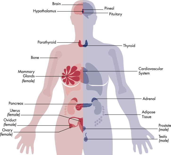
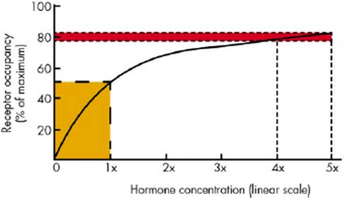
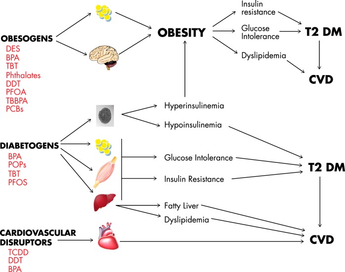
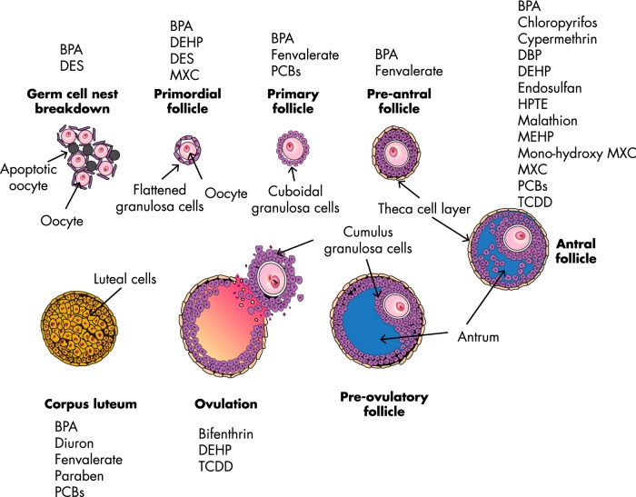
The Endocrine Society's first Scientific Statement in 2009 provided a wake-up call to the scientific community about how environmental endocrine-disrupting chemicals (EDCs) affect health and disease. Five years later, a substantially larger body of literature has solidified our understanding of plausible mechanisms underlying EDC actions and how exposures in animals and humans-especially during development-may lay the foundations for disease later in life. At this point in history, we have much stronger knowledge about how EDCs alter gene-environment interactions via physiological, cellular, molecular, and epigenetic changes, thereby producing effects in exposed individuals as well as their descendants. Causal links between exposure and manifestation of disease are substantiated by experimental animal models and are consistent with correlative epidemiological data in humans. There are several caveats because differences in how experimental animal work is conducted can lead to difficulties in drawing broad conclusions, and we must continue to be cautious about inferring causality in humans. In this second Scientific Statement, we reviewed the literature on a subset of topics for which the translational evidence is strongest: 1) obesity and diabetes; 2) female reproduction; 3) male reproduction; 4) hormone-sensitive cancers in females; 5) prostate; 6) thyroid; and 7) neurodevelopment and neuroendocrine systems. Our inclusion criteria for studies were those conducted predominantly in the past 5 years deemed to be of high quality based on appropriate negative and positive control groups or populations, adequate sample size and experimental design, and mammalian animal studies with exposure levels in a range that was relevant to humans. We also focused on studies using the developmental origins of health and disease model. No report was excluded based on a positive or negative effect of the EDC exposure. The bulk of the results across the board strengthen the evidence for endocrine health-related actions of EDCs. Based on this much more complete understanding of the endocrine principles by which EDCs act, including nonmonotonic dose-responses, low-dose effects, and developmental vulnerability, these findings can be much better translated to human health. Armed with this information, researchers, physicians, and other healthcare providers can guide regulators and policymakers as they make responsible decisions.


Figures

Figure 1.
Figure 1.
Diagram of many of the body's endocrine glands in females (left) and males (right).
Figure 2.
Figure 2.
Schematic example of the relationship between receptor occupancy and hormone concentration. In this theoretical example, at low concentrations, an increase in hormone concentration from 0 to 1× causes an increase in receptor occupancy of approximately 50% (from 0 to 50%; see yellow box). Yet the same increment in hormone concentration at higher doses (from 4× to 5×) causes an increase in receptor occupancy of only approximately 4% (from 78 to 82%; see red box). However, it is important to recognize that receptor occupancy is not linearly related to hormone effect, and low receptor occupancy (1 to 10%) can be associated with maximal effects. [Reprinted from L. N. Vandenberg et al: Hormones and endocrine-disrupting chemicals: low-dose effects and nonmonotonic dose responses. Endocr Rev. 2012;33:378–455 (133), with permission. © Endocrine Society.]
Figure 3.
Figure 3.
EDCs can act as obesogens, diabetogens, and/or cardiovascular disruptors. For obesogenic effects, EDCs act upon adipocytes and the brain to induce obesity, which generates insulin resistance, glucose intolerance, and dyslipidemia and greatly increases the susceptibility to T2D and CVDs. Additionally, EDCs work as diabetogens that directly affect the islet of Langerhans and increase or decrease normal insulin biosynthesis and release, generating hyper- or hypoglycemia. An excess of insulin signaling, as well as insulin resistance, can result in metabolic syndrome. In animal models, EDCs induce insulin resistance, glucose intolerance, fatty liver, and dyslipidemia on WAT, liver, and skeletal muscle. This generates T2D and CVDs. The EDC BPA has been shown to act directly on the heart, increasing the probability of CVDs in animal models.
Figure 4.
Figure 4.
The effects of EDCs on the ovary. This schematic shows the normal developmental stages of ovarian follicles beginning with germ cell nest breakdown around birth, formation of primordial follicles, and their growth to primary follicles, preantral follicles, antral follicles, and finally, preovulatory follicles. This schematic also shows ovulation and the formation of the corpus luteum. Examples of EDCs that adversely affect the ovary are listed in red font above or below their likely site of action.
Figure 5.
Figure 5.
Steroidogenic pathways leading to estradiol biosynthesis. The red Xs indicate the hormones or enzymes that have been shown to be affected by EDCs. Each hormone in the pathway is derived from cholesterol. Reactions are catalyzed by steroidogenic enzymes as they metabolize each hormone to a different hormone down the pathway. Estradiol is the major hormone produced by antral follicles.
Figure 6.
Figure 6.
The effects of EDCs on the uterus, vagina, and anterior pituitary gland. This schematic shows the normal structure of the uterus, vagina, and anterior pituitary, with a list of EDCs (in red text) that have been shown to perturb the development and function of these structures.
Figure 7.
Figure 7.
The multiple influences of the environment on mammary development. Environmental factors may affect breast development and later-life risk for disease or dysfunction via indirect and direct mechanisms. Environmental exposures may change endogenous signals (hormones and growth factors) that affect endocrine organs, as well as tissues near the mammary gland (ie, fat). Those nearby tissues can send atypical messages through the vascular system, culminating in perturbed mammary development. Mammary tissue may also be a direct target of environmental exposures; epithelia, fibroblasts, fat cells, and inflammatory cells express unique and shared receptors that are targets for environmental chemicals. The tight cellular junctions signal across cell types, affecting neighboring cells as well as the target cells (ie, epithelial cells in the terminal end bud). These various endocrine/paracrine/autocrine signaling mechanisms may affect the status of mammary epithelia over the lifetime of the individual. [Reprinted from Figure 2 in S. E. Fenton et al: Perinatal environmental exposures affect mammary development, function, and cancer risk in adulthood. Annu Rev Pharmacol Toxicol. 2012;52:455–479 (1276), with permission. © Annual Reviews.]
Figure 8.
Figure 8.
Stages of normal rat mammary gland (MG) development and effects of environment on subsequent events. Different effects and outcomes after EDC exposure are strongly dependent upon the age of exposure (neonatal period, puberty, pregnancy) and time of analysis. Early-life effects such as altered thelarche or gynecomastia present themselves in adolescents, whereas effects on lactation or mammary tumorigenesis become evident during adulthood. Arrows indicate plausible (gray) or more certain (white) mechanistic pathways. Photomicrographs for early life and puberty were all taken at 16× magnification on a macroscope. [Adapted from R. R. Enoch et al: Mammary gland development as a sensitive end point after acute prenatal exposure to an atrazine metabolite mixture in female Long-Evans rats. Environ Health Perspect. 2007;115:541–547 (884), with permission.] Photomicrographs for pregnancy/lactation and adulthood were taken at 10× magnification on a standard microscope (from S.E.F.). [Reprinted from Figure 1 in R. A. Rudel et al: Environmental exposures and mammary gland development: state of the science, public health implications, and research recommendations. Environ Health Perspect. 2011;119:1053–1061 (801), with permission.]
Figure 9.
Figure 9.
The HPT axis can be disrupted at several points of regulation. The control of thyroid hormone delivery to the site of action in tissues is a highly complex process that includes synthesis in the thyroid gland, transport through blood, selective uptake into tissues and cells, and metabolism at the site of action. These elements are represented as follows. 1) TRH is produced in parvocellular neurons of the hypothalamic PVN. Only a subset of these neurons appears to control pituitary TSH release (ie, are hypophysiotropic). These neurons are controlled by a combination of neuronal afferents and thyroid hormone negative feedback. This feedback is mediated principally by T4 from serum, which is converted to T3 by tanycytes and delivered to TRH neurons through the cellular transporter MCT8. Negative feedback itself is mediated selectively by the ThRB2 receptor. TRH stimulates the synthesis and release of TSH in pituitary by the action of membrane receptors that signal through protein kinase C. 2) Thyroid hormone synthesis requires the active uptake of iodide through the NIS, the production of thyroglobulin, and its iodination by the TPO enzyme. TSH stimulates the thyroid cell through cAMP, which increases thyroid hormone synthesis and release simultaneously. Thyroid hormone release requires the endocytosis of iodinated thyroglobulin in the colloid, vesicular transport through the thyrocyte, during which time iodotyrosyl residues are coupled and excised from the protein backbone before exocytotic release. Once in blood, 3) thyroid hormones are carried on binding proteins (so-called “distributor proteins”). Most T4 (75%) is carried on T4 binding protein (TBG), about 25% on transthyretin (TTR), and a small proportion on albumin. This leaves about 0.01% “free” (unbound). 4) T4 in serum gains entry into tissues and cells through selective transporters such as the organic anion transport protein 1C1 (OATP1c1) or the monocarboxylate transporter 8 (MCT8). The delivery of biologically active T3 is complex especially in the nervous system. T4 is taken up by glial cells and converted to T3 by the action of Dio2. Then, T3 is transported actively to neurons and acts on the ThR α or β. 5) The half-life of T4 in serum is 7–10 days in humans and 24 hours in rodents. This is controlled by the liver, which expresses enzymes (glucuronidases or sulfotransferases) that modify T4 and T3 such that they are eliminated in bile.
Figure 10.
Figure 10.
Representative environmental chemicals with structures similar to that of thyroid hormone. A) Thyroid hormone (T4) is metabolized by a series of enzymes to form more a potent (outer ring deiodination by Dio1 and Dio2) or less potent (inner ring deiodination by Dio3) hormone. These metabolic events are important steps in the control of thyroid hormone action in tissues and take place in specific cells as described in the text. It is important to recognize that some environmental chemicals may interfere with specific enzymes, but little is known of this pathway. Some of these chemicals are: B, PCBs; C, triclosan; D, PBDEs; and E, BPA. [Modified from M. E. Gilbert et al: Developmental thyroid hormone disruption: prevalence, environmental contaminants and neurodevelopmental consequences. Neurotoxicology. 2012;33:842–852 (1322), with permission. © Elsevier.]
Figure 11.
Figure 11.
The HPG and HPA neuroendocrine axes are shown. For each system, a group of hypothalamic neurons synthesizes and secretes a neuropeptide, CRH (HPA axis) or GnRH (HPG axis). These hormones travel through the portal capillary vasculature between the base of the hypothalamus and the anterior pituitary. There, CRH activates the synthesis and secretion of ACTH from corticotropes. Similarly, GnRH stimulates LH and FSH release. These pituitary hormones, when they reach their target glands (adrenal or gonad), regulate steroidogenic processes involved in biosynthesis of the glucocorticoids and sex steroid hormones. These hormones activate stress and reproductive responses, respectively, in the body. They also exert feedback actions on the hypothalamus and pituitary to enable adaptation of the body and to modulate homeostasis of these neuroendocrine systems.

Comment in

References

    1. Diamanti-Kandarakis E, Bourguignon JP, Giudice LC, et al. Endocrine-disrupting chemicals: an Endocrine Society Scientific Statement. Endocr Rev. 2009;30:293–342. - PMC - PubMed
    1. Gore AC, Chappell VA, Fenton SE, et al. Executive summary to EDC-2: The Endocrine Society's second scientific statement on endocrine-disrupting chemicals. Endocr Rev. 2015;36:••••. - PMC - PubMed
    1. Zoeller RT, Brown TR, Doan LL, et al. Endocrine-disrupting chemicals and public health protection: a statement of principles from the Endocrine Society. Endocrinology. 2012;153:4097–4110. - PMC - PubMed
    1. Dodds EC, Lawson W. Synthetic oestrogenic agents without the phenanthrene nucleus. Nature. 1936;137:996.
    1. vom Saal FS, Welshons WV. Evidence that bisphenol A (BPA) can be accurately measured without contamination in human serum and urine, and that BPA causes numerous hazards from multiple routes of exposure. Mol Cell Endocrinol. 2014;398:101–113. - PMC - PubMed

MeSH terms