. 2015 Nov 6;10(11):e0141432.
doi: 10.1371/journal.pone.0141432. eCollection 2015.

Characterization of Heterotopic Ossification Using Radiographic Imaging: Evidence for a Paradigm Shift

Affiliations

Characterization of Heterotopic Ossification Using Radiographic Imaging: Evidence for a Paradigm Shift

R Cameron Brownley et al. PLoS One. .

Abstract

Heterotopic ossification (HO) is the growth of extra-skeletal bone which occurs following trauma, burns, and in patients with genetic bone morphogenetic protein (BMP) receptor mutations. The clinical and laboratory evaluation of HO is dependent on radiographic imaging to identify and characterize these lesions. Here we show that despite its inadequacies, plain film radiography and single modality microCT continue to serve as a primary method of HO imaging in nearly 30% of published in vivo literature. Furthermore, we demonstrate that detailed microCT analysis is superior to plain film and single modality microCT radiography specifically in the evaluation of HO formed through three representative models due to its ability to 1) define structural relationships between growing extra-skeletal bone and normal, anatomic bone, 2) provide accurate quantification and growth rate based on volume of the space-occupying lesion, thereby facilitating assessments of therapeutic intervention, 3) identify HO at earlier times allowing for evaluation of early intervention, and 4) characterization of metrics of bone physiology including porosity, tissue mineral density, and cortical and trabecular volume. Examination of our trauma model using microCT demonstrated two separate areas of HO based on anatomic location and relationship with surrounding, normal bone structures. Additionally, microCT allows HO growth rate to be evaluated to characterize HO progression. Taken together, these data demonstrate the need for a paradigm shift in the evaluation of HO towards microCT as a standard tool for imaging.


Conflict of interest statement

Competing Interests: The authors have declared that no competing interests exist.

Figures

Fig 1
Fig 1. Literature review of current HO imaging modalities and the role of positioning on HO identification in three experimental models.
(A) Comparison of plain film (n = 17; 43%) and microCT (n = 29; 73%) radiographic imaging in published manuscripts studying HO between 2005–2015. (B) Imaging outcomes from HO studies using plain film radiographs vs. microCT. (C) Plain film radiographs of HO in two separate orientations for burn/tenotomy (9 weeks after injury), Ad.cre/cardiotoxin-induced (17 days post-injection), and genetic (day 50) models. (D) MicroCT cross-sections demonstrating areas of HO in burn/tenotomy, Ad.cre/cardiotoxin-induced, and genetic HO models. (E) 3D microCT reconstructions (800 HU) of HO sites with 90° clockwise rotations of same limb in all 3 HO models (red: bone-associated HO; yellow: soft tissue HO; orange: Ad.cre/cardiotoxin-induced HO; green: genetic HO; red arrows: HO between toes).
Fig 2
Fig 2. MicroCT allows for superior volume and growth quantification of HO.
(A) Representative 3D reconstructions of normal, anatomic bone, bone-associated HO (red outline), and soft tissue HO (yellow outline) of burn/tenotomy model at 5 and 9 weeks; HO visual rendered at three density-dependent thresholds. (B) Total and density-dependent volumes of HO in burn/tenotomy model at 9 weeks after injury. (C) HO volume following trauma in two separate anatomic sites (bone-associated or soft tissue) at separate time points. (D) Mean daily growth rate of HO in burn/tenotomy model based on longitudinal, total volume quantification, (E) Representative 3D reconstructions (800 HU) of normal, anatomic bone and genetic HO (green outline) at four timepoints. (F) Density-dependent volumes of day 40 genetic HO model. (G) Genetic HO 800HU volume at separate timepoints. (H) Mean daily growth rate of HO in genetic model based on longitudinal, 800HU volume quantification. (In graphs: * = p<0.05; # = p<0.07).
Fig 3
Fig 3. MicroCT allows clear delineation of HO from normal anatomic bone.
(A) Plain film radiograph of 9-week burn/tenotomy of mouse hindlimb to demonstrate obscuring effect by normal anatomic bone. (B) High resolution microCT image of burn/tenotomy mouse hindlimb 9 weeks post-op (red arrow: axial plane of cross section; red outline: bone-associated HO growth at calcaneus). (C) Plain film radiograph of day 22 Ad.cre/cardiotoxin-induced mouse hindlimb to demonstrate obscuring effect by normal anatomic bone (D) High resolution microCT image of day 22 Ad.cre/cardiotoxin-induced hindlimb with representative serial cross sections (orange outline: Ad.cre/cardiotoxin-induced HO). (E) Histologic cross sections of uninjured, contralateral mouse hindlimb stained with aniline blue with comparable microCT cross sections depicting normal tibia, talus, and calcaneal bones. (F) Experimental burn/tenotomy hindlimb histologic cross sections stained with aniline blue with comparable microCT cross sections depicting normal tibia, talus, and calcaneal bones with HO sites. (G) MicroCT evaluation of HO with blanking technique demonstrated to remove normal bone (orange dotted circles) from cross-sections of burn/tenotomy model. H) MicroCT evaluation of HO with blanking technique demonstrated to remove normal bone (orange dotted circles) from cross-sections of Ad.cre/cardiotoxin-induced model.
Fig 4
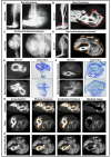
Fig 4. MicroCT can evaluate HO metrics in addition to volume.
(A) Average tissue mineral density (TMD) of tibia, soft tissue HO and bone-associated HO evaluated at 5 and 9 weeks post-op in burn/tenotomy model. (B) Representative cross sectional images of soft tissue HO (top two rows) and bone-associated HO (bottom row) at 5 and 9 weeks in burn/tenotomy model. (C) Tibial bone, soft tissue HO, and bone-associated HO in burn/tenotomy model evaluated for average porosity using 1250 HU threshold. (D) Burn/tenotomy HO shell and marrow space average volumes isolated with manual, trabecular contours (white) or by automated 800 HU threshold (blue). (E) Average TMD of soft tissue and bone-associated HO in burn/tenotomy mice that were: untreated, celecoxib-injected, or apyrase-injected. (F) Representative cross sectional images of soft tissue HO (top two rows) and bone-associated HO (bottom row) of untreated and treated burn/tenotomy groups at 9 weeks after injury. (G) Average porosity of soft tissue (left) and calcaneal (right) HO in burn/tenotomy untreated and treated groups.
Fig 5
Fig 5. MicroCT allows for earlier identification of HO.
(A) Plain film radiographs taken of injured limb in burn/tenotomy model 24 days after injury (red boxes: zoomed region of high risk HO site). (B) High resolution microCT image of injured limb and representative cross sections depicting mineral deposition in calcaneal (bone-associated) (left images) and tibial (right images) regions of burn/tenotomy mouse 24 days after injury. (C) Plain film radiographs taken of two Ad.cre/cardiotoxin-induced mice with minimal HO growth (red boxes: zoomed regions of injection sites) at day 22. (D) High resolution microCT image of both injected limbs and representative cross sections depicting mineral deposition in day 22 Ad.cre/cardiotoxin-induced mouse (red box: imaged for cross sectional detection of HO from proximal (top image) to distal (bottom image) regions of growth; red arrow: plane of injected limb imaged and magnified to view minimal HO growth).

References

    1. Shore EM, Xu M, Feldman GJ, Fenstermacher DA, Cho TJ, Choi IH, et al. A recurrent mutation in the BMP type I receptor ACVR1 causes inherited and sporadic fibrodysplasia ossificans progressiva. Nat Genet. 2006. 38: 525–527. - PubMed
    1. van Kuijk AA, Geurts AC, van Kuppevelt HJ. Neurogenic heterotopic ossification in spinal cord injury. Spinal Cord. 2002. 40: 313–326. - PubMed
    1. Peterson JR, De La Rosa S, Eboda O, Cilwa KE, Agarwal S, Buchman SR. et al. Treatment of heterotopic ossification through remote ATP hydrolysis. Sci Transl Med. 2014. 6: 255ra132 10.1126/scitranslmed.3008810 - DOI - PMC - PubMed
    1. Thomas BJ, Amstutz HC. Results of the administration of diphosphonate for the prevention of heterotopic ossification after total hip arthroplasty. J Bone Joint Surg Am. 1985. 67: 400–403. - PubMed
    1. Banovac K, Williams JM, Patrick LD, Haniff YM. Prevention of heterotopic ossification after spinal cord injury with indomethacin. Spinal Cord. 2001. 39: 370–374. - PubMed

Publication types

LinkOut - more resources