. 2015 Nov 6;10(11):e0142329.
doi: 10.1371/journal.pone.0142329. eCollection 2015.

Merkel Cell Polyomavirus Small T Antigen Induces Cancer and Embryonic Merkel Cell Proliferation in a Transgenic Mouse Model

Affiliations

Merkel Cell Polyomavirus Small T Antigen Induces Cancer and Embryonic Merkel Cell Proliferation in a Transgenic Mouse Model

Masahiro Shuda et al. PLoS One. .

Abstract

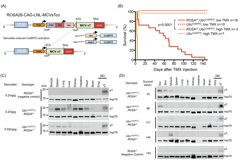
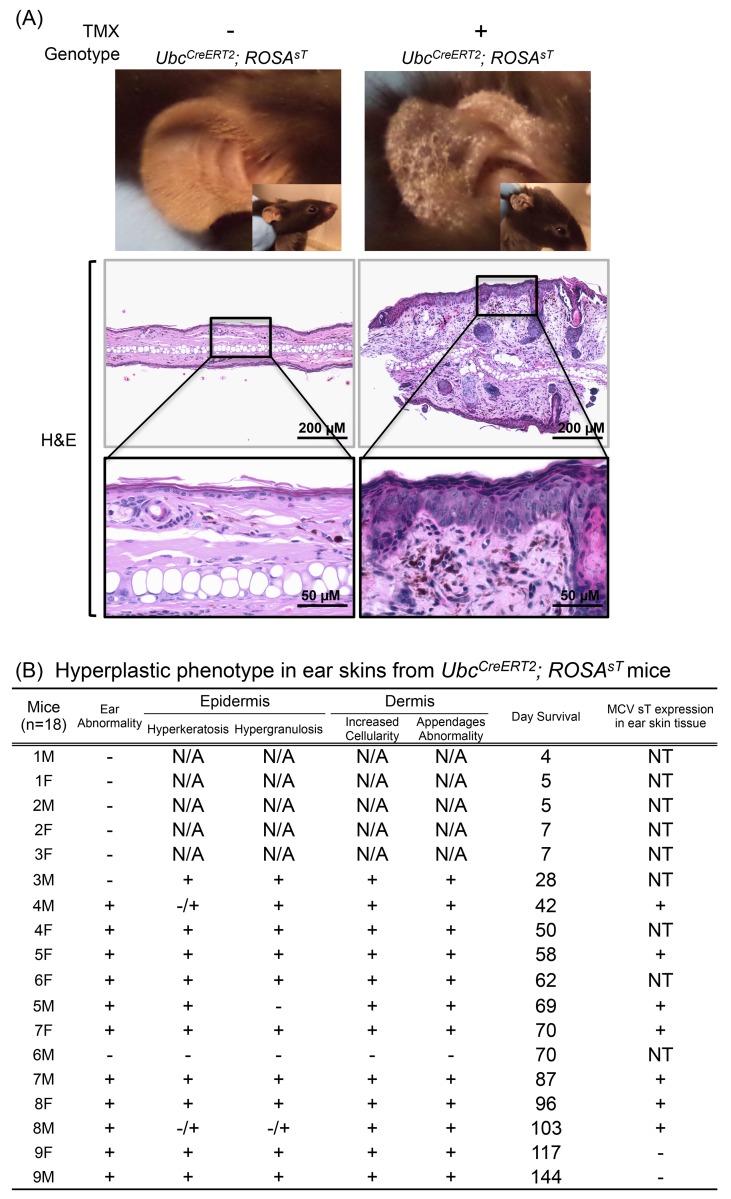
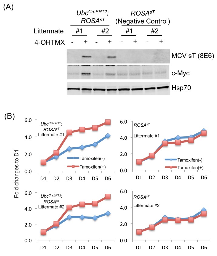
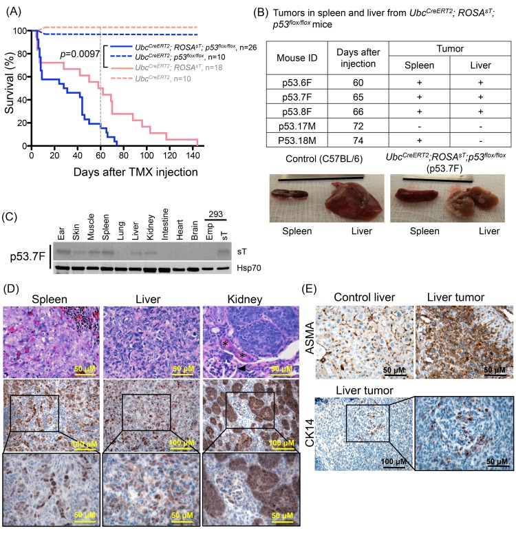
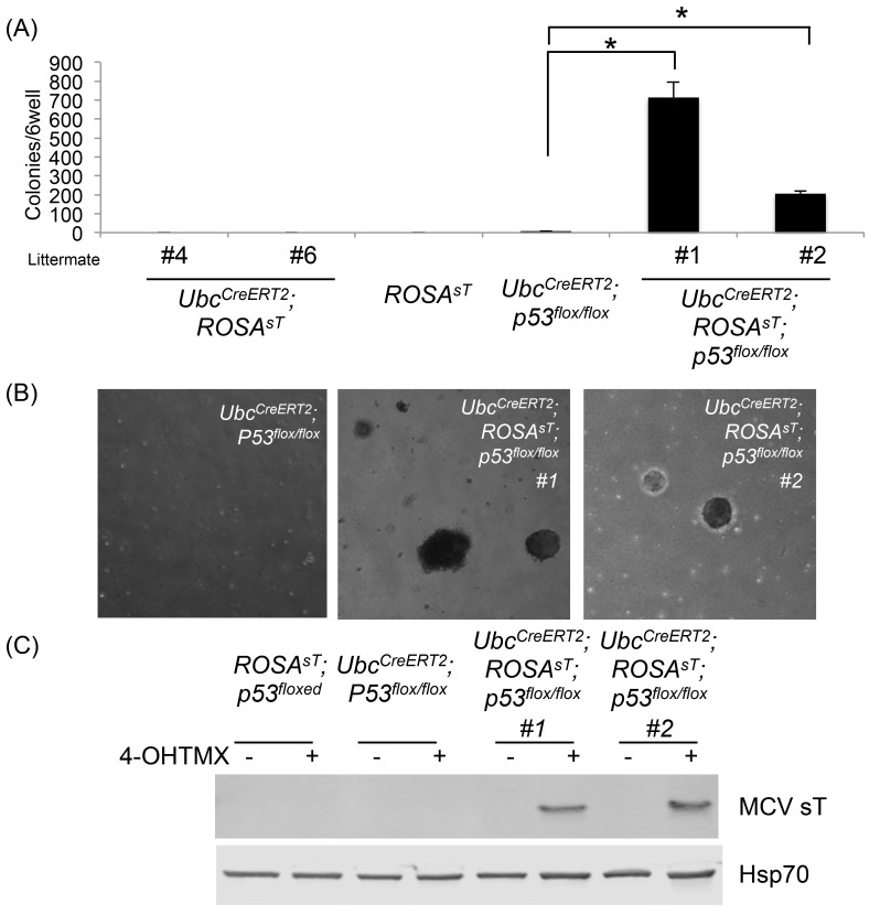
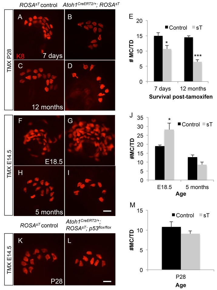
Merkel cell polyomavirus (MCV) causes the majority of human Merkel cell carcinomas (MCC) and encodes a small T (sT) antigen that transforms immortalized rodent fibroblasts in vitro. To develop a mouse model for MCV sT-induced carcinogenesis, we generated transgenic mice with a flox-stop-flox MCV sT sequence homologously recombined at the ROSA locus (ROSAsT), allowing Cre-mediated, conditional MCV sT expression. Standard tamoxifen (TMX) administration to adult UbcCreERT2; ROSAsT mice, in which Cre is ubiquitously expressed, resulted in MCV sT expression in multiple organs that was uniformly lethal within 5 days. Conversely, most adult UbcCreERT2; ROSAsT mice survived low-dose tamoxifen administration but developed ear lobe dermal hyperkeratosis and hypergranulosis. Simultaneous MCV sT expression and conditional homozygous p53 deletion generated multi-focal, poorly-differentiated, highly anaplastic tumors in the spleens and livers of mice after 60 days of TMX treatment. Mouse embryonic fibroblasts from these mice induced to express MCV sT exhibited anchorage-independent cell growth. To examine Merkel cell pathology, MCV sT expression was also induced during mid-embryogenesis in Merkel cells of Atoh1CreERT2/+; ROSAsT mice, which lead to significantly increased Merkel cell numbers in touch domes at late embryonic ages that normalized postnatally. Tamoxifen administration to adult Atoh1CreERT2/+; ROSAsT and Atoh1CreERT2/+; ROSAsT; p53flox/flox mice had no effects on Merkel cell numbers and did not induce tumor formation. Taken together, these results show that MCV sT stimulates progenitor Merkel cell proliferation in embryonic mice and is a bona fide viral oncoprotein that induces full cancer cell transformation in the p53-null setting.


Conflict of interest statement

Competing Interests: The authors have declared that no competing interests exist.

Figures

Fig 1
Fig 1. MCV sT expression is lethal in mice.
(A) Design of the ROSA26-CAG-LNL-MCVsTco, a ROSA26 knock-in construct encoding codon-optimized MCV sT with flanking loxP-STOP-Neo-loxP (LNL) sequence, recombined with the ROSA26 genomic locus. ROSA26-CAG-LNL-MCVsTco crossed with Ubc CreERT2 or Atoh1 CreERT2 mice allows for TMX-induced Cre recombinase excision of the loxP-STOP-Neo-loxP sequence that results in MCV sT expression under the CAG promoter. (B) MCV sT expression is lethal in Ubc CreERT2 ; ROSA sT transgenic mice. Kaplan-Meier curve of high dose (0.2 mg/g) and low dose (0.02 mg/g) TMX injected mice. High dose TMX injection caused rapid weight loss and mice reached the euthanasia criterion (20% loss of body weight) within 5 days of the injection (n = 4, solid orange). Survival of the control Ubc CreERT2 mice (n = 7, dotted orange) was not affected by TMX injection. Ubc CreERT2 ; ROSA sT mice injected with lower dose TMX exhibited significantly prolonged survival compared to high dose TMX (n = 18, solid red) and control mice were unaffected (n = 10, dotted red) (C) Multi-tissue MCV sT protein expression in Ubc CreERT2 ; ROSA sT mice that died immediately after high dose TMX injection (< 5 days). Lower dose TMX injection induces less MCV sT protein expression in a mouse that died at day 7 after the injection as detected by immunoblotting using an antibody raised against MCV sT, CM8E6 [22]. MCV sT vector or empty vector transfected HEK293 cells were used as a positive and a negative control, respectively. Equal amounts of sT-transfected HEK293 cell lysates were loaded for normalization across different blots. Hsp70/Hsc70 was detected as a protein quality control. (D) MCV sT protein expression was maintained in tissues of Ubc CreERT2 ; ROSA sT mice that survived over 70 days after low dose TMX injection. Immunoblots were performed as in (C). Tissue lysates from ROSA sT were used as a negative control.
Fig 2
Fig 2. MCV sT induces hyperproliferaton of acral skin.
(A) Hyperplastic phenotype in ear skin from Ubc CreERT2 ; ROSA sT mice. Ear skin tissues from Ubc CreERT2 ; ROSA sT mice were subjected to H&E staining. (B) Hyperproliferative phenotype in ear skin of Ubc CreERT2 ; ROSA sT mice. H&E stained ear skin sections from low-dose TMX injected Ubc CreERT2 ; ROSA sT and ROSA sT mice were randomized and evaluated in a blinded manner by two pathologists for hyperkeratosis/hypergranulosis in the epidermis and increased cellularity/appendage abnormalities. N/A: not available since ear tissues were normal and not harvested. NT: not tested.
Fig 3
Fig 3. MCV sT induces hyperproliferaton in MEF cells.
(A) Induction of MCV sT in multiple mouse embryonic fibroblast (MEF) cells from littermate by 4-OHTMX. MEFs extracted from Ubc CreERT2 ; ROSA sT and ROSA sT embryos were cultured in the presence or absence of 500 nM 4-OHTMX for 7 days. MCV sT and c-Myc protein expression was detected by immunoblot. Hsp/Hsc70 protein expression was detected as a loading control. (B) MCV sT expression accelerates cell proliferation in MEF cells. Proliferation of MEF cells treated with or without 4-OHTMX was evaluated by Wst1 assays.
Fig 4
Fig 4. MCV sT transgenic mice develop tumors in a p53 null setting.
(A) p53 ablation does not rescue mice from MCV sT-induced lethality. Kaplan-Meier curve of low dose (0.02 mg/g) TMX injected Ubc CreERT2 ; ROSA sT ; p53 flox/flox mice (Solid blue line, n = 26) and Ubc CreERT2 ; p53 flox/flox mice (dotted blue line, n = 10). Pink lines indicate the survival of Ubc CreERT2 ; ROSA sT (solid line) and Ubc CreERT2 (dotted line) mice with wild type p53 as shown in Fig 1B for comparison with p53 knockout background (blue lines). (B) MCV sT expression produces tumors in vivo in p53 null setting. 80% (4/5) of mice that survived over 60 days after TMX injection develop grossly visible tumor in the spleen and liver. Representative spleen and liver tissues with tumor nodules from p53.7F are shown with corresponding normal tissues from a C57BL/6 control mouse. (C) Immunoblotting of MCV sT protein expression in liver and spleen tissues from mouse p53.7F) with macroscopic tumors. Spleen, muscle and ear tissues consistently maintained sT expression over 60 days after TMX injection. sT expression was detectable in liver tissues by immunoblotting only from liver with visible nodules (mouse p53.7F). MCV sT protein was detected using CM8E6 antibody, and Hsp/Hsc70 expression was used as a loading control. (D) The top panel shows anaplastic neoplasia in the spleen and liver. A range of proliferative changes was observed in the kidney, where distal tubular epithelia are most severely affected, but glomeruli (black arrowhead), proximal tubular epithelia (*), and interstitial tissues are relatively spared of proliferative changes. The middle panel shows a representative immunohistochemical staining of sT protein expression in liver tumor, spleen tumor and kidney tissues from Ubc CreERT2 ; ROSA sT ; p53 flox/flox mice. Tissue samples were immunostained with MCV sT (CM5E1) antibody. (E) The top panel shows a sT-induced liver tumor with immunoreactivity to α-smooth muscle actin (ASMA) and bottom panel shows K14 positivity in scattered tumor cells.
Fig 5
Fig 5. p53 ablation is critical for MCV sT transformation of MEF cells.
(A) MCV sT induces soft agar colony formation in the absence of p53 in MEF cells. MEF cells isolated form Ubc CreERT2 ; ROSA sT, ROSA sT, Ubc CreERT2 ; p53 flox/flox and two Ubc CreERT2 ; ROSA sT ; p53 flox/flox embryos from littermate were treated with 500nM 4OHTMX over 7 days, and cells were subjected to soft agar colony formation assay. Asterisks(*) indicate statistical significance p<0.05. (B) Soft agar colonies induced by p53 ablation. p53 ablation alone did not lead to colony formation. (C) Expression of MCV sT in MEFs from Ubc CreERT2 ; ROSA sT ; p53 flox/flox.
Fig 6
Fig 6. MCV sT expression increases Merkel cell numbers only during embryogenesis.
Wholemount, K8-immunostained skin showing touch domes from control littermate (A, C, F, H, K), Atoh1 CreERT2/+; ROSA sT (B, D, G, I) and Atoh1 CreERT2/+; ROSA sT; p53 flox/flox (L) mice given TMX at P28 (A-D) or E14.5 (F-I, K, L). Age at tissue harvest is shown for each pair of panels. (E, J, M) Graphs showing Merkel cell counts per touch dome for accompanying panels. *p < 0.05, ***p<0.001. Scale bar: 10μm.

References

    1. Feng H, Shuda M, Chang Y, Moore PS. Clonal integration of a polyomavirus in human Merkel cell carcinoma. Science. 2008;319(5866):1096–100. 10.1126/science.1152586 - DOI - PMC - PubMed
    1. DeCaprio JA, Garcea RL. A cornucopia of human polyomaviruses. Nature reviews Microbiology. 2013;11(4):264–76. 10.1038/nrmicro2992 - DOI - PMC - PubMed
    1. Feng H, Taylor JL, Benos PV, Newton R, Waddell K, Lucas SB, et al. Human transcriptome subtraction by using short sequence tags to search for tumor viruses in conjunctival carcinoma. Journal of virology. 2007;81(20):11332–40. 10.1128/JVI.00875-07 - DOI - PMC - PubMed
    1. Borozan I, Watt SN, Ferretti V. Evaluation of alignment algorithms for discovery and identification of pathogens using RNA-Seq. PloS one. 2013;8(10):e76935 10.1371/journal.pone.0076935 - DOI - PMC - PubMed
    1. Shuda M, Feng H, Kwun HJ, Rosen ST, Gjoerup O, Moore PS, et al. T antigen mutations are a human tumor-specific signature for Merkel cell polyomavirus. Proc Natl Acad Sci U S A. 2008;105(42):16272–7. 10.1073/pnas.0806526105 - DOI - PMC - PubMed

Publication types

MeSH terms

Substances