. 2015 Nov 6;10(11):e0142424.
doi: 10.1371/journal.pone.0142424. eCollection 2015.

Pretreatment with Resveratrol Prevents Neuronal Injury and Cognitive Deficits Induced by Perinatal Hypoxia-Ischemia in Rats

Affiliations

Pretreatment with Resveratrol Prevents Neuronal Injury and Cognitive Deficits Induced by Perinatal Hypoxia-Ischemia in Rats

Olatz Arteaga et al. PLoS One. .

Abstract

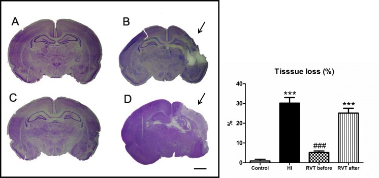
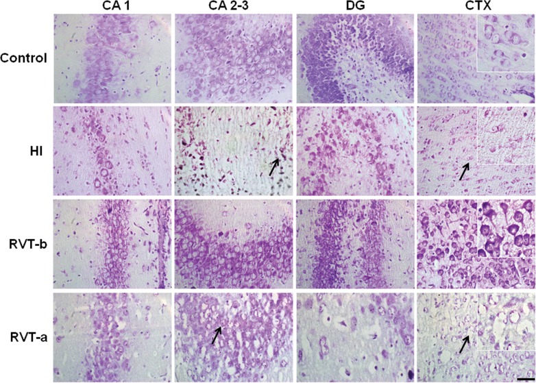
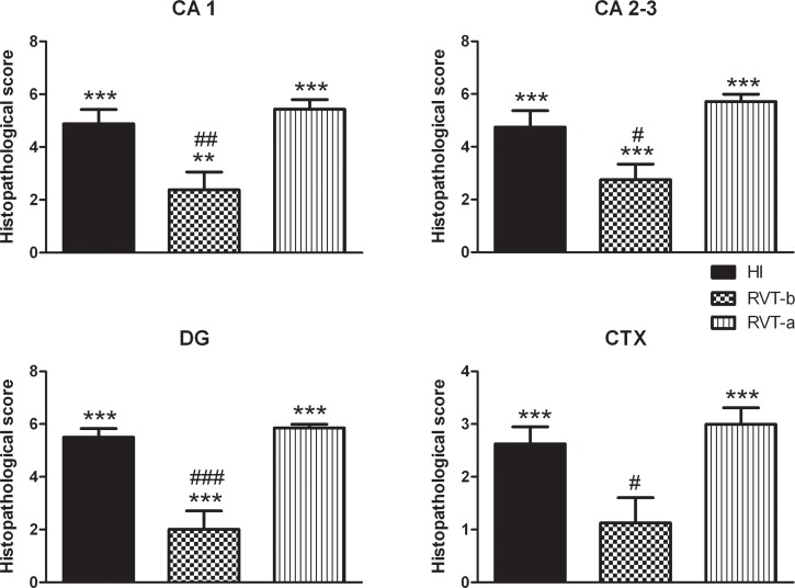
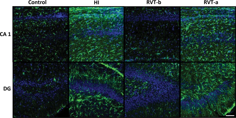
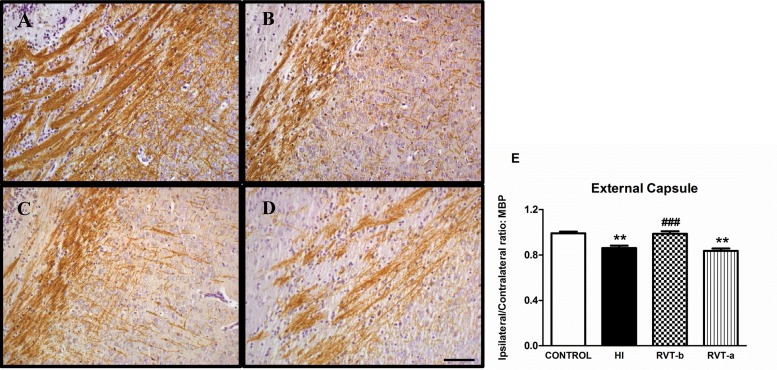
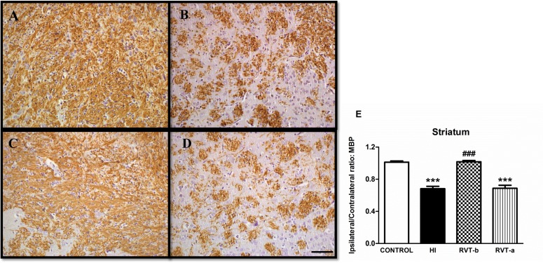
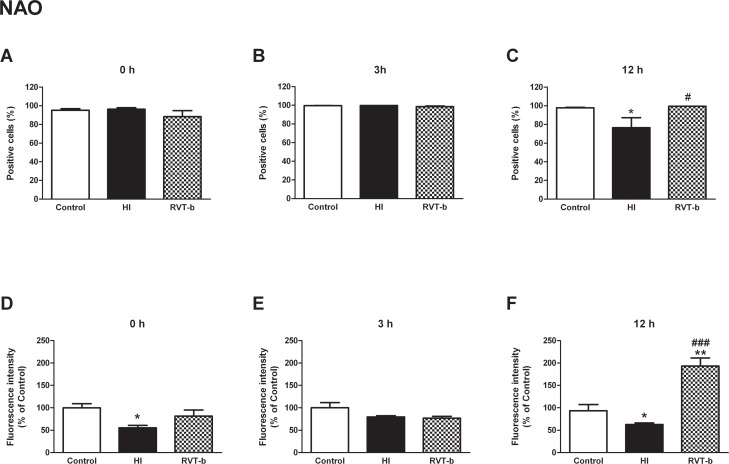
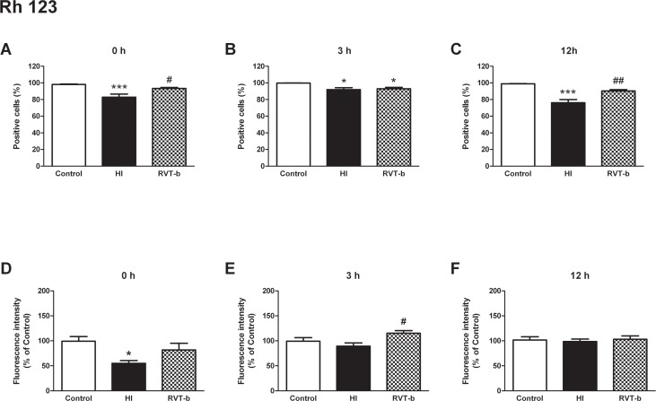
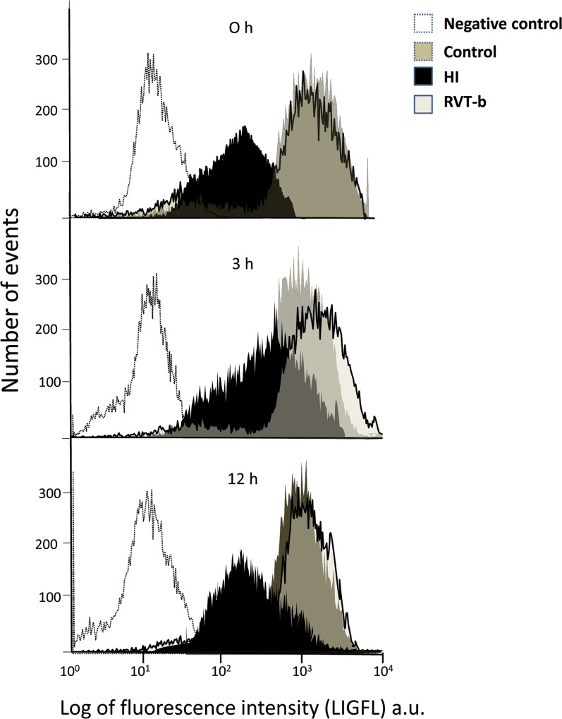
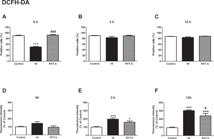
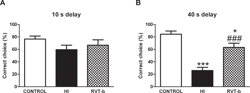
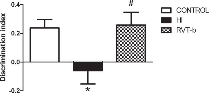
Despite advances in neonatal care, hypoxic-ischemic brain injury is still a serious clinical problem, which is responsible for many cases of perinatal mortality, cerebral palsy, motor impairment and cognitive deficits. Resveratrol, a natural polyphenol with important anti-oxidant and anti-inflammatory properties, is present in grapevines, peanuts and pomegranates. The aim of the present work was to evaluate the possible neuroprotective effect of resveratrol when administered before or immediately after a hypoxic-ischemic brain event in neonatal rats by analyzing brain damage, the mitochondrial status and long-term cognitive impairment. Our results indicate that pretreatment with resveratrol protects against brain damage, reducing infarct volume, preserving myelination and minimizing the astroglial reactive response. Moreover its neuroprotective effect was found to be long lasting, as behavioral outcomes were significantly improved at adulthood. We speculate that one of the mechanisms for this neuroprotection may be related to the maintenance of the mitochondrial inner membrane integrity and potential, and to the reduction of reactive oxygen species. Curiously, none of these protective features was observed when resveratrol was administered immediately after hypoxia-ischemia.


Conflict of interest statement

Competing Interests: The authors have declared that no competing interests exist.

Figures

Fig 1
Fig 1. Brain tissue loss induced by hypoxia-ischemia in 7 day-old rats and evaluated at P14.
Representative stereomicroscopic photographs of 5 μm Nissl-stained brain sections (interaural distance 5.40 mm and bregma -3.60 mm) are shown. (A) Control group with normal morphological brain (n = 8). (B) HI brain with evident loss of tissue in the ipsilateral side of the cortex, and with obvious damage to the hippocampus (region indicated by arrows) (n = 10). (C) Brain treated with resveratrol 10 minutes before hypoxia, which is similar to the control brain (n = 10). (D) HI brain treated with resveratrol immediately after hypoxia, with obvious signs of infarct, as denoted by the arrow (n = 10). Scale bar: 2.5 mm. The histogram illustrates percentage of tissue loss is expressed. Asterisks denote the significance levels when compared to the control group (*** P<0.0001). The hash denotes the significance levels when compared to the HI group (### P<0.0001).
Fig 2
Fig 2. Representative microphotographs of Nissl-stained brain sections in animals exposed to hypoxia-ischemia at P7 and evaluated at P14.
Individual fields represent different experimental groups and different areas of the hippocampus (CA 1, CA 2–3 and DG) and from the parietal cortex (CTX), with high magnification insets in CTX. Cell loss was especially evident in the DG and parietal cortex (arrows) in the hypoxia-ischemia and resveratrol post-treatment groups. In contrast, the resveratrol pre-treated group showed a remarkable conservation in the cellularity of the different studied areas with respect to the HI group. Scale bar: 50 μm.
Fig 3
Fig 3. Histopathological score of damage in P14 rat brains of different groups expressed as the mean ± SEM.
Asterisks denote the significance levels when compared to the control group (*** P<0.0001). The control group (n = 11) is not shown, because it histopathological score for all areas was 0. The hash symbols denote the significance levels when compared to the HI group (### P<0.0001). It can be clearly seen that the group pretreated with resveratrol (n = 8) had a lower histopathological score compared with the HI group (n = 8). This was clearly not the case for the post-resveratrol treated group (n = 8).
Fig 4
Fig 4. Representative confocal microphotographs of glial fibrillary acidic protein (GFAP)-immunoreactivity in brain sections counterstained with DAPI.
On postnatal day 14, GFAP immunoreactivity (green) was particularly pronounced in the vicinity of damaged areas and this reactivity was substantially reduced in animals pre-treated with resveratrol. Scale bar: 40 μm.
Fig 5
Fig 5. Representative light microphotographs of myelin basic protein (MBP)-stained brain sections and comparison of loss of MBP immunostaining in the external capsule of different groups: 14-day-old control (n = 5) (A), HI (n = 14) (B), RVT-b (n = 14) (C) and RVT-a (n = 7) (D) (scale bar: 40 μm).
In the histogram (E), the extent of tissue injury, expressed as a ratio of left-to-right hemispheric MBP immunostaining is represented. Asterisks denote the significance levels when compared to the control group (** P<0.0001). Hashes denote the significance level when compared to the HI group (### P<0.0001).
Fig 6
Fig 6. Representative light microphotographs of myelin basic protein (MBP)-stained brain sections and comparison of loss of MBP immunostaining in the striatum of different groups: 14-day-old control (n = 6) (A), HI (n = 15) (B), RVT-b (n = 13) (C) and RVT-a (n = 7) (D) (scale bar: 40 μm).
The histogram (E) illustrates the extent of tissue injury associated with the various treatments and values are expressed as a ratio of left-to-right hemispheric MBP immunoreactivity levels. Asterisks denote the significance levels when compared to the control group (***P<0.0001). Hashes denote the significance levels when compared to the HI group (### P<0.0001).
Fig 7
Fig 7. Mitochondrial inner membrane integrity evaluation in suspension of acutely isolated cells using nonyl acridine orange (NAO).
Percentage of NAO-positive cells at different time points after hypoxia-ischemia: (A) 0 h, (B) 3 h and (C) 12 h. Relative fluorescence intensity of cells with in vivo marker NAO at different time points after hypoxia-ischemia: (D) 0 h, (E) 3 h and (F) 12 h, in control (n≥5), HI (n≥5) and animals pretreated with resveratrol (n≥5). Asterisk denotes the significance levels when compared to the control group (* P<0.05). The hash symbol denotes the significance levels when compared to the HI group (# P<0.05).
Fig 8
Fig 8. Analysis of mitochondrial transmembrane potential by Rhodamine 123 in suspension of acutely isolated cells.
Percentage of cells labeled with the in vivo marker Rh 123 at different time points after hypoxia-ischemia: (A) 0 h, (B) 3 h and (C) 12 h. Relative fluorescence intensity of cells exhibiting Rh 123 fluorescence at different time points after hypoxia-ischemia: (D) 0 h, (E) 3 h and (F) 12 h, in control (n≥5), HI (n≥5) and animals pretreated with resveratrol (n≥5) groups. Asterisks denote the significance levels when compared to the control group (*** P<0.0001 or * P<0.05). The hash symbols denote the significance levels when compared to the HI group (## P<0.005 or # P<0.05).
Fig 9
Fig 9. Representative fluorograms obtained after flow cytometry analysis showing mitochondrial transmembrane potential measured as Rh 123 fluorescence at different points of time after hypoxia-ischemia (0 h, 3 h and 12 h) in control, HI and animals pretreated with resveratrol.
The x-axis represents the number of events and the y-axis represents the values of fluorescence intensity in logarithm values. The negative control consisted of unstained samples from each animal to remove the autofluorescence.
Fig 10
Fig 10. Effect of brain hypoxia-ischemia on the production of reactive oxygen species in suspension of acutely isolated cells, measured using DCFH-DA.
Percentage of DCFH-DA positive cells at different time points after hypoxia-ischemia: (A) 0 h, (B) 3 h and (C) 12 h. Relative fluorescence intensity of cells with in vivo marker DCFH-DA at different time points after hypoxia-ischemia; (D) 0 h, (E) 3 h and (F) 12 h, in control (n≥5), HI (n≥5) and animals pretreated with resveratrol (n≥5) groups. Asterisks denote the significance levels when compared to the control group (*** P<0.0001 * P<0.05). The hash symbols denote the significance levels when compared to the hypoxia-ischemia group (### P<0.0001 or # P<0.05).
Fig 11
Fig 11. Evaluation of spontaneous locomotor activity in the open field test performed at P90 in control (n = 16), HI (n = 14) and RVT (20 mg/kg) treated animals (n = 10).
Evaluated parameters were (A) % time in the periphery, (B) % time in the center, (C) total distance travelled and (D) speed in the open field.
Fig 12
Fig 12. Effect of neonatal hypoxia-ischemia and resveratrol pretreatment on the hole-board test performed at P90.
(A) The frequency and (B) the time spent head-dipping into the holes were recorded in control (n = 16), HI (n = 14) and RVT (20 mg/kg) treated animals (n = 10). Asterisks denote the significance levels when compared to the control group (**P < 0.005).
Fig 13
Fig 13. Effect of neonatal hypoxia-ischemia and pretreatment with resveratrol on choice accuracy in the discrete-trial delayed spatial alternation task (T-maze) in adult animals on P90.
(A) Control (n = 16) and HI rats (n = 14), as well as RVT pretreated animals (n = 10) made a similar number of correct choices in the T-maze at 10 s delay. (B) In contrast, HI animals made significantly fewer correct choices after the 40 s delay. Impaired memory performance (percentage of correct trials) due to hypoxia-ischemia was reverted by RVT pre-administration. Asterisks denote the significance levels when compared to the control group (*** P<0.0001). The hash symbols denote the significance levels when compared to the HI group (### P<0.0001).
Fig 14
Fig 14. Effect of neonatal hypoxia-ischemia and treatment with resveratrol on the results of a novel object recognition test.
On P90, HI adult animals (n = 14) displayed a decrease in discrimination index when compared to control animals (n = 16) that was fully reversed by resveratrol pretreatment (n = 10). Asterisk denotes the significance levels when compared to the control group (* P<0.05). The hash symbol denotes the significance levels when compared to the HI group (# P<0.05).

References

    1. du Plessis AJ, Volpe JJ. Perinatal brain injury in the preterm and term newborn. Curr Opin Neurol 2002. April;15(2):151–157. - PubMed
    1. Azra Haider B, Bhutta ZA. Birth asphyxia in developing countries: current status and public health implications. Curr Probl Pediatr Adolesc Health Care 2006. May-Jun;36(5):178–188. - PubMed
    1. Jiang H, Lei JJ, Zhang YH. Protective effect of topiramate on hypoxic-ischemic brain injury in neonatal rat. Asian Pac J Trop Med 2014. June;7(6):496–500. 10.1016/S1995-7645(14)60082-1 - DOI - PubMed
    1. Volpe JJ. Perinatal brain injury: from pathogenesis to neuroprotection. Ment Retard Dev Disabil Res Rev 2001;7(1):56–64. - PubMed
    1. Low JA. Determining the contribution of asphyxia to brain damage in the neonate. J Obstet Gynaecol Res 2004. August;30(4):276–286. - PubMed

Publication types

MeSH terms

LinkOut - more resources