. 2015 Nov 6;10(11):e0141126.
doi: 10.1371/journal.pone.0141126. eCollection 2015.

Identification of Small-Molecule Inhibitors against Meso-2, 6-Diaminopimelate Dehydrogenase from Porphyromonas gingivalis

Affiliations

Identification of Small-Molecule Inhibitors against Meso-2, 6-Diaminopimelate Dehydrogenase from Porphyromonas gingivalis

Victoria N Stone et al. PLoS One. .

Abstract

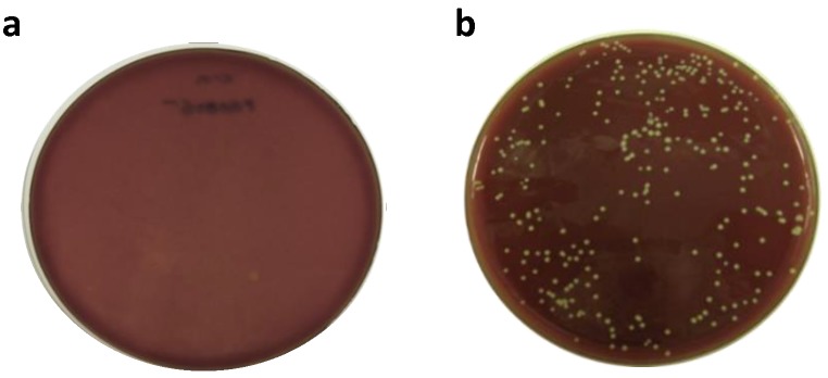
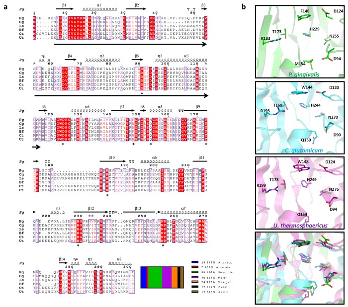
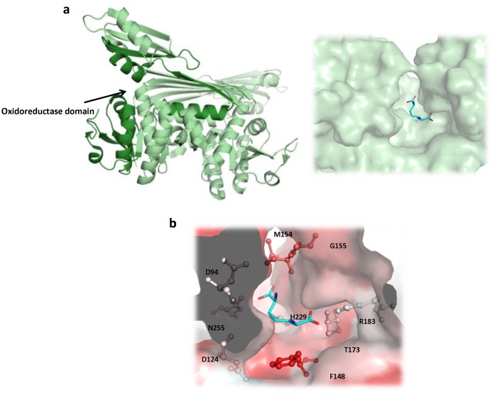
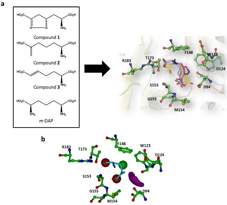
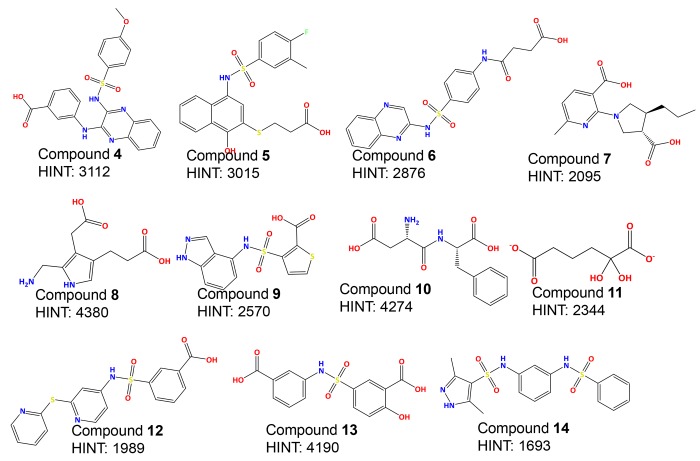
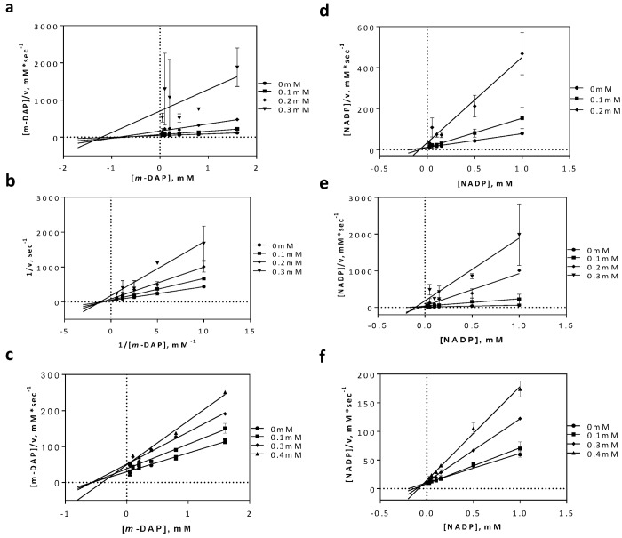
Species-specific antimicrobial therapy has the potential to combat the increasing threat of antibiotic resistance and alteration of the human microbiome. We therefore set out to demonstrate the beginning of a pathogen-selective drug discovery method using the periodontal pathogen Porphyromonas gingivalis as a model. Through our knowledge of metabolic networks and essential genes we identified a "druggable" essential target, meso-diaminopimelate dehydrogenase, which is found in a limited number of species. We adopted a high-throughput virtual screen method on the ZINC chemical library to select a group of potential small-molecule inhibitors. Meso-diaminopimelate dehydrogenase from P. gingivalis was first expressed and purified in Escherichia coli then characterized for enzymatic inhibitor screening studies. Several inhibitors with similar structural scaffolds containing a sulfonamide core and aromatic substituents showed dose-dependent inhibition. These compounds were further assayed showing reasonable whole-cell activity and the inhibition mechanism was determined. We conclude that the establishment of this target and screening strategy provides a model for the future development of new antimicrobials.


Conflict of interest statement

Competing Interests: The authors have declared that no competing interests exist.

Figures

Fig 1
Fig 1. Allelic replacement mutagenesis for predicted essential and non-essential gene.
(a) Predicted essential gene PG0806 was transformed and plated on antibiotic selective media resulting in no colony formation, validating prediction that the gene was essential. (b) Predicted non-essential gene PG0027 was transformed and plated on antibiotic selective media resulting in colony formation, validating prediction that the gene was not essential. Transformations were performed two independent times.
Fig 2
Fig 2. Protein sequence and structural alignment of m-Ddh.
(a) Sequence alignment of m-Ddh from other bacterial organisms. Sequences were aligned using T-Coffee and the multiple alignment was then created in Espript 3.0. The putative binding sites of Corynebacterium glutamicum (Cg), Lysinibacillus sphaericus (Ls), Bacteroides fragilis (Bf), Clostridium thermocellum (Ct) and Ureibacillus thermosphaericus (Ut) cited in the sequence annotations in UniProtKB/Swiss-Prot and P. gingivalis (Pg) predicted based on homology are indicated by astericks. The oxidoreductase domain for P. gingivalis is indicated by arrows below sequence. Secondary structure for P. gingivalis is annotated above the sequence. Relative percentage of characterized amino acid residues are shown below. (b) Secondary structure alignment of m-Ddh’s putative binding site from P. gingivalis (green), C. glutamicum (cyan) and U. thermosphaericus (purple). Key residues are labeled, side chains are displayed as sticks and colored corresponding to atom type. Hydrogens were omitted for clarity.
Fig 3
Fig 3. Cartoon representation of m-Ddh protein structure.
(a) Ribbon based structure of m-Ddh with a zoomed in view of the substrate m-DAP binding cavity (b) and the hydrophobicity of binding site protein surface (red = hydrophobic).
Fig 4
Fig 4. Generation of pharmacophore model for the high-throughput virtual screen.
(a) Structure of m-DAP and inhibitor analogs that were previously shown to be active against m-Ddh in C. glutamicum and B. sphaericus. (b) Compounds docked into m-Ddh binding site and conserved interactions were identified. (c) Pharmacophore model with selected core features for inhibitor identification during virtual screen. The model focused on four features: first, a hydrophobic region complementary to amino acid residues Trp123 and Phe148 (green), second, a ligand donor atom complementary to residues Asp94 and Asp124 (purple), third, a negative center complementary to the side chain of residue Ser153 and the backbone of residues Met154 and Gly155 (red) and fourth, a negative center complementary to the side chain of residues Arg183 and Thr173 (red). The interaction was also restricted for an area 12Å in distance for Arg183. Key residues are labeled, displayed as ball and sticks and colored corresponding to atom type. Hydrogens were omitted for clarity.
Fig 5
Fig 5. Structure and scoring of top-ranking inhibitors.
Fig 6
Fig 6. Time-kill analysis of P. gingivalis treated with Compound 4 and 5.
P. gingivalis cells were treated with 5x the previously determined MIC for either Compound 4 (triangle) or Compound 5 (square) and bacterial cell counts were assessed at 0, 0.25, 0.5, 1, 2, 3, 4, 6 and 24 hours. The mean plus the standard deviation is shown for each time point from a minimum of n = 3 independent experiments. For cell counts equal to 0 CFU/mL, 1 was used for the log transformation.
Fig 7
Fig 7. SEM analysis of P. gingivalis cells treated with Compound 4 and 5.
(a) Untreated cells. (b) Compound 4 treated cells at 5x the previously determined MIC concentration. (c) Compound 5 treated cells at 5x the previously determined MIC concentration. (d) Ampicillin treated cells.
Fig 8
Fig 8. Docking and binding interaction of three active compounds in complex with m-Ddh.
(a) Compound 4 (b) Compound 5 and (c) Compound 6. Key residues are labeled, displayed as ball and sticks and colored corresponding to atom type. Hydrogens were omitted for clarity. Potential hydrogen bonding interactions between m-Ddh residues and inhibitors are shown by yellow dashed lines.
Fig 9
Fig 9. Inhibition mechanism of active compounds in regards to substrate, m-DAP and co-substrate, NADP+.
(a) Compound 4 (b) Compound 5 and (c) Compound 6 inhibition mechanisms against m-DAP. (d) Compound 4 (e) Compound 5 and (f) Compound 6 inhibition mechanisms against NADP+.

References

    1. Koul A, Arnoult E, Lounis N, Guillemont J, Andries K. The challenge of new drug discovery for tuberculosis. Nature. 2011;469(7331):483–90. 10.1038/nature09657 - DOI - PubMed
    1. DiMasi JA, Hansen RW, Grabowski HG. The price of innovation: new estimates of drug development costs. Journal of health economics. 2003;22(2):151–85. doi: S0167-6296(02)00126-1 [pii]. - PubMed
    1. Gwynn MN, Portnoy A, Rittenhouse SF, Payne DJ. Challenges of antibacterial discovery revisited. Annals of the New York Academy of Sciences. 2010;1213:5–19. 10.1111/j.1749-6632.2010.05828.x - DOI - PubMed
    1. Human Microbiome Project C. Structure, function and diversity of the healthy human microbiome. Nature. 2012;486(7402):207–14. 10.1038/nature11234 - DOI - PMC - PubMed
    1. Johnson CL, Versalovic J. The human microbiome and its potential importance to pediatrics. Pediatrics. 2012;129(5):950–60. 10.1542/peds.2011-2736 - DOI - PMC - PubMed

Publication types

MeSH terms