Review
. 2015 Nov 5;163(4):811-28.
doi: 10.1016/j.cell.2015.10.044.

Hippo Pathway in Organ Size Control, Tissue Homeostasis, and Cancer

Affiliations
Review

Hippo Pathway in Organ Size Control, Tissue Homeostasis, and Cancer

Fa-Xing Yu et al. Cell. .

Abstract

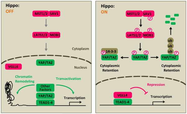
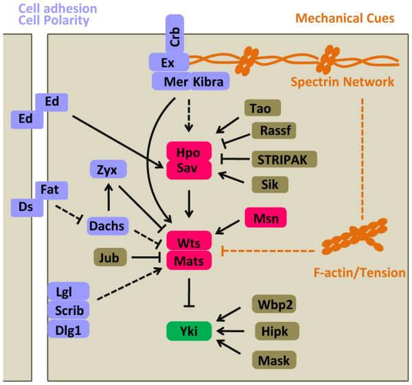
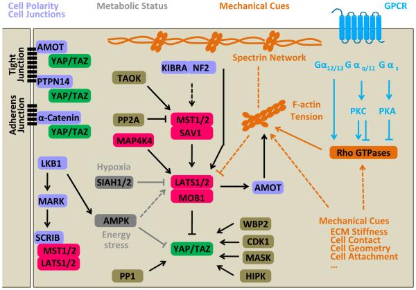
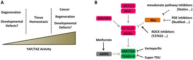
Two decades of studies in multiple model organisms have established the Hippo pathway as a key regulator of organ size and tissue homeostasis. By inhibiting YAP and TAZ transcription co-activators, the Hippo pathway regulates cell proliferation, apoptosis, and stemness in response to a wide range of extracellular and intracellular signals, including cell-cell contact, cell polarity, mechanical cues, ligands of G-protein-coupled receptors, and cellular energy status. Dysregulation of the Hippo pathway exerts a significant impact on cancer development. Further investigation of the functions and regulatory mechanisms of this pathway will help uncovering the mystery of organ size control and identify new targets for cancer treatment.


Figures

Figure 1
Figure 1
Inhibition of YAP/TAZ transcriptional coactivators by LATS1/2. (A) When Hippo signaling is off, YAP/TAZ enters the nucleus, competes with VGLL4 for TEADs, and recruits other factors to induce gene transcription. YAP/TAZ may bind proximal promoters or distal enhancers of target genes to induce transcription. (B) When Hippo signaling is on, YAP/TAZ is phosphorylated by LATS1/2 on multiple sites, resulting interaction with 14-3-3 and cytoplasmic retention; phosphorylation also leads to YAP/TAZ poly-ubiquitination and degradation. VGLL4 interacts with TEADs and represses target gene transcription.
Figure 2
Figure 2
Regulation of the Hippo pathway in Drosophila. The Hippo pathway is regulated by cell adhesion molecules (Ed), determinants of cell polarity (Crb, Fat/Ds, Scrib complex), and mechanical cues (spectrin, F-actin, or cellular tension). In addition, Hpo is regulated by Tao, salt induced kinase (Sik), Ras-associated factor (Rassf), striatin-interacting phosphatase and kinase (STRIPAK) complex; Wts is regulated by Zyxin (Zyx) and Jub; Yki is regulated by WW Domain Binding Protein 2 (Wbp2), Hipk, and Multiple ankyrin repeats single KH domain (Mask) (refer to (Varelas, 2014)). Arrows, blunt ends, and dashed lines indicate activation, inhibition, and indirect regulation, respectively.
Figure 3
Figure 3
Regulation of the Hippo pathway in mammals. The Hippo pathway is regulated by diverse signals: 1) Determinants of cell polarity and cell-cell junctions, such as SCRIB that interacts with MST1/2 and LATS1/2, AMOT, PTPN14, and α-Catenin, which can sequester YAP/TAZ to cell junctions; 2) Mechanical cues, such as stiffness, cell contact, cell geometry, and cell attachment status that regulate the Hippo pathway by modulating activity of Rho GTPases, remodeling the actin cytoskeleton, or altering cellular tension. Both apical and basolateral spectrin networks may function as sensors for mechanical cues in Hippo pathway regulation; 3) Soluble factors, especially ligands for GPCRs, regulate LATS1/2 likely through Rho GTPases and actin dynamics; 4) Metabolic status, such as cellular energy and oxygen stress, also regulate Hippo signaling. Many other proteins such as protein phosphatase 2A (PP2A), protein phosphatase 1 (PP1), WBP2, CDK1, MASK, and HIPK can also regulate activities of different Hippo pathway components (refer to (Varelas, 2014)). Arrows, blunt ends, and dashed lines indicate activation, inhibition, and indirect regulation, respectively.
Figure 4
Figure 4
Therapeutic targeting of the Hippo pathway. (A) Potential roles of YAP/TAZ activity in tissue development and diseases. A confined window of YAP/TAZ activity is required for normal tissue development and homeostasis. (B) Strategies for targeting YAP/TAZ activity. Inhibitors for MST1/2, MAP4K4, and LATS1/2 can activate YAP/TAZ. YAP/TAZ-TEAD interaction may be disrupted by small molecules directly (Verteporfin) or AMPK activators (Metformin). Small molecules inhibiting Rho family GTPases or ROCK can indirectly activate LATS1/2, leading to YAP/TAZ inhibition.

References

    1. Antonescu CR, Le Loarer F, Mosquera JM, Sboner A, Zhang L, Chen CL, Chen HW, Pathan N, Krausz T, Dickson BC, et al. Novel YAP1-TFE3 fusion defines a distinct subset of epithelioid hemangioendothelioma. Gene Chromosome Canc. 2013;52:775–784. - PMC - PubMed
    1. Aragona M, Panciera T, Manfrin A, Giulitti S, Michielin F, Elvassore N, Dupont S, Piccolo S. A mechanical checkpoint controls multicellular growth through YAP/TAZ regulation by actin-processing factors. Cell. 2013;154:1047–1059. - PubMed
    1. Azzolin L, Panciera T, Soligo S, Enzo E, Bicciato S, Dupont S, Bresolin S, Frasson C, Basso G, Guzzardo V, et al. YAP/TAZ Incorporation in the beta-Catenin Destruction Complex Orchestrates the Wnt Response. Cell. 2014;158:157–170. - PubMed
    1. Azzolin L, Zanconato F, Bresolin S, Forcato M, Basso G, Bicciato S, Cordenonsi M, Piccolo S. Role of TAZ as mediator of Wnt signaling. Cell. 2012;151:1443–1456. - PubMed
    1. Bai H, Zhang N, Xu Y, Chen Q, Khan M, Potter JJ, Nayar SK, Cornish T, Alpini G, Bronk S, et al. Yes-associated protein regulates the hepatic response after bile duct ligation. Hepatology. 2012;56:1097–1107. - PMC - PubMed

Publication types

Substances