. 2015 Nov 5;163(4):960-74.
doi: 10.1016/j.cell.2015.10.034.

Estrogen Receptor β Modulates Apoptosis Complexes and the Inflammasome to Drive the Pathogenesis of Endometriosis

Affiliations

Estrogen Receptor β Modulates Apoptosis Complexes and the Inflammasome to Drive the Pathogenesis of Endometriosis

Sang Jun Han et al. Cell. .

Abstract

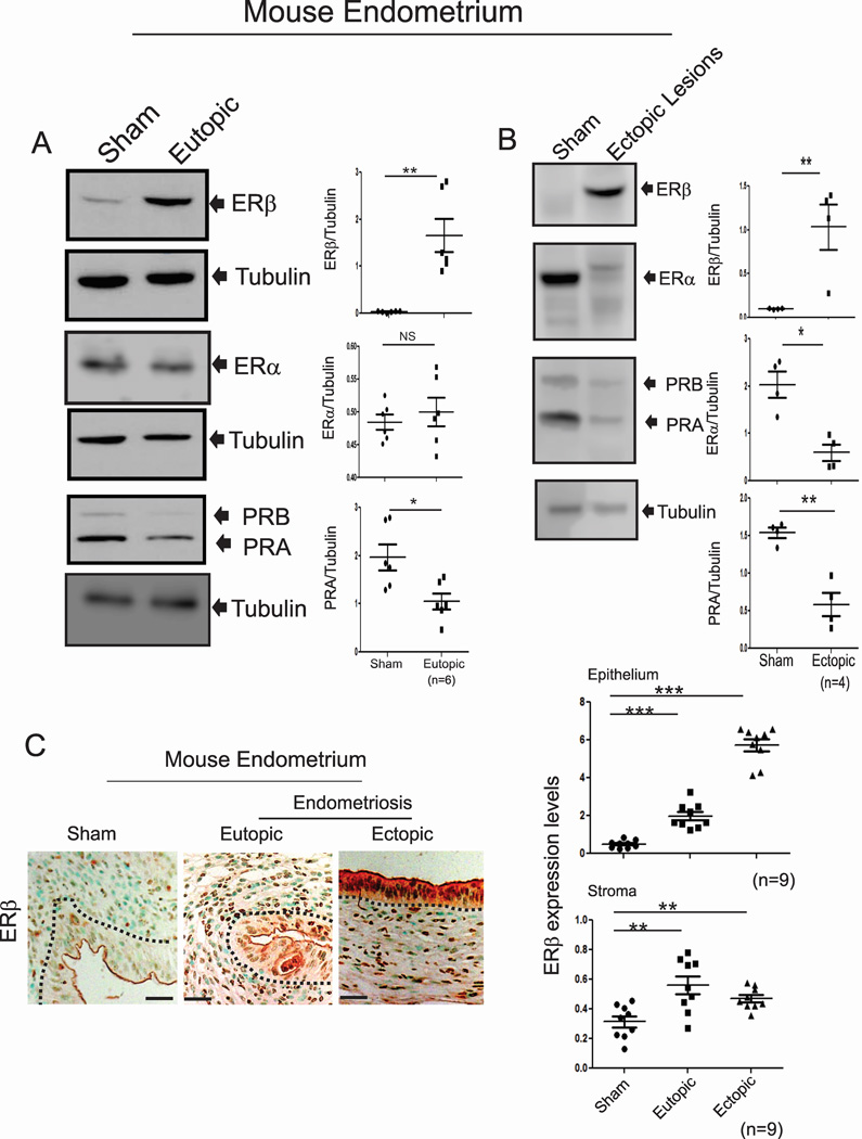
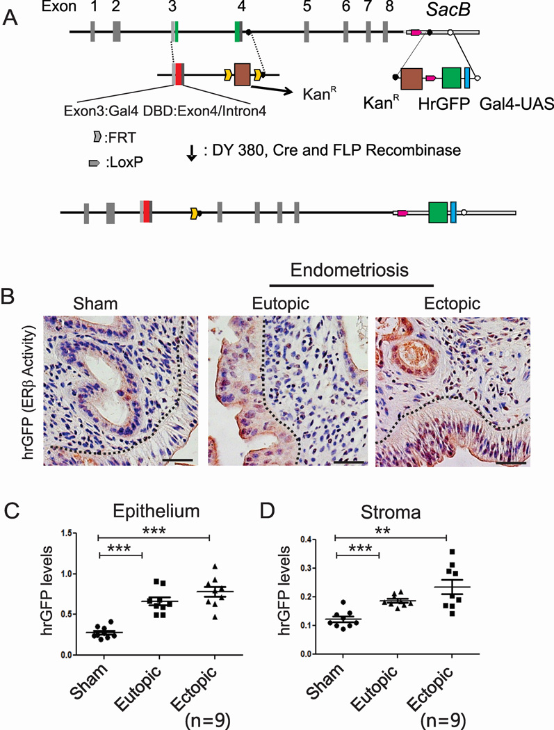
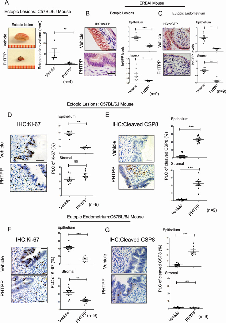
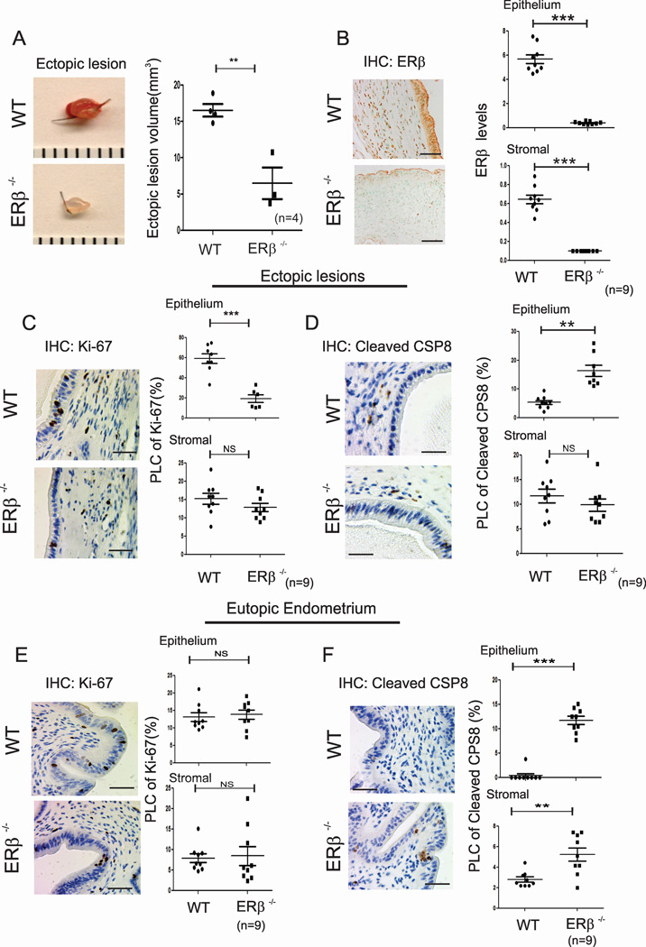
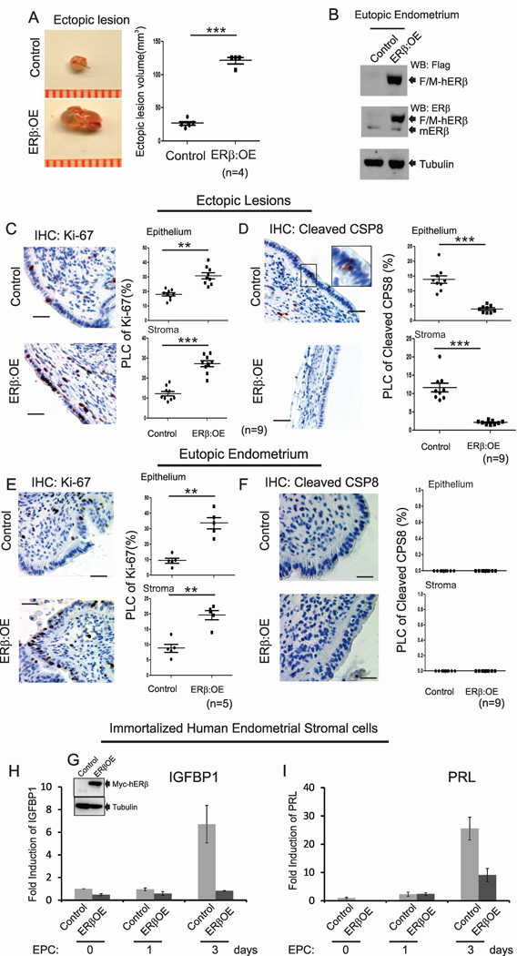
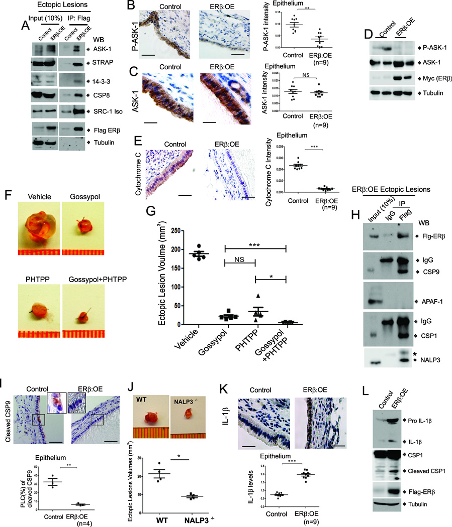
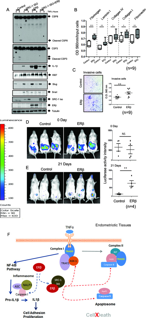
Alterations in estrogen-mediated cellular signaling play an essential role in the pathogenesis of endometriosis. In addition to higher estrogen receptor (ER) β levels, enhanced ERβ activity was detected in endometriotic tissues, and the inhibition of enhanced ERβ activity by an ERβ-selective antagonist suppressed mouse ectopic lesion growth. Notably, gain of ERβ function stimulated the progression of endometriosis. As a mechanism to evade endogenous immune surveillance for cell survival, ERβ interacts with cellular apoptotic machinery in the cytoplasm to inhibit TNF-α-induced apoptosis. ERβ also interacts with components of the cytoplasmic inflammasome to increase interleukin-1β and thus enhance its cellular adhesion and proliferation properties. Furthermore, this gain of ERβ function enhances epithelial-mesenchymal transition signaling, thereby increasing the invasion activity of endometriotic tissues for establishment of ectopic lesions. Collectively, we reveal how endometrial tissue generated by retrograde menstruation can escape immune surveillance and develop into sustained ectopic lesions via gain of ERβ function.


Figures

Figure 1
Figure 1. Mouse Endometriotic Tissues Have Elevated Levels of ERβ
(A–B) The expression levels of ERβ, ERα, PR and tubulin in the uteri of sham-treated C57BL/6J mice and the eutopic endometria (A) and ectopic lesions (B) of C57BL/6J mice with endometriosis. (C) IHC and quantitative analyses of ERβ levels in the uteri of sham-treated C57BL/6J mice and ectopic and eutopic endometria of C57BL/6J mice with endometriosis.
Figure 2
Figure 2. Enhanced ERβ Activity is Detected in the Endometriotic Tissues of Mice with Endometriosis Compared to Normal Endometrium
(A) Generation of a modified ERβ bacterial artificial clone that has a Gal4 DNA-binding domain and the Gal4-UAS-hrGFP reporter. DY380, Bacterial recombination strain. KanR, kanamycin-resistant gene. DBD, DNA-binding domain. Gal4-UAS, Gal4-upstream activating sequence. FLP, flippase. hrGFP, humanized renilla GFP. (B) IHC analyses of hrGFP levels in the uteri of sham-treated ERBAI mice and ectopic and eutopic endometria of ERBAI mice with endometriosis. (C and D) The quantification of hrGFP levels in the epithelial (C) and stromal compartment (D) of each type of endometrium in panel B. See also Figures S1.
Figure 3
Figure 3. ERβ-Specific Antagonist Regresses Ectopic Lesion Growth
(A) Ectopic lesions isolated from C57BL/6J mice with endometriosis subcutaneously treated with vehicle or PHTPP. (B and C) IHC and quantitative analyses of hrGFP levels in ectopic lesions (B) and eutopic endometria (C) of ERBAI mice with endometriosis subcutaneously treated with vehicle or PHTPP. (D and E) IHC and quantitative analyses of the expression patterns of Ki-67 (D) and cleaved CSP8 (E) in ectopic lesions of C57BL/6J mice with endometriosis subcutaneously treated with vehicle or PHTPP. (F and G) IHC and quantitative analyses of the levels of Ki-67 (F) and cleaved CSP8 (G) in the eutopic endometria of C57BL/6J mice with endometriosis subcutaneously treated with vehicle or PHTPP. PLC, percentage of labeled cells. CSP8, caspase 8. See also Figures S2.
Figure 4
Figure 4. The Loss of ERβ Function Prevents Ectopic Lesion Growth
(A) Ectopic lesions isolated from C57BL/6J (WT) and ERβ−/− mice with endometriosis. (B) IHC analyses and quantification of the ERβ levels in ectopic lesions isolated from WT and ERβ−/− mice with endometriosis. (C–F) IHC and quantitative analyses of Ki-67 (C and E) and cleaved CSP8 (D and F) in the epithelial and stromal compartments of ectopic lesions (C and D) and eutopic endometrium (E and F) of WT and ERβ−/− mice with endometriosis.
Figure 5
Figure 5. The Gain of ERβ Function Stimulates Ectopic Lesion Growth
(A) Ectopic lesions isolated from control and ERβ:OE mice with endometriosis. (B) Exogenous Flag/Myc-tagged human ERβ (F/M-hERβ) protein levels in the eutopic endometria of control and ERβ:OE mice with endometriosis. mERβ, endogenous mouse ERβ. (C–F) IHC and quantitative analyses Ki-67 (C and E) and cleaved CSP8 (D and F) in the epithelial and stromal compartments of ectopic lesions (C and D) and eutopic endometrium (E and F) of control and ERβ:OE mice with endometriosis. Higher magnification views of the boxed regions. (G) Exogenous Myc-tagged human ERβ (Myc-hERβ) protein levels in iHESCs/ERβ as determined with a Myc antibody. (H and I) The quantification of relative changes in the mRNA levels of decidualization marker genes, IGFBP1 (H) and PRL (I), in iHESCs (Control) and iHESCs/ERβ (ERβOE) upon estrogen/medroxyprogesterone/db-cAMP (ECP) treatment at the indicated day. See also Figures S3.
Figure 6
Figure 6. ERβ Interacts with TNFα-induced Apoptosis Complexes and the Inflammasome in Endometriotic Tissues of Mice with Endometriosis
(A) Flag-ERβ complexes immunoprecipitated (IPed) with a Flag antibody from ectopic lesions of Control and ERβ:OE mice with endometriosis followed by western blotting (WB) with antibodies against ASK-1, STRAP, 14-3-3, CSP8, SRC-1, Flag and tubulin. (B–C) IHC and quantitative analyses of phospho-Thr845-ASK-1 (P-ASK-1) (B) and total ASK-1 (C) in Control and ERβ:OE ectopic lesions. (D) Western blot analyses of phospho- Thr845-ASK-1 (P-ASK-1), total ASK-1, ERβ and tubulin in Control and ERβ:OE ectopic lesions. (E) IHC and quantitative analyses of cytochrome C levels in Control and ERβ:OE ectopic lesions. (F and G) Regression of ectopic lesion growth in endometriosis-induced C57BL/6J mice subcutaneously treated with Gossypol, PHTPP or their combination compared to vehicle (F). Quantification of ectopic lesion volume in panel F is shown in the graph (G). (H) The IPed Flag-ERβ complex from ERβ:OE ectopic lesions with a Flag antibody or IgG followed by western blotting with antibodies against Flag, CSP 9, APAF1, CSP1 and NRLP3. *, Non-Specific Protein. (I) IHC and quantitative analyses of cleaved CSP9 levels in Control and ERβ:OE ectopic lesions. Higher magnification views of the boxed regions. (J) Ectopic lesions isolated from C57BL/6J (WT) and NALP3−/− mice with endometriosis. (K) IHC and quantitative analyses of IL-1β levels in Control and ERβ:OE ectopic lesions. (L) Western blot analyses of levels of IL-1β, CSP1, Flag-tagged ERβ and tubulin (as a protein loading control) in ectopic lesions of control and ERβ:OE mice with surgically induced endometriosis. See also Figure S4 and S5.
Figure 7
Figure 7. Gain of ERβ Function Prevents TNFα-Induced Apoptosis Signaling but Stimulates Proliferation, Adhesion and Invasion Activities of Human Endometriotic Cells
(A) Levels of cleaved CSP8, cleaved CSP3, IL-1β, Ki67, Slug, Snail, and SRC-1 isoform (determined by a Flag antibody), ERβ (determined using a Myc antibody) and tubulin in iHEECs (Control), iHEECs/SRC-1Iso (SRC-1ISO), iHEECs/ERβ (ERβ), or iHEECs/SRC-1Iso/ERβ (SRC-1ISO+ERβ) upon 50 ng/ml TNFα plus 10 µg/ml cycloheximide treatment for 0 and 8 hours. (B) Cell adhesion activities of paternal iHEECs (Control) and iHEECs/ERβ (ERβ) against various extracellular matrices in the presence of 50 ng/ml TNFα. (C) Invasion activities of iHEECs (Control) and iHEECs/ERβ (ERβ) for 2 days using a Transwell plate assay. The amounts of invasive cells in each group were determined using a crystal violet staining protocol and are shown in the graph. (D–E) Bioluminescence and quantitative analyses of iHEECs/Luc (Control) and iHEECs/ERβ/Luc (ERβ) in SCID mice at 0(D) and 21(E) days after the induction of endometriosis. (F) Working model for the non-genomic action of ERβ in endometriosis progression. See also Figures S6 and S7.

Comment in

References

    1. Acien P, Velasco I, Gutierrez M, Martinez-Beltran M. Aromatase expression in endometriotic tissues and its relationship to clinical and analytical findings. Fertil Steril. 2007;88:32–38. - PubMed
    1. Ahn SH, Monsanto SP, Miller C, Singh SS, Thomas R, Tayade C. Pathophysiology and Immune Dysfunction in Endometriosis. BioMed research international. 2015;2015:795976. - PMC - PubMed
    1. Bono Y, Kyo S, Takakura M, Maida Y, Mizumoto Y, Nakamura M, Nomura K, Kiyono T, Inoue M. Creation of immortalised epithelial cells from ovarian endometrioma. British journal of cancer. 2012;106:1205–1213. - PMC - PubMed
    1. Bratton SB, Salvesen GS. Regulation of the Apaf-1-caspase-9 apoptosome. Journal of cell science. 2010;123:3209–3214. - PMC - PubMed
    1. Bulun SE. Endometriosis. The New England journal of medicine. 2009;360:268–279. - PubMed

Publication types