. 2015 Nov 5;163(4):1011-25.
doi: 10.1016/j.cell.2015.10.025.

The Molecular Taxonomy of Primary Prostate Cancer

Collaborators

The Molecular Taxonomy of Primary Prostate Cancer

Cancer Genome Atlas Research Network. Cell. .

Abstract

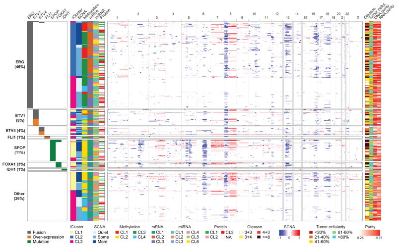
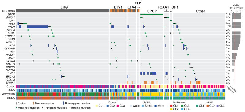
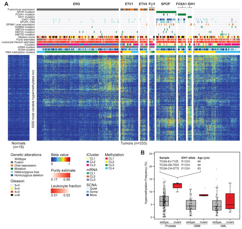
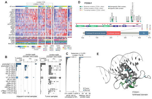
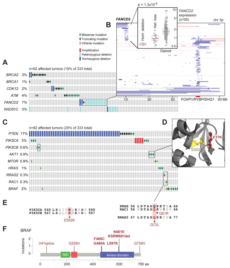
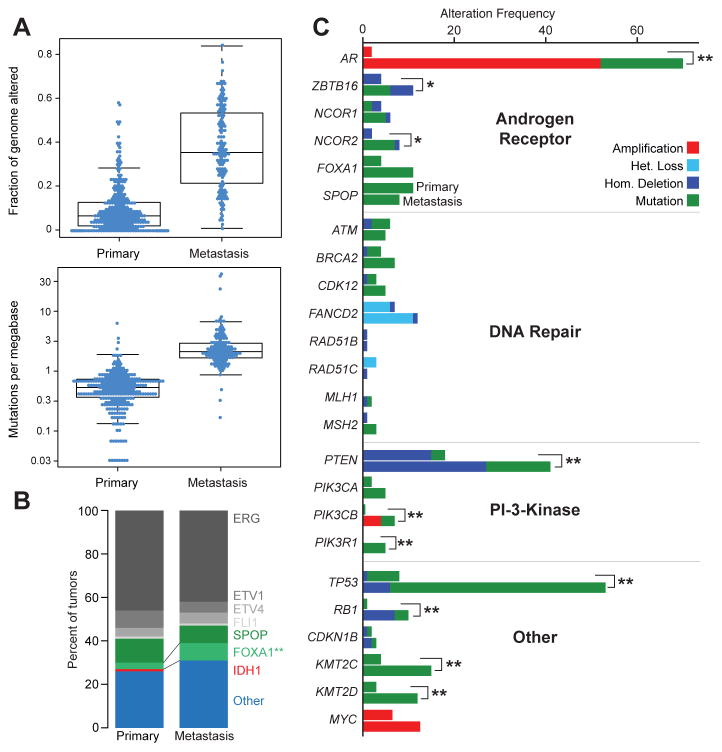
There is substantial heterogeneity among primary prostate cancers, evident in the spectrum of molecular abnormalities and its variable clinical course. As part of The Cancer Genome Atlas (TCGA), we present a comprehensive molecular analysis of 333 primary prostate carcinomas. Our results revealed a molecular taxonomy in which 74% of these tumors fell into one of seven subtypes defined by specific gene fusions (ERG, ETV1/4, and FLI1) or mutations (SPOP, FOXA1, and IDH1). Epigenetic profiles showed substantial heterogeneity, including an IDH1 mutant subset with a methylator phenotype. Androgen receptor (AR) activity varied widely and in a subtype-specific manner, with SPOP and FOXA1 mutant tumors having the highest levels of AR-induced transcripts. 25% of the prostate cancers had a presumed actionable lesion in the PI3K or MAPK signaling pathways, and DNA repair genes were inactivated in 19%. Our analysis reveals molecular heterogeneity among primary prostate cancers, as well as potentially actionable molecular defects.


Figures

Figure 1
Figure 1. The molecular taxonomy of primary prostate cancer
Comprehensive molecular profiling of 333 primary prostate cancer samples revealed seven genomically distinct subtypes, defined (top to bottom) by ERG fusions (46%), ETV1/ETV4/FLI1 fusions or overexpression (8%, 4%, 1%, respectively), or by SPOP (11%), FOXA1 (3%), and IDH1 (1%) mutations. A subset of these subtypes was correlated with clusters computationally derived from the individual characterization platforms (somatic copy-number alterations, methylation, mRNA, microRNA, and protein levels from reverse phase protein arrays). The heatmap shows DNA copy-number for all cases, with chromosomes shown from left to right. Regions of loss are indicated by shades of blue, and gains by shades of red. See also Figures S1, S2, S3, S4, S5, S6, S7, and Tables S1A, S1B, S1E, S2.
Figure 2
Figure 2. Recurrent alterations in primary prostate cancer
The spectrum and type of recurrent alterations and genes (mutations, fusions, deletions, and overexpression) in the cohort are shown (left to right) grouped by the molecular subtypes defined in Figure 1. On the right, the statistical significance of individual mutant genes (MutSig q-value) is shown. Mutations in IDH1, PIK3CA, RB1, KMT2D, CHD1, BRCA2, and CDK12 are also shown, despite their not being statistically significant. SPINK1 overexpression is shown for reference. See also Tables S1B, S1C, S1D, S1E.
Figure 3
Figure 3. Hypermethylation is common across primary prostate cancer
A. Primary prostate cancers show diverse methylation changes compared to normal prostate samples (left). Unsupervised clustering was performed on the beta-values of the 5,000 most hypermethylated loci, and the results mapped to the genomic subtypes. ERG-positive tumors had a high diversity of methylation changes, with a distinct subgroup (cluster 1) nearly unique to this group. SPOP and FOXA1 mutant tumors also exhibited global hypermethylation. B. IDH1-mutant prostate cancers, which are associated with younger age, are among the most hypermethylated tumors, as in Glioblastoma (GBM) and Acute Myeloid Leukemia (AML). See also Figure S4 and Table S1F.
Figure 4
Figure 4. The diversity of androgen receptor activity in primary prostate cancer
A. Androgen receptor activity, as inferred by the induction of AR target genes, was significantly increased in SPOP and FOXA1 mutant tumors when compared to normal prostate or ERG-positive tumors. This increase in activity cannot be fully explained by AR mRNA or protein levels. B. Multiple known AR splice variants were detected in benign prostate (left) and primary prostate cancer (right), with the AR-V7 variant detected in 50% of tumors. C. Real-time qPCR comparison of AR-V7 in 74 tumor samples (grey) and 5 adjacent-normal samples (blue). D. FOXA1 missense mutations were clustered in the forkhead domain, mostly in residues that do not form contacts with DNA (see also the 3-D structure in panel E).
Figure 5
Figure 5. Alterations in clinically relevant pathways
A. Alterations in DNA repair genes were common in primary prostate cancer, affecting almost 20% of samples through mutations or deletions in BRCA2, BRCA1, CDK12, ATM, FANCD2, or RAD51C. B. Focal deletions of FANCD2 were found in 7% of samples and were associated with reduced mRNA expression of FANCD2. C. The RAS or PI-3-Kinase pathways were altered in about a quarter of tumors, mostly through deletion or mutation of PTEN, but also through rare mutations in other pathway members. D. AKT1 mutations were found in three samples. Two of them were the known activating E17K, and the third one affected the D323 residue, which is adjacent to E17 in the protein structure. E. One of the observed PIK3CB mutations, E552K, is paralogous to the known activating E545K mutation in PIK3CA, and the RAC1 Q61 and RRAS2 Q72 mutations are paralogous to the Q61 mutations in KRAS. F. BRAF mutations were found in 2% of samples, mostly in known non-V600E hotspots in the kinase domain. See also Figure S3.
Figure 6
Figure 6. Comparison of primary with metastatic prostate cancer
A. Metastatic prostate cancer samples have more copy-number alterations (top panel, measured as fraction of genome altered) and mutations (bottom panel). B. The relative distribution of main subtypes (ERG, ETV1/4, FLI1, SPOP, FOXA1, IDH1, other) is similar in primary and metastatic samples. C. The alteration frequencies of several genes and pathways are higher in metastatic samples. The upper bar for each gene indicates the alteration frequency in primary samples, the lower bar for metastatic samples. The most notable differences in alteration frequencies involve the Androgen Receptor pathway, the PI3K pathway, and TP53. See also Table S3.

Comment in

References

    1. Ahn J, Yuan Y, Parmigiani G, Suraokar MB, Diao L, Wistuba II, Wang W. DeMix: deconvolution for mixed cancer transcriptomes using raw measured data. Bioinformatics. 2013 Aug 1;29(15):1865–71. - PMC - PubMed
    1. Alexandrov LB, Nik-Zainal S, Wedge DC, Aparicio SA, Behjati S, Biankin AV, Bignell GR, Bolli N, Borg A, Børresen-Dale AL, et al. Signatures of mutational processes in human cancer. Nature. 2013 Aug 22;500(7463):415–21. - PMC - PubMed
    1. Al Olama AA, Kote-Jarai Z, Berndt SI, Conti DV, Schumacher F, Han Y, Benlloch S, Hazelett DJ, Wang Z, Saunders E, et al. A meta-analysis of 87,040 individuals identifies 23 new susceptibility loci for prostate cancer. Nat Genet. 2014 Oct;46(10):1103–9. - PMC - PubMed
    1. An J, Wang C, Deng Y, Yu L, Huang H. Destruction of full-length androgen receptor by wild-type SPOP, but not prostate-cancer-associated mutants. Cell Rep. 2014 Feb 27;6(4):657–69. - PMC - PubMed
    1. Antonarakis ES, Lu C, Wang H, Luber B, Nakazawa M, Roeser JC, Chen Y, Mohammad TA, Chen Y, Fedor HL, et al. AR-V7 and resistance to enzalutamide and abiraterone in prostate cancer. N Engl J Med. 2014 Sep 11;371(11):1028–38. - PMC - PubMed

Publication types

Grants and funding