. 2015 Nov 5;60(3):398-407.
doi: 10.1016/j.molcel.2015.10.030.

Single-Stranded DNA Cleavage by Divergent CRISPR-Cas9 Enzymes

Affiliations

Single-Stranded DNA Cleavage by Divergent CRISPR-Cas9 Enzymes

Enbo Ma et al. Mol Cell. .

Abstract

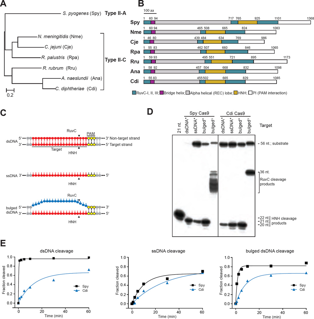
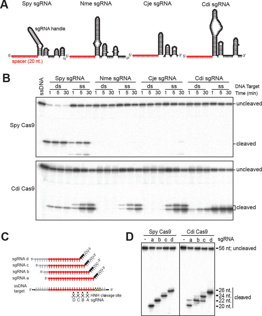
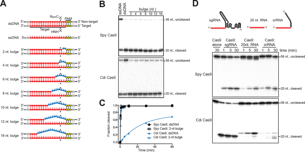
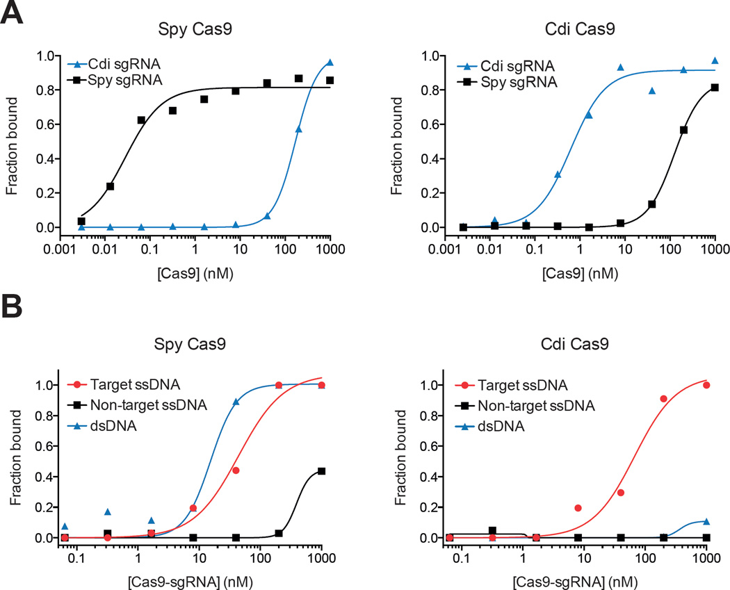
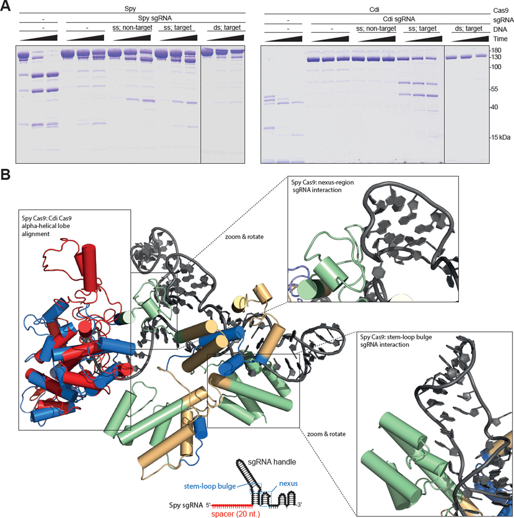
Double-stranded DNA (dsDNA) cleavage by Cas9 is a hallmark of type II CRISPR-Cas immune systems. Cas9-guide RNA complexes recognize 20-base-pair sequences in DNA and generate a site-specific double-strand break, a robust activity harnessed for genome editing. DNA recognition by all studied Cas9 enzymes requires a protospacer adjacent motif (PAM) next to the target site. We show that Cas9 enzymes from evolutionarily divergent bacteria can recognize and cleave single-stranded DNA (ssDNA) by an RNA-guided, PAM-independent recognition mechanism. Comparative analysis shows that in contrast to the type II-A S. pyogenes Cas9 that is widely used for genome engineering, the smaller type II-C Cas9 proteins have limited dsDNA binding and unwinding activity and promiscuous guide RNA specificity. These results indicate that inefficiency of type II-C Cas9 enzymes for genome editing results from a limited ability to cleave dsDNA and suggest that ssDNA cleavage was an ancestral function of the Cas9 enzyme family.


Figures

Figure 1
Figure 1. Diverse DNA cleavage activity of divergent Cas9 enzymes
(A). Phylogenetic tree of the seven Cas9 proteins used in this study, generated using MAFFT (Katoh, 2002). (B). Secondary structures and sizes of divergent Cas9 enzymes in this study. Domains are colored and drawn proportionally to the full length of its protein sequence. (C). Schematic presentation of three DNA substrates used for the figures of D and E. Target sequence is presented in red and underlined. PAM (AGG) is shown in yellow. (D). In vitro cleavage of DNA substrates. Cleavages of single-stranded DNA (ssDNA), double-strand DNA (dsDNA) and bulged DNA (bulged) mediated by Spy and Cdi Cas9 proteins are shown. * denotes that the target strand is labeled; Δ indicates the non-target strand is labeled. Substrate and product sizes are labeled on the right. Vertical lines indicate the border between two separate gels (E). Quantification of DNA cleavage activity on dsDNA, ssDNA and bulged substrates with the 5′- end of the target strand radiolabeled. Cleavage assays were conducted in at least triplicate, as described in the Methods, and the quantified data were fitted with single-exponential decays to obtain pseudo first-order rate constants (kcleave) for each reaction. kcleave ± SD values for dsDNA cleavage by Spy and Cdi Cas9 are 3.5 ± 0.9 min−1 and 0.041 ± 0.003 min−1, respectively. kcleave ± SD values for ssDNA cleavage by Spy and Cdi Cas9 are 0.27 ± 0.09 min−1 and 0.16 ± 0.1 min−1, respectively. kcleave ± SD values for bulged dsDNA cleavage by Spy and Cdi Cas9 are 1.0 ± 0.5 min−1 and 0.59 ± 0.4 min−1, respectively. See also Figures S1, S2 and Table S1.
Figure 2
Figure 2. Programmable and PAM-independent Cas9-catalyzed DNA cutting
(A). Schematic representation of the five sgRNAs used in this study. All of the sgRNAs were drawn based on their mfold predictions (Zuker, 2003) and contain the same 20 nt. ‘spacer’ sequence (shown in red), which is complementary to the target sequence in the DNA substrates. (B). In vitro cleavage assay of ssDNA (ss) and dsDNA (ds) using various combinations of Cas9 proteins and sgRNAs. The 5′-end of the target strand is radiolabeled. Cas9 proteins are shown on the left. (C). Schematic presentation of the four sgRNAs (sgRNAs A–D) used in D. All four sgRNAs have a same Spy sgRNA handle and contain different 20-nt. spacer sequence that is able to base pair to the target sequence in the DNA substrate. (D). Programmable ssDNA cleavage by divergent Cas9 enzymes. The product sizes are labeled on the right; solid lines indicate borders between separate gels. See also Figure S3.
Figure 3
Figure 3. Substrate recognition varies among divergent Cas9 enzymes
(A). Schematic representation of DNA substrates used in this experiment. Target sequence is same in all of the substrates (red). For bulged dsDNAs, the bulge sizes are given on the left and mismatches are colored in blue. The PAM is indicated in yellow. (B). In vitro cleavage of bulged DNA substrates. Bulged substrates are indicated by the number of mismatches (2 to 16). Cas9 proteins are labeled on the left of each panel, and sizes of the substrate and cleaved products are labeled on the right. (C). Kinetic analysis of 2-nt. bulged substrate versus perfectly matched dsDNA. Cleavage assays were conducted in at least triplicate, as described in the Methods, and the quantified data were fitted with single-exponential decays to obtain pseudo first-order rate constants (kcleave) for each reaction. kcleave ± SD values for dsDNA cleavage by Spy and Cdi Cas9 are 2.9 ± 0.3 min−1 and 0.041 ± 0.002 min−1, respectively. kcleave ± SD values for 2-nt bulge dsDNA cleavage by Spy and Cdi Cas9 are 2.3 ± 0.1 min−1 and 0.99 ± 0.6 min−1, respectively. (D). DNA cleavage by Cas9 proteins can be guided by short RNAs. Guide RNAs (sgRNA, 20-nt. and crRNA) used in this study are represented schematically above each lane. Cas9 proteins are labeled on the left of each panel, and sizes of the substrate and cleaved products are labeled on the right. See also Figure S4 and Table S2.
Figure 4
Figure 4
Binding affinity of Cas9 proteins for sgRNA and DNA. (A). Binding affinity of Cas9 proteins for cognate and non-cognate guides as determined by filter binding assays. Measurements were made in at least triplicate to determine KD and a representative replicate is shown. Data were fit to a binding isotherm. (KD ± SD for Spy Cas9 to Spy sgRNA, 28 pM ± 6 pM; Spy Cas9 to Cdi sgRNA, 146 ± 11nM; Cdi Cas9 to Spy sgRNA, 115 ± 51nM; Cdi Cas9 to Cdi sgRNA, 0.56 ± 0.13nM) (B). Binding affinity of Cas9 proteins for dsDNA, target ssDNA and non-target ssDNA as measured by electrophoretic mobility shift assay (EMSA). Each Cas9 protein was incubated with its cognate guide. Measurements were made in at least triplicate to determine KD and a representative replicate is shown. Data were fit to a binding isotherm. (KD ± SD [where appropriate] for Spy Cas9 to dsDNA, 25 ± 11 nM; Spy Cas9 to target ssDNA, 40 ± 11 nM; Spy Cas9 to non-target ssDNA, >1000 nM; Cdi Cas9 to dsDNA, >1000 nM; Cdi Cas9 to target ssDNA, 58 ± 32 nM; Cdi Cas9 for non-target ssDNA, >1000 nM. See also Figure S5 and Table S2
Figure 5
Figure 5
Limited proteolysis and structural comparison of Spy and Cdi Cas9 proteins. (A). Each Cas9 (5 µM) was incubated with its preferred guide RNA before the addition of DNA. Trypsin was added to the preformed complexes and allowed to digest the protein for 5, 15 or 30min. Degradation products were analyzed on 10% SDS-PAGE gels. ds: dsDNA; ss: ssDNA. Vertical lines indicate the border between two separate gels and approximate molecular weights are indicated on the right. (B). Structural alignment between Spy Cas9 alpha-helical (REC) lobe (PDB ID: 4ZT9) and the homology model of the alpha-helical lobe of Cdi Cas9 (based on the crystal structure of Ana Cas9 PDB ID: 4OGE); all atom RMSD: 3.9Å. The alpha-helical lobe of Cdi Cas9 is shown in red, sgRNA in shown in grey, and Spy Cas9 is colored according to primary sequence alignment with Cdi Cas9: green represents regions not present in the Cdi Cas9 protein sequence, blue represents regions present and conserved in the Cdi Cas9 sequence, and straw represents regions that are loosely conserved in the Cdi protein sequence. Spy sgRNA schematic is shown below with regions of interest boxed and labeled in blue. See also Figure S5.

References

    1. Briner AE, Donohoue PD, Gomaa AA, Selle K, Slorach EM, Nye CH, Haurwitz RE, Beisel CL, May AP, Barrangou R. Guide RNA Functional Modules Direct Cas9 Activity and Orthogonality. Mol. Cell. 2014;56:333–339. - PubMed
    1. Chase JW, Williams KR. Single-stranded DNA binding proteins required for DNA replication. Annu. Rev. Biochem. 1986;55:103–136. - PubMed
    1. Chylinski K, Makarova KS, Charpentier E, Koonin EV. Classification and evolution of type II CRISPR-Cas systems. Nucleic Acids Res. 2014;42:6091–6105. - PMC - PubMed
    1. Deng L, Garrett RA, Shah SA, Peng X, She Q. A novel interference mechanism by a type IIIB CRISPR-Cmr module in Sulfolobus. Mol. Microbiol. 2013;87:1088–1099. - PubMed
    1. Doudna JA, Charpentier E. The new frontier of genome engineering with CRISPR-Cas9. Sci. 2014;346 - PubMed

Publication types

MeSH terms