. 2016 Feb;17(2):190-202.
doi: 10.1016/j.jpain.2015.10.009. Epub 2015 Nov 3.

Disruption of Spinal Noradrenergic Activation Delays Recovery of Acute Incision-Induced Hypersensitivity and Increases Spinal Glial Activation in the Rat

Affiliations

Disruption of Spinal Noradrenergic Activation Delays Recovery of Acute Incision-Induced Hypersensitivity and Increases Spinal Glial Activation in the Rat

Vipin Arora et al. J Pain. 2016 Feb.

Abstract

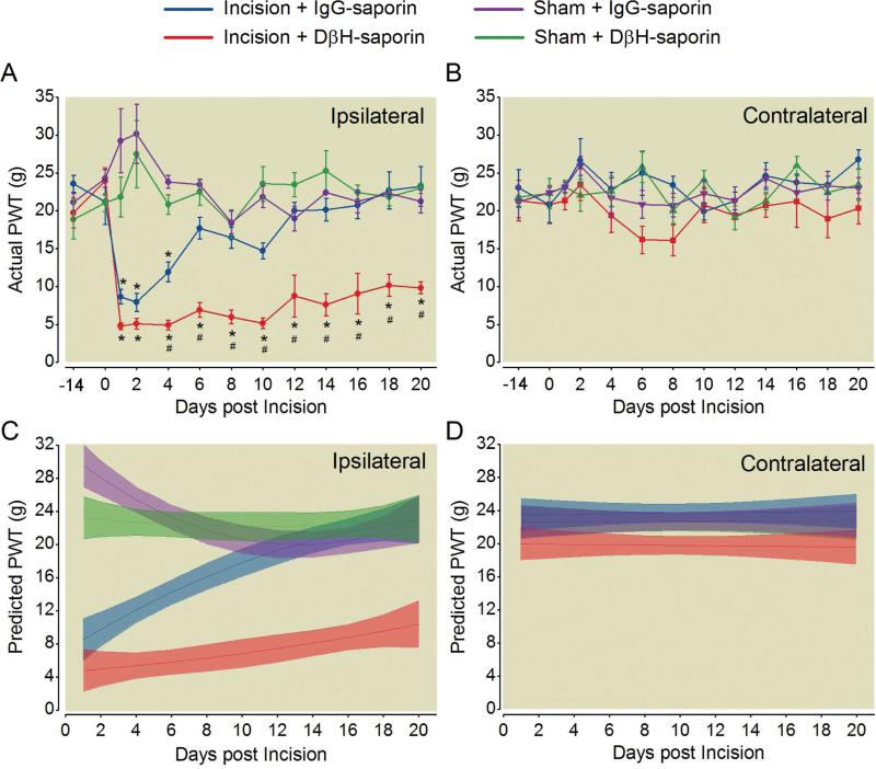
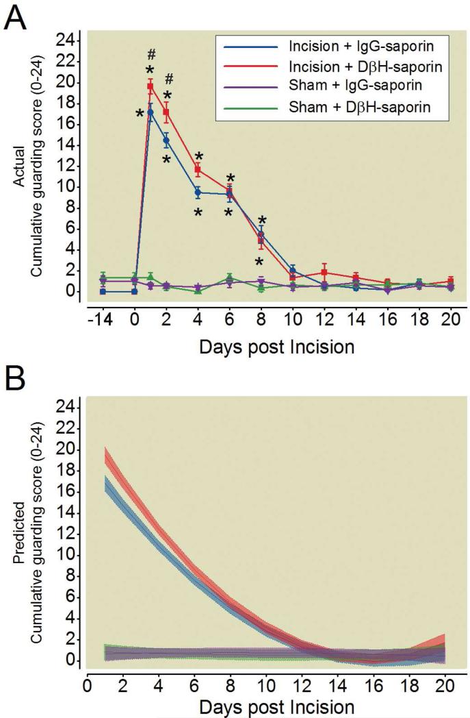
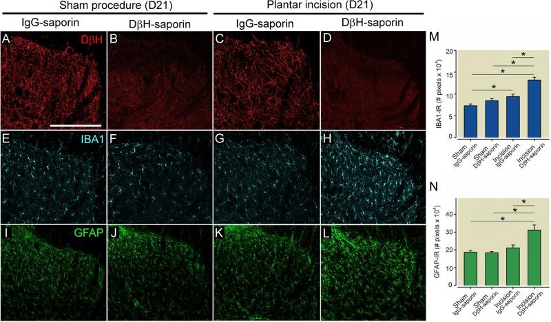
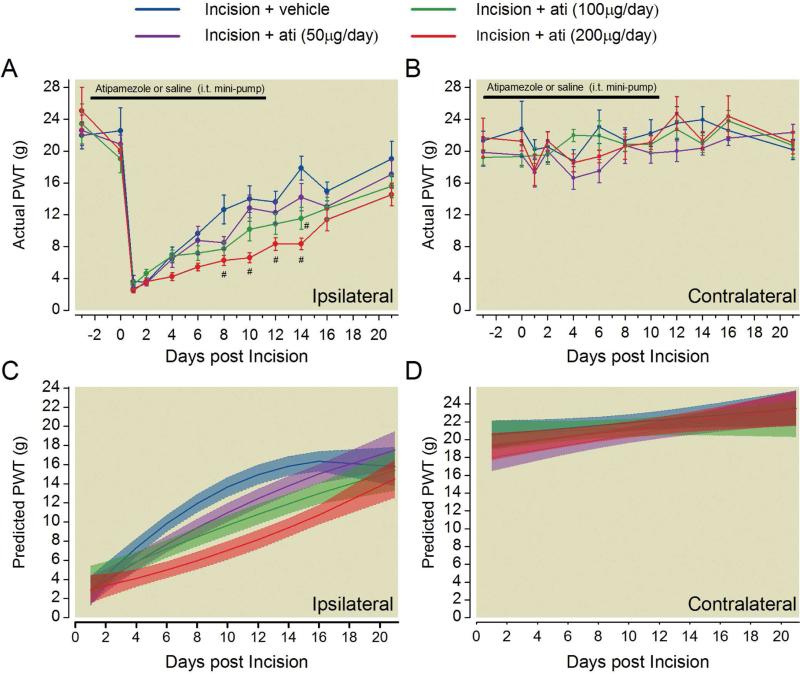
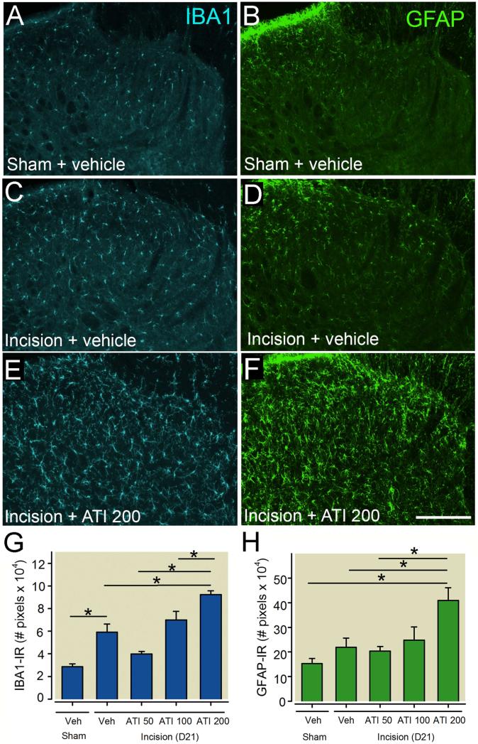
Results of clinical studies suggest that descending inhibitory controls from the brainstem are important for speeding recovery from pain after surgery. We examined the effects of destroying spinally projecting noradrenergic neurons via intrathecally administered antibody to dopamine β-hydroxylase conjugated to saporin (DβH-saporin) on recovery in an acute incisional pain model. Mechanical and thermal paw withdrawal thresholds and nonevoked spontaneous guarding scores were tested for several weeks postoperatively and analyzed using mixed effects growth curve modeling. DβH-saporin treatment resulted in a significant prolongation in the duration of mechanical and to a lesser degree thermal hypersensitivity in the ipsilateral paw of incised rats but did not increase the duration of spontaneous guarding. DβH-saporin treatment was also associated with increased microglial and astrocyte activation in the ipsilateral spinal cord 21 days after incision compared with immunoglobulin G-saporin treated controls. Chronic intrathecal administration of the α2 adrenergic receptor antagonist atipamezole (50-200 μg/d) produced similar effects. These data suggest that spinally projecting noradrenergic pathways and spinal α2 adrenergic receptor activation are important for speeding recovery from hypersensitivity after surgical incision possibly by reducing spinal glial activation. Interventions that augment the noradrenergic system might be important to speed recovery from pain after surgery.

Perspective: Endogenous descending spinal noradrenergic activation promotes resolution of incision-induced hypersensitivity and inhibits spinal microglial and astrocyte activation in part through α2 adrenergic receptors.

Keywords: Descending inhibition; chronification; glial plasticity; growth curve; postoperative pain.


Figures

Figure 1
Figure 1
Spinal depletion of noradrenergic fibers prior to plantar incision delays resolution of ipsilateral mechanical hypersensitivity. Rats received intrathecal treatment with dopamine β-hydroxylase (DβH)-saporin or control immunoglobulin G (IgG)-saporin 14 days before plantar incision or sham procedure and were assessed for mechanical hypersensitivity with von Frey filaments in the ipsilateral (A) and contralateral (B) hindpaw. Data is expressed as Mean ± SEM. Two-way repeated-measures ANOVA with Bonferroni multiple comparisons. # P < 0.05 for within time point comparison to Incision + IgG-saporin value; * P < 0.001 for within treatment group comparisons to pre-incision (D0) baseline value. Modeled group trajectories of postoperative mechanical withdrawal thresholds in the ipsilateral (C) and contralateral (D) paw of treated rats. Group averaged trajectories depict the mean fit for all the animals within each treatment group (n = 6 per group) with 95% CIs indicated by shading.
Figure 2
Figure 2
Spinal depletion of noradrenergic fibers prior to plantar incision delays resolution of ipsilateral thermal hypersensitivity. Rats received intrathecal treatment with dopamine β-hydroxylase (DβH)-saporin or control immunoglobulin G (IgG)-saporin 14 days before plantar incision or sham procedure and were assessed for thermal response latency with a radiant heat device in the ipsilateral (A) and contralateral (B) hindpaw. Data is expressed as Mean ± SEM. Two-way repeated-measures ANOVA with Bonferroni multiple comparisons. # P < 0.001 for within time point comparison to Incision + IgG-saporin value,* P < 0.003 for within treatment group comparison to pre-incision (D0) baseline value. Modeled group trajectories of postoperative thermal withdrawal latencies in the ipsilateral (C) and contralateral (D) paw of treated rats. Group averaged trajectories depict the mean fit for all the animals within each treatment group (n = 6 per group) with 95% CIs indicated by shading.
Figure 3
Figure 3
Spinal depletion of noradrenergic fibers prior to plantar incision does not alter duration of spontaneous guarding behavior. Rats received intrathecal treatment with dopamine β-hydroxylase (DβH)-saporin or control immunoglobulin G (IgG)-saporin 14 days before plantar incision or sham procedure and were assessed for spontaneous guarding behavior in the ipsilateral hindpaw (A). Data is expressed as Mean ± SEM. Kruskal-Wallis ANOVA on ranks followed by Tukey test . # P < 0.05 for within time point comparison to Incision + IgG-saporin value. Friedman repeated measures ANOVA on ranks followed by Dunnett test, * P < 0.05 for within treatment group comparison to pre-incision (D0) baseline value. Modeled group trajectories of cumulative guarding scores in the ipsilateral (B) paw of treated rats. Group averaged trajectories depict the mean fit for all the animals within each treatment group (n = 6 per group) with 95% CIs indicated by shading.
Figure 4
Figure 4
Spinal depletion of noradrenergic fibers increases microglial and astrocyte activation following plantar incision in rats. Rats were administered DβH-saporin (i.t., 5μg) to deplete spinal noradrenergic fibers or control IgG saporin 14 days prior to surgery. Representative images of ipsilateral dorsal medial spinal cord of rats 21 days following plantar incision or sham procedure labeled with antibodies against dopamine β hydroxylase (DβH, red, A-D) for noradrenergic fibers, ionized calcium binding adaptor molecule 1 (IBA1,blue, E-H) for microglia and glial fibrillary acid protein (GFAP, green, I-L) for astrocytes. Note loss of noradrenergic fibers in spinal cord of DβH-saporin treated rats accompanied by increased IBA1-IR and GFAP-IR in rats following plantar incision but not sham procedure. Immunofluorescence levels of IBA1 (M) and GFAP (N) in the dorsal medial spinal cord were quantified. Data is presented as mean ± SEM. n = 6 per group. Groups were analysis by Two-way ANOVA, IBA1: p (Surgery × Treatment) = 0.015, GFAP: p (Surgery × Treatment) = 0.005, Bonferroni Multiple Comparison Test * p < 0.001. Scale bar in A = 250 μms.
Figure 5
Figure 5
Blockade of spinal α2 adrenergic receptors delays the resolution of mechanical hypersensitivity after plantar incision. Rats were chronically treated with varying doses of the α2 adrenergic receptor antagonist atipamezole or saline beginning three days prior to until eleven days after plantar incision. Mechanical withdrawal thresholds in the ipsilateral (A) and contralateral (B) hindpaw of treated rats prior to and following incision surgery. Data is expressed as Mean ± SEM. Two-way repeated-measures ANOVA with Bonferroni multiple comparisons. # P < 0.05 for within time point comparison to Incision + vehicle treated value. Modeled group trajectories of postoperative mechanical withdrawal thresholds in the ipsilateral (C) and contralateral (D) paw. The highest dose of atipamezole (200μg/day, red) had significantly lower slope of recovery evident as non-overlapping 95% confidence intervals compared to vehicle treated rats (blue) from 4 to 18 days postoperatively. Group averaged trajectories depict the mean fit for all the animals within each treatment group (n=12-14 per group) with 95% CIs indicated by shading.
Figure 6
Figure 6
Blockade of spinal α2 adrenergic receptor increases microglial and astrocyte activation in the ipsilateral spinal cord 21 days following plantar incision in rats. Representative images of ionized calcium binding adaptor molecule 1 (IBA1, blue, A,C,E) for microglia and glial fibrillary acidic protein (GFAP, green, B,D,F) for astrocytes in rats without surgery (A,B) or in rats with plantar incision administered chronic intrathecal vehicle solution (C,D) or atipamezole (200μg/day, E, F) beginning three days prior to until eleven days following plantar incision. Immunofluorescence levels of IBA1 (G) and GFAP (H) in the dorsal medial spinal cord. Data are presented as mean ± SEM. n = 5 per group. Groups were analysis by one-way ANOVA, IBA1: p (Treatment) = 0.002, GFAP: p (Treatment) <0.001, Bonferroni Multiple Comparison Test * P < 0.05. Scale bar in A = 250 μms.

References

    1. Alkaitis MS, Solorzano C, Landry RP, Piomelli D, DeLeo JA, Romero-Sandoval EA. Evidence for a role of endocannabinoids, astrocytes and p38 phosphorylation in the resolution of postoperative pain. PloS one. 2010;5:e10891. - PMC - PubMed
    1. Amr YM, Yousef AA. Evaluation of efficacy of the perioperative administration of Venlafaxine or gabapentin on acute and chronic postmastectomy pain. The Clinical Journal of Pain. 2010;26:381–385. - PubMed
    1. Aschenbrenner CA, Houle TT, Gutierrez S, Eisenach JC. Modeling Individual Recovery after Peripheral Nerve Injury in Rats and the Effects of Parturition. Anesthesiology. 2014 - PMC - PubMed
    1. Baba H, Goldstein PA, Okamoto M, Kohno T, Ataka T, Yoshimura M, Shimoji K. Norepinephrine facilitates inhibitory transmission in substantia gelatinosa of adult rat spinal cord (part 2): effects on somatodendritic sites of GABAergic neurons. Anesthesiology. 2000;92:485–492. - PubMed
    1. Bourgoin S, Pohl M, Mauborgne A, Benoliel JJ, Collin E, Hamon M, Cesselin F. Monoaminergic control of the release of calcitonin gene-related peptide- and substance P-like materials from rat spinal cord slices. Neuropharmacology. 1993;32:633–640. - PubMed

Publication types

MeSH terms

LinkOut - more resources