. 2016 Jan;15(1):164-76.
doi: 10.1074/mcp.M115.052449. Epub 2015 Nov 6.

Novel N-terminal and Lysine Methyltransferases That Target Translation Elongation Factor 1A in Yeast and Human

Affiliations

Novel N-terminal and Lysine Methyltransferases That Target Translation Elongation Factor 1A in Yeast and Human

Joshua J Hamey et al. Mol Cell Proteomics. 2016 Jan.

Abstract

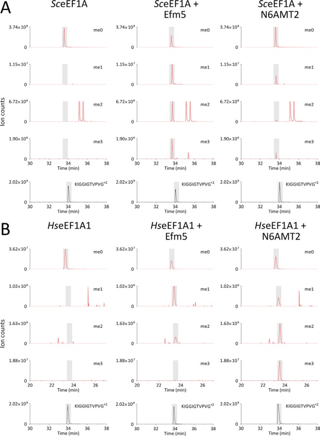
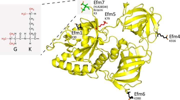
Eukaryotic elongation factor 1A (eEF1A) is an essential, highly methylated protein that facilitates translational elongation by delivering aminoacyl-tRNAs to ribosomes. Here, we report a new eukaryotic protein N-terminal methyltransferase, Saccharomyces cerevisiae YLR285W, which methylates eEF1A at a previously undescribed high-stoichiometry N-terminal site and the adjacent lysine. Deletion of YLR285W resulted in the loss of N-terminal and lysine methylation in vivo, whereas overexpression of YLR285W resulted in an increase of methylation at these sites. This was confirmed by in vitro methylation of eEF1A by recombinant YLR285W. Accordingly, we name YLR285W as elongation factor methyltransferase 7 (Efm7). This enzyme is a new type of eukaryotic N-terminal methyltransferase as, unlike the three other known eukaryotic N-terminal methyltransferases, its substrate does not have an N-terminal [A/P/S]-P-K motif. We show that the N-terminal methylation of eEF1A is also present in human; this conservation over a large evolutionary distance suggests it to be of functional importance. This study also reports that the trimethylation of Lys(79) in eEF1A is conserved from yeast to human. The methyltransferase responsible for Lys(79) methylation of human eEF1A is shown to be N6AMT2, previously documented as a putative N(6)-adenine-specific DNA methyltransferase. It is the direct ortholog of the recently described yeast Efm5, and we show that Efm5 and N6AMT2 can methylate eEF1A from either species in vitro. We therefore rename N6AMT2 as eEF1A-KMT1. Including the present work, yeast eEF1A is now documented to be methylated by five different methyltransferases, making it one of the few eukaryotic proteins to be extensively methylated by independent enzymes. This implies more extensive regulation of eEF1A by this posttranslational modification than previously appreciated.


Figures

Fig. 1.
Fig. 1.
Yeast eEF1A is trimethylated at its N terminus before being mono- and dimethylated at lysine three. (A) Q Exactive Plus MS/MS spectrum of a doubly-charged AspN peptide of 843.51 m/z identifies trimethylation at the N terminus of eEF1A as well as dimethylation at lysine 3 (for mass-annotated spectrum see Supplemental Fig. S14). (B) Transitions from the di-, tri-, tetra- and pentamethylated AspN peptide (GKEKSHINVVVIGHV+2) to the un-, mono- and dimethylated forms of the y14 ion (KEKSHINVVVIGHV+1) indicate that y14 is unmethylated in the di- and trimethylated AspN peptide, monomethylated in the tetramethylated AspN peptide and dimethylated in the pentamethylated AspN peptide. This indicates that the N terminus is trimethylated before Lys3 is partially mono- and dimethylated. The monomethylated AspN peptide was not abundant enough for this analysis. (C) MS/MS/MS (MS3) spectra of MS2 precursors of 1,559.06 (i), 1,572.99 (ii) and 1,587.14 m/z (iii) confirm their identities as the y14 ion in its un-, mono- and dimethylated state, respectively (for mass-annotated spectra see Supplemental Figs. S15-S17).
Fig. 2.
Fig. 2.
YLR285W is responsible for N-terminal and Lys3 methylation of yeast eEF1A in vivo and can N-terminally methylate eEF1A in vitro. (A) Deletion of YLR285W abolishes in vivo eEF1A methylation at the N terminus and Lys3 in yeast. Whole cell lysates from wild-type yeast (WT) and the single gene knockout of YLR285W were separated by SDS-PAGE and bands corresponding to eEF1A were analyzed by LC-MS/MS. The N terminus and Lys3 were both found to be unmethylated in the knockout of YLR285W, with the unmethylated form of the peptide (me0) being the only form of the peptide present. Trimethylation of the N terminus was estimated to be of high stoichiometry (>85%) by measuring the area under the curve for each methylation state. This does not, however, consider differences in ionization efficiency between the different methylation states. (B) Overexpression of YLR285W results in increased methylation at both the N-terminal Gly2 and Lys3 of eEF1A, as evidenced by the disappearance of the dimethylated peptide and the increase in abundance of the penta- and hexamethylated peptides. BG1805-YLR285W or the empty BG1805 vector were overexpressed in WT yeast and lysates were separated by SDS-PAGE and then bands corresponding to eEF1A were analyzed by LC-MS/MS. Un- and monomethylated eEF1A were below the limit of detection in both conditions. (C) YLR285W methylates eEF1A at its N terminus in vitro. Yeast eEF1A purified from E. coli was incubated with or without YLR285W in the presence of AdoMet. eEF1A was found to be mono- and dimethylated at the N terminus when incubated with YLR285W (see Supplemental Fig. S5 for MS/MS spectrum). For (A), (B), and (C), the methylation status of the N terminus/Lys3 was analyzed by taking mass windows (±10 ppm) corresponding to all relevant methylation states of the AspN peptide GKEKSHINVVVIGHV+2 (green). Peaks were normalized to the most abundant ion for each methylation state. The abundance of the eEF1A AspN peptide DAIEQPSRPT+2 is shown as an internal standard (black). Elution times of peptides are shaded; peaks outside shading are unrelated, near-isobaric ions.
Fig. 3.
Fig. 3.
Methylation of eEF1A by Efm7 is affected by the structure and conformation of eEF1A. (A) In vitro methylation of eEF1A by Efm7 is enhanced by the addition of either GDP or GTP. Recombinant yeast eEF1A was in vitro methylated by Efm7 in the presence or absence of GDP or a GTP analog. eEF1A was found to be more dimethylated, and trimethylated to a small degree, in the presence of GDP or the GTP analog, indicating that the ability of Efm7 to methylate eEF1A is affected by its conformation. (B) In vitro methylation of recombinant full length and C-terminal truncations of eEF1A indicates that Efm7 does not require domains 2 or 3 in order to methylate eEF1A. For both (A) and (B), the graphs represents the relative amounts of each methylation state as determined by the area under the curve of the extracted ion chromatograms, which are shown as Supplemental Figs. S7 and S8 for (A) and (B), respectively.
Fig. 4.
Fig. 4.
Human eEF1A is trimethylated at its N terminus. (A) Q Exactive Plus MS/MS spectrum of a doubly charged AspN peptide of 843.51 m/z identifies trimethylation at the N terminus of human eEF1A (for mass-annotated spectrum see Supplemental Fig. S18). (B) Transitions from the trimethylated AspN peptide (GKEKTHINIVVIGHV+2) to the un-, mono-, di- and trimethylated forms of the y14 ion (KEKTHINIVVIGHV+1) indicate that y14 is completely unmethylated in the trimethylated peptide. This indicates that the N terminus is trimethylated. The mono- and dimethylated AspN peptides were not abundant enough for this analysis. (C) MS/MS/MS (MS3) spectrum of an MS2 precursor of 1586.99 m/z confirms its identity as the unmethylated y14 ion (for mass-annotated spectrum see Supplemental Fig. S19).
Fig. 5.
Fig. 5.
Sequence alignment of Efm5 and its human ortholog, N6AMT2. Efm5 and N6AMT2 were aligned using EMBOSS Stretcher (EMBL-EBI), indicating 36.1% identity and 53.4% similarity. Vertical lines (∣) indicate identical residues; double dots (:) indicate chemically similar residues; single dots (.) indicate dissimilar residues; dashes (-) indicate missing residues. The characteristic seven-beta-strand methyltransferase motifs I and Post-I are indicated according to (53), as well as the [D/N]XX[Y/F] motif proposed to be defining of nitrogen methylation (18). A BLASTp search of Efm5 against human proteins in SwissProt returns N6AMT2 as the best match with an expect value of 3 × 10−41; the reciprocal search returns Efm5 as the best match with an expect value of 7 × 10−42.
Fig. 6.
Fig. 6.
N6AMT2 is the human ortholog of Efm5. N6AMT2 or Efm5 was incubated with either SceEF1A (A) or HseEF1A1 (B), both purified from E. coli, in the presence of AdoMet. The methylation status of Lys79 was analyzed by taking mass windows (±10 ppm) corresponding to all relevant methylation states of the LysargiNase peptides KFETPKYQVTVIDAPGH+3 (SceEF1A) or KFETS+1 (HseEF1A1) (red). Peaks were normalized to the most abundant ion for each methylation state. The abundance of the eEF1A LysargiNase peptide KIGGIGTVPVG+2 is shown as an internal standard (black). Elution times of peptides are shaded; peaks outside shading are unrelated, near-isobaric ions. Lys79 from both SceEF1A (A) and HseEF1A1 (B) was found to be mono-, di- and trimethylated only when incubated with Efm5 or N6AMT2, with the exception that trimethylation of Lys79 was not detected on HseEF1A1 when incubated with Efm5. Therefore, Efm5 and N6AMT2 were able to methylate both yeast eEF1A and human eEF1A1 at lysine 79 in vitro. Trimethylation of lysine 79 on eEF1A is therefore a highly conserved modification catalyzed by a methyltransferase that is conserved from yeast to human. We propose N6AMT2 be renamed eEF1A-KMT1.
Fig. 7.
Fig. 7.
Methylation of yeast eEF1A by five methyltransferases. The structure of yeast eEF1A (PDB ID: 1F60) showing the five methyltransferases that act on it and their substrate residues. eEF1A is shown as a yellow ribbon structure; methylated residues are shown as stick structures (N-terminal Gly2 and Lys3 (K3): green; Lys30 (K30), Lys316 (K316), and Lys390 (K390): black; Lys79 (K79): red), with added methyl groups shown in orange. Inset: the chemical structure of the trimethylated N terminus and dimethylated Lys3 is shown, showing the local chemical similarity between the N-terminal α-amine of the glycine and the sidechain ε-amine of the lysine. eEF1A was visualized in PyMOL (The PyMOL Molecular Graphics System, Version 1.3 Schrödinger, LLC.).

References

    1. Khoury G. A., Baliban R. C., and Floudas C. A. (2011) Proteome-wide post-translational modification statistics: Frequency analysis and curation of the Swiss-prot database. Sci. Rep. 1, Article number: 90 - PMC - PubMed
    1. Erce M. A., Pang C. N., Hart-Smith G., and Wilkins M. R. (2012) The methylproteome and the intracellular methylation network. Proteomics 12, 564–586 - PubMed
    1. Winter D. L., Erce M. A., and Wilkins M. R. (2014) A web of possibilities: Network-based discovery of protein interaction codes. J. Proteome Res. 13, 5333–5338 - PubMed
    1. Jenuwein T., and Allis C. D. (2001) Translating the histone code. Science 293, 1074–1080 - PubMed
    1. Gu B., and Zhu W. G. (2012) Surf the post-translational modification network of p53 regulation. Int. J. Biol. Sci. 8, 672–684 - PMC - PubMed

Publication types

MeSH terms

Associated data