. 2015 Nov 1;29(21):2287-97.
doi: 10.1101/gad.267609.115.

LEDGF/p75 interacts with mRNA splicing factors and targets HIV-1 integration to highly spliced genes

Affiliations

LEDGF/p75 interacts with mRNA splicing factors and targets HIV-1 integration to highly spliced genes

Parmit Kumar Singh et al. Genes Dev. .

Abstract

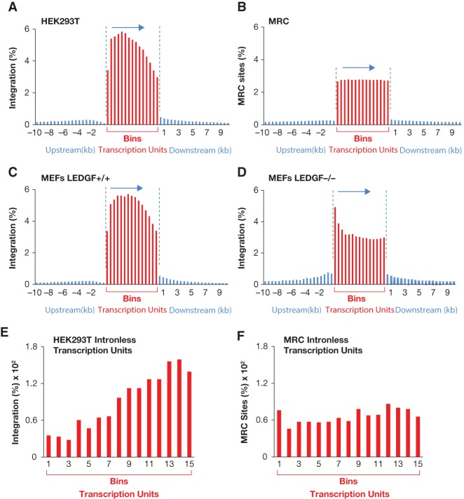
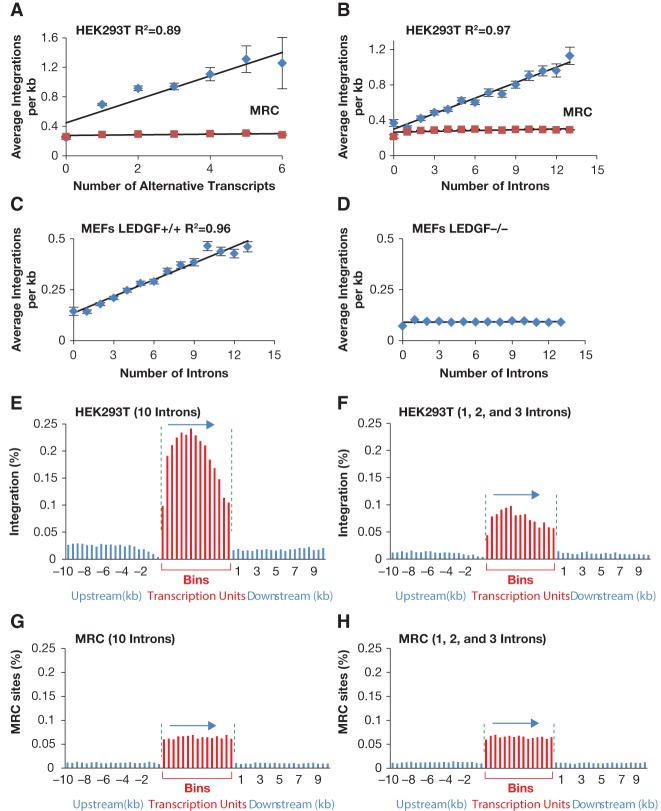
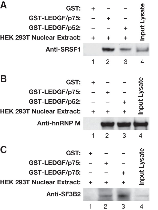
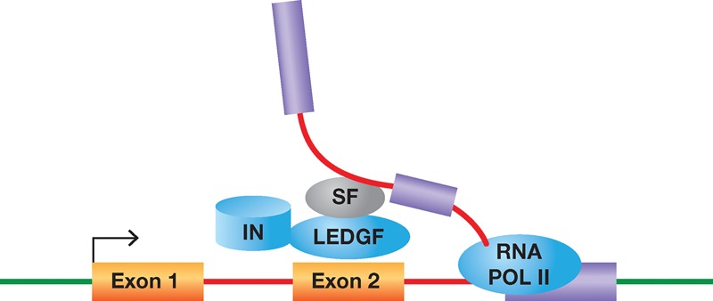
The host chromatin-binding factor LEDGF/p75 interacts with HIV-1 integrase and directs integration to active transcription units. To understand how LEDGF/p75 recognizes transcription units, we sequenced 1 million HIV-1 integration sites isolated from cultured HEK293T cells. Analysis of integration sites showed that cancer genes were preferentially targeted, raising concerns about using lentivirus vectors for gene therapy. Additional analysis led to the discovery that introns and alternative splicing contributed significantly to integration site selection. These correlations were independent of transcription levels, size of transcription units, and length of the introns. Multivariate analysis with five parameters previously found to predict integration sites showed that intron density is the strongest predictor of integration density in transcription units. Analysis of previously published HIV-1 integration site data showed that integration density in transcription units in mouse embryonic fibroblasts also correlated strongly with intron number, and this correlation was absent in cells lacking LEDGF. Affinity purification showed that LEDGF/p75 is associated with a number of splicing factors, and RNA sequencing (RNA-seq) analysis of HEK293T cells lacking LEDGF/p75 or the LEDGF/p75 integrase-binding domain (IBD) showed that LEDGF/p75 contributes to splicing patterns in half of the transcription units that have alternative isoforms. Thus, LEDGF/p75 interacts with splicing factors, contributes to exon choice, and directs HIV-1 integration to transcription units that are highly spliced.

Keywords: HIV-1; LEDGF; integration; mRNA splicing; p75; retrovirus.


Figures

Figure 1.
Figure 1.
Distribution of HIV-1 integration sites within transcription units. Each transcription unit was divided into 15 equal parts (bins), and HIV-1 integration sites or MRC insertion sites were counted for each bin (shown as red bars). Outside of transcription units, the genome was divided into 500-bp bins, and the HIV-1 integration sites or MRC insertion sites were counted in each bin (shown as blue bars). The percentages of all of the HIV-1 integration sites and MRC insertion sites are shown on the Y-axis. The horizontal arrow shows the direction of transcription. The green vertical lines separate the transcription units from the upstream and downstream regions to indicate that the bins within the transcription units are not equivalent in size to the 500-bp bins outside of the transcription units. (A) Distribution of HIV-1 integration sites in human HEK293T cells. (B) Distribution of MRC insertion sites in human transcription units. (C) Distribution of HIV-1 integration sites in transcription units in mouse fibroblast cells that contain the wild-type LEDGF gene. (D) Distribution of HIV-1 integration sites in mouse fibroblast cells that lack the LEDGF gene. (E,F) Distribution of HIV-1 integration sites (E) and MRC insertion sites (F) within intronless transcription units.
Figure 2.
Figure 2.
Integration density correlates strongly with the level of splicing. (A) Correlation between HIV-1 integration density and the number of alternative transcripts produced by transcription units in human HEK293T cells. Each transcription unit was assigned to a group based on the number of alternatively spliced transcripts that originated from it. The X-axis shows the number of alternative transcripts per transcription unit, and the Y-axis shows the average HIV-1 integration density for all transcription units that produce the same number of alternative transcripts. The blue diamonds represent data for HIV-1 integration sites in HEK293T, whereas the red rectangles are for the MRC insertion sites. The vertical bar for each data point is the standard error. R2 is the Pearson correlation. (BD) Correlation between average HIV-1 integration density and the number of introns in the transcription units. The X-axis shows the number of introns per transcription unit, and the Y-axis shows the average HIV-1 integration density (integration sites per kilobase) for all transcription units that have the same number of introns. The blue diamonds represent data for HIV-1 integrations, and the red rectangles are for the MRC insertion sites. (B) HIV-1 integration sites in human HEK293T cells. C and D are based on HIV-1 integration sites in mouse fibroblast cells that either have or lack the LEDGF gene, respectively. (EH) Distribution of HIV-1 integration sites (E,F) or MRC insertion sites (G,H) within transcription units that contain either 10 introns (E,G) or one to three introns (F,H), based on the data from HEK293T cells. For each transcription unit with 10 introns, a partner transcription unit of equal size was selected from the group that contains transcription units with one to three introns, so the total length of all transcription units with 10 introns is equal to the total length of all transcription units with one to three introns. As in Figure 1, each transcription unit was divided into 15 equal parts (shown in red), and HIV-1 integration sites or MRC sites were counted in each segment.
Figure 3.
Figure 3.
The interactions of representative splicing proteins with LEDGF/p75 and LEDGF/p52. GST pull-down of HEK293T nuclear lysate with GST, GST-LEDGF/p75, or GST–LEDGF/p52 followed by Western blotting to detect splicing factors SRSF1 (A), hnRNP M (B), and SF3B2 (C).
Figure 4.
Figure 4.
Model for HIV-1 integration in highly spliced transcription units. RNA Pol II transcription and splicing of introns are concurrent. Splicing factors (SF) interact with LEDGF/p75, which in turn binds HIV-1 integrase (IN). These interactions help direct HIV-1 integration to transcription units with a large number of introns.

References

    1. Berry C, Hannenhalli S, Leipzig J, Bushman FD. 2006. Selection of target sites for mobile DNA integration in the human genome. PLoS Comput Biol 2: e157. - PMC - PubMed
    1. Brody Y, Neufeld N, Bieberstein N, Causse SZ, Bohnlein EM, Neugebauer KM, Darzacq X, Shav-Tal Y. 2011. The in vivo kinetics of RNA polymerase II elongation during co-transcriptional splicing. PLoS Biol 9: e1000573. - PMC - PubMed
    1. Busschots K, Vercammen J, Emiliani S, Benarous R, Engelborghs Y, Christ F, Debyser Z. 2005. The interaction of LEDGF/p75 with integrase is lentivirus-specific and promotes DNA binding. J Biol Chem 280: 17841–17847. - PubMed
    1. Cherepanov P, Maertens G, Proost P, Devreese B, Van Beeumen J, Engelborghs Y, De Clercq E, Debyser Z. 2003. HIV-1 integrase forms stable tetramers and associates with LEDGF/p75 protein in human cells. J Biol Chem 278: 372–381. - PubMed
    1. Cherepanov P, Devroe E, Silver PA, Engelman A. 2004. Identification of an evolutionarily conserved domain in human lens epithelium-derived growth factor/transcriptional co-activator p75 (LEDGF/p75) that binds HIV-1 integrase. J Biol Chem 279: 48883–48892. - PubMed

Publication types

Substances

Associated data

LinkOut - more resources