. 2015 Nov;27(11):3143-59.
doi: 10.1105/tpc.15.00562. Epub 2015 Nov 6.

SMAX1-LIKE/D53 Family Members Enable Distinct MAX2-Dependent Responses to Strigolactones and Karrikins in Arabidopsis

Affiliations

SMAX1-LIKE/D53 Family Members Enable Distinct MAX2-Dependent Responses to Strigolactones and Karrikins in Arabidopsis

Ishwarya Soundappan et al. Plant Cell. 2015 Nov.

Abstract

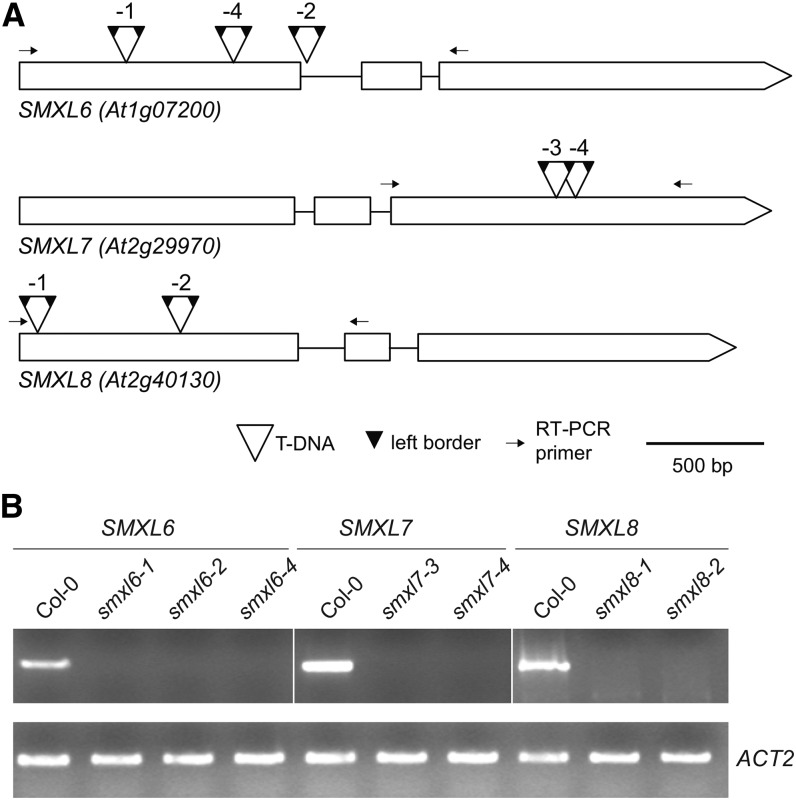
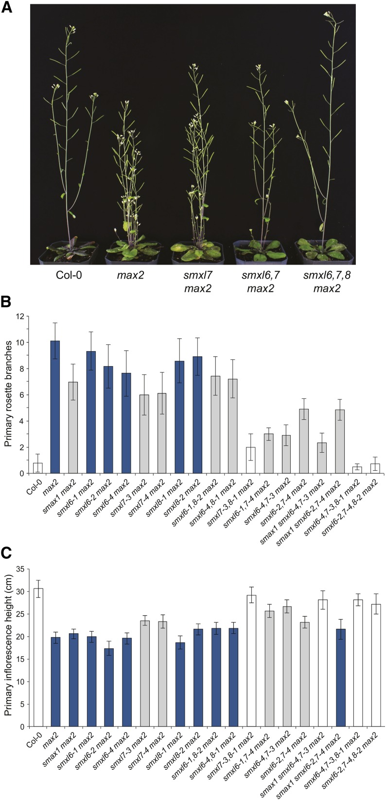
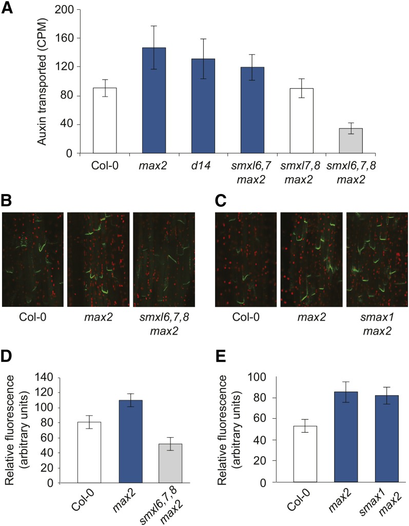
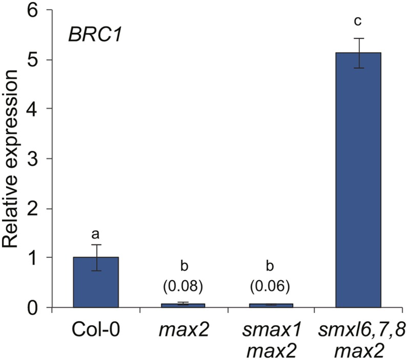
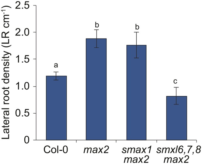
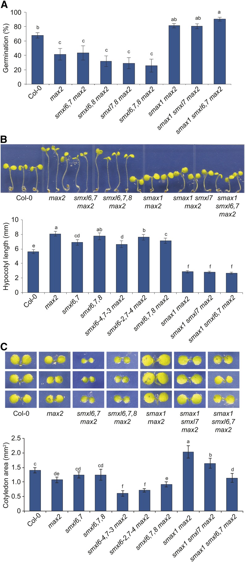
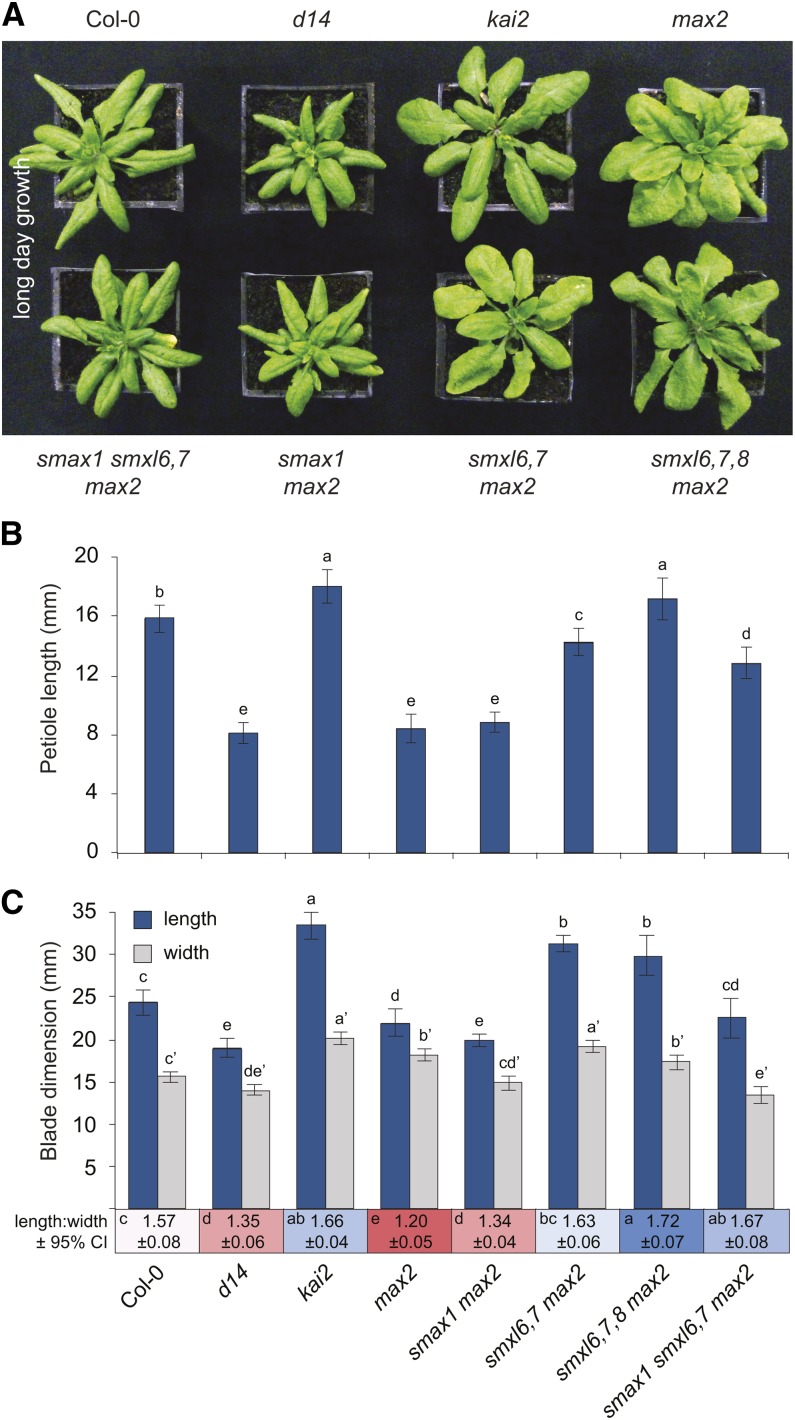
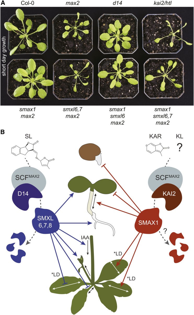
The plant hormones strigolactones and smoke-derived karrikins are butenolide signals that control distinct aspects of plant development. Perception of both molecules in Arabidopsis thaliana requires the F-box protein MORE AXILLARY GROWTH2 (MAX2). Recent studies suggest that the homologous SUPPRESSOR OF MAX2 1 (SMAX1) in Arabidopsis and DWARF53 (D53) in rice (Oryza sativa) are downstream targets of MAX2. Through an extensive analysis of loss-of-function mutants, we demonstrate that the Arabidopsis SMAX1-LIKE genes SMXL6, SMXL7, and SMXL8 are co-orthologs of rice D53 that promote shoot branching. SMXL7 is degraded rapidly after treatment with the synthetic strigolactone mixture rac-GR24. Like D53, SMXL7 degradation is MAX2- and D14-dependent and can be prevented by deletion of a putative P-loop. Loss of SMXL6,7,8 suppresses several other strigolactone-related phenotypes in max2, including increased auxin transport and PIN1 accumulation, and increased lateral root density. Although only SMAX1 regulates germination and hypocotyl elongation, SMAX1 and SMXL6,7,8 have complementary roles in the control of leaf morphology. Our data indicate that SMAX1 and SMXL6,7,8 repress karrikin and strigolactone signaling, respectively, and suggest that all MAX2-dependent growth effects are mediated by degradation of SMAX1/SMXL proteins. We propose that functional diversification within the SMXL family enabled responses to different butenolide signals through a shared regulatory mechanism.


Figures

Figure 1.
Figure 1.
Mutant Alleles of SMXL6, SMXL7, and SMXL8. (A) T-DNA insertion positions for smxl6-1 (SAIL_86_H04), smxl6-2 (SAIL_1285_H05), smxl6-4 (SALK_049115), smxl7-3 (WiDsLox339_C04), smxl7-4 (SALK_082032), smxl8-1 (SALK_025338C), and smxl8-2 (SALK_126406C). Boxes indicate exons, open triangles indicate the positions of T-DNAs, and small dark triangles indicate left borders. Most insertions had two outward-facing left borders, implying T-DNA concatamers. Left border insertion positions in the gene sequence were defined by Sanger sequencing and are described in Supplemental Table 2. (B) End-point RT-PCR analysis of SMXL6 (30 cycles), SMXL7 (30 cycles), and SMXL8 (40 cycles) expression in respective T-DNA mutant lines. ACT2 (30 cycles) was used as an internal reference gene. RT-PCR primers span the T-DNA insertion sites, as indicated in (A).
Figure 2.
Figure 2.
SMXL6, SMXL7, and SMXL8 Promote Shoot Branching Downstream of MAX2. (A) Representative images of adult Col-0 (wild type), max2-1, smxl7-3 max2-1, smxl6-4,7-3 max2-1, and smxl6-4,7-3,8-1 max2-1 plants. (B) Primary rosette branch number. (C) Height of the primary inflorescence. Plants were grown under a LD photoperiod (16 h light/8 h dark). Images and data were collected for each plant at 10 d postanthesis. For (B) and (C), graphs show mean ± 99% CI, n ≥ 17. Bar colors indicate no significant difference to Col-0 (white), no significant difference to max2 (dark blue), and significant differences to both Col-0 and max2 (gray). P < 0.001 (P = 0.02, with Bonferroni correction for 19 comparisons), Student’s t test.
Figure 3.
Figure 3.
SMXL6,7,8 Promote Auxin Transport and PIN1 Accumulation. (A) Auxin transport in stem segments from basal internodes. Mean ± 95% CI, n = 15 to 20. (B) Confocal laser scanning fluorescence microscopy of PIN1-GFP in stem segments from basal internodes of Col-0, max2, and smxl6,7,8 max2. (C) Confocal laser scanning fluorescence microscopy of PIN1-GFP in stem segments from basal internodes of Col-0, max2, and smax1 max2. (D) PIN1-GFP fluorescence intensity at the basal plasma membrane in stem segments, as exemplified in (B). Mean ± 99% CI, n = 40 (five basal membranes in eight plants per genotype). (E) PIN1-GFP fluorescence intensity at the basal plasma membrane in stem segments, as exemplified in (C). Mean ± 99% CI, n = 40 (five basal membranes in eight plants per genotype). Bar colors indicate no significant difference to Col-0 (white), no significant difference to max2 (dark blue), and significant differences to both Col-0 and max2 (gray). Student’s t test, P < 0.05 for (A) and P < 0.01 for (D) and (E). PIN1-GFP is from Xu et al. (2006).
Figure 4.
Figure 4.
SMXL6,7,8 Repress BRC1 Expression in Axillary Buds. RT-qPCR analysis of BRC1/TCP18 gene expression in nonelongated axillary buds of Col-0, max2-1, smax1-2 max2-1, and smxl6-4,7-3,8-1 max2-1 collected 7 d after anthesis. Expression of BRC1 is relative to CACS internal reference gene. Expression values are scaled to the Col-0 level of expression. Expression for max2-1 and smax1-2 max2-1 is indicated in parentheses. Mean ± se; n = 3 to 4 pooled tissue samples, four plants per pool. ANOVA with post-hoc Fisher’s LSD, P < 0.01.
Figure 5.
Figure 5.
Lateral Root Formation Is Suppressed by smxl6,7,8. Lateral root density of 10-d-old seedlings. Mean ± se; n = 6 experiments, ≥10 roots measured per genotype in each experiment. ANOVA with post-hoc Student’s paired t test, P < 0.05.
Figure 6.
Figure 6.
rac-GR24 Triggers Rapid Degradation of SMXL7. (A) and (B) Col-0 35Spro:SMXL7-YFP roots after 0 and 20 min of treatment with 1 μM rac-GR24 (A) or 1 μM KAR1 (B). (C) Col-0 35Spro:SMXL7-YFP after 0, 5, 10, and 20 min treatment with 10 μM rac-GR24. (D) max2-1 35Spro:SMXL7-YFP after 0 and 20 min of treatment with 10 μM rac-GR24. (E) d14-1 35Spro:SMXL7-YFP after 0 and 20 min of treatment with 10 μM rac-GR24. (F) Col-0 35Spro:SMXL7-YFP after 20 min of treatment with 10 μM rac-GR24, without and with a 1-h pretreatment with 50 μM MG132, a 26S proteasome inhibitor. (G) An eight-amino acid sequence (FRGKTVVD) containing a putative P-loop was removed from SMXL7-YFP. Col-0 35Spro:SMXL7ΔP-loop-YFP after 0 and 20 min of treatment with 10 μM rac-GR24.
Figure 7.
Figure 7.
Distinct Roles for SMXL6,7,8 and SMAX1 during Germination and Seedling Growth. (A) Germination of primary dormant seed after 66 h imbibition at 21°C in LD photoperiod on 0.8% (w/v) Bacto-agar media. Mean ± se; n = 6 to 7 batches of seed, 75 seed tested per batch. ANOVA with post-hoc Fisher’s LSD, P < 0.025. (B) Hypocotyl lengths of 5-d-old seedlings grown under continuous red light (30 μE) for 4 d at 21°C. Mean ± 99% CI; n = 45 seedlings (15 seedlings from three replicate plates). ANOVA with post-hoc Fisher’s LSD, P < 0.01. (C) Cotyledon surface area of seedlings grown as described in (B). Mean ± 99% CI; n = 36 cotyledons from 18 seedlings. ANOVA with post-hoc Fisher’s LSD, P < 0.01.
Figure 8.
Figure 8.
SMXL6,7,8 and SMAX1 Have Different Effects on Leaf Morphology. (A) Rosettes of 28-d-old plants grown under LD photoperiod (16 h light/8 h dark). d14-1, kai2-1, max2-1, smax1-2, smxl6-4, smxl7-3, and smxl8-1 alleles were used. kai2-1, first identified in Landsberg erecta, was backcrossed twice into the Col-0 background. (B) Petiole length of the 7th leaf of 35-d-old plants grown under LD photoperiod. Mean ± 95% CI; n = 11 to 12. ANOVA with post-hoc Fisher’s LSD, P < 0.05. (C) Blade length (blue), not including the petiole, and width (gray) of the 7th leaf of 35-d-old plants grown under LD photoperiod. Mean ± 95% CI; n = 11 to 12. ANOVA with post-hoc Fisher’s LSD, P < 0.05. Prime symbol (e.g., a′) differentiates statistical tests of width measurements from length measurements. Mean length:width ratio ± 95% CI is shown below the x axis. Statistical groups for length:width ratios are indicated in each colored box.
Figure 9.
Figure 9.
Tissue-Specific GUS Expression from SMAX1, SMXL6, SMXL7, and SMXL8 Promoters. Expression of promoter:GUS-GFP fusions in the primary rosette buds and young leaves of 4.5-week-old plants (A), shoot apex and developing leaves (B), hypocotyl (C), cotyledonary veins (D), stomata on first true leaves (E), vasculature in the maturation zone of primary roots (F), and primary root cap (G). (B) to (G) are tissues of 10-d-old light-grown seedlings. For each tissue, the different SMAX1 and SMXL transcriptional reporters were stained at the same time, for the same duration.
Figure 10.
Figure 10.
EAR Motif-Dependent Interaction of SMAX1 and SMXL7 with TOPLESS Family Proteins. (A) Yeast two-hybrid interaction tests of SMAX1 and SMXL7 with TPL and TPR proteins TPR1-TPR4. SMAX1/SMXL7 proteins were bait (N-terminal GAL4 DNA binding domain fusions), and TPL/TPR proteins were prey (N-terminal GAL4 activation domain fusions). RalGDS was a negative control for interactions with SMAX1 and SMXL7. RalGDS + Krev1 was a strong positive control. SMAX1 and SMXL7 variants with a mutated EAR domain (mEAR, sequence FDLNQ or LDLNL modified to ADANA) were also tested. Growth is shown for equally diluted colonies grown for 3 d at 30°C on -Leu/Trp/His (-LTH) dropout media with 10 mM 3-amino-1,2,4-triazole (3-AT). Qualitative LacZ assays were performed by growing colonies on -Leu/Trp (-LT) double dropout media with 80 μg/mL X-α-Gal for 30 h. (B) BiFC tests for interaction between SMAX1, SMAX1mEAR, SMXL7, and SMXL7mEAR with TPR2. The N-terminal portion of GFP was fused to the N terminus of TPR2, and the C-terminal portion of GFP was fused to the N terminus of SMAX1/SMXL7 proteins. Confocal fluorescence microscopy images of transiently transformed N. benthamiana leaves were taken with the same exposure settings across construct comparisons. A representative field of view is shown (left). Representative nuclei (right) are shown with GFP signal (green) overlaid onto 4′,6-diamidino-2-phenylindole (DAPI) signal (blue).
Figure 11.
Figure 11.
SMAX1 and SMXL6,7,8 Have Complementary Roles in MAX2-Regulated Development. (A) Rosettes of 37-d-old plants grown under a short-day photoperiod (8 h light/16 h dark). The kai2/htl allele is htl-3 (Toh et al., 2014). (B) Model of SL and KAR/KL signaling. SMXL6,7,8 act in processes controlled by D14, and SMAX1 acts in processes controlled by KAI2. SMAX1 represses germination and seedling responses to light (e.g., reduces cotyledon expansion and promotes hypocotyl elongation) and promotes medio-lateral blade expansion (i.e., blade width) and elongation in long days. SMXL6,7,8 promote branching, auxin transport, PIN1 accumulation at the basal plasma membrane (data not shown), and lateral root density but inhibit petiole elongation and proximo-distal blade expansion in long days (i.e., blade elongation). *LD denotes control in a long-day photoperiod; under short-day conditions, SMAX1 and SMXL6,7,8 have different effects on leaf growth but are still antagonistic (A). Loss of SMXL6,7 inhibits cotyledon expansion, although this is countered somewhat by loss of SMXL8. D53/SMXL7 interacts with D14 after SL perception and is targeted for degradation in a MAX2-dependent manner (Figure 6; Jiang et al., 2013; Zhou et al., 2013; Umehara et al., 2015). A similar mechanism for SMAX1 degradation after KAR/KL perception by KAI2 is hypothesized.

References

    1. Aguilar-Martínez J.A., Poza-Carrión C., Cubas P. (2007). Arabidopsis BRANCHED1 acts as an integrator of branching signals within axillary buds. Plant Cell 19: 458–472. - PMC - PubMed
    1. Agusti J., Herold S., Schwarz M., Sanchez P., Ljung K., Dun E.A., Brewer P.B., Beveridge C.A., Sieberer T., Sehr E.M., Greb T. (2011). Strigolactone signaling is required for auxin-dependent stimulation of secondary growth in plants. Proc. Natl. Acad. Sci. USA 108: 20242–20247. - PMC - PubMed
    1. Akiyama K., Hayashi H. (2006). Strigolactones: chemical signals for fungal symbionts and parasitic weeds in plant roots. Ann. Bot. (Lond.) 97: 925–931. - PMC - PubMed
    1. Al-Babili S., Bouwmeester H.J. (2015). Strigolactones, a novel carotenoid-derived plant hormone. Annu. Rev. Plant Biol. 66: 161–186. - PubMed
    1. Alder A., Jamil M., Marzorati M., Bruno M., Vermathen M., Bigler P., Ghisla S., Bouwmeester H., Beyer P., Al-Babili S. (2012). The path from β-carotene to carlactone, a strigolactone-like plant hormone. Science 335: 1348–1351. - PubMed

Publication types

MeSH terms