. 2016 Mar 15;22(6):1520-30.
doi: 10.1158/1078-0432.CCR-15-1259. Epub 2015 Nov 6.

Combined Tumor Suppressor Defects Characterize Clinically Defined Aggressive Variant Prostate Cancers

Affiliations

Combined Tumor Suppressor Defects Characterize Clinically Defined Aggressive Variant Prostate Cancers

Ana M Aparicio et al. Clin Cancer Res. .

Abstract

Purpose: Morphologically heterogeneous prostate cancers that behave clinically like small-cell prostate cancers (SCPC) share their chemotherapy responsiveness. We asked whether these clinically defined, morphologically diverse, "aggressive variant prostate cancer (AVPC)" also share molecular features with SCPC.

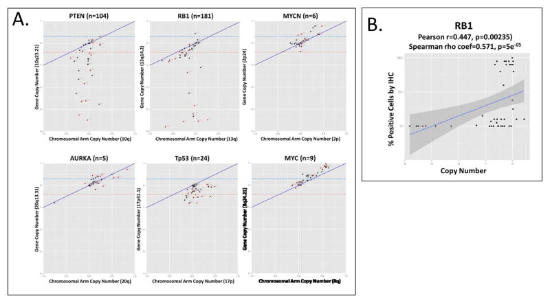
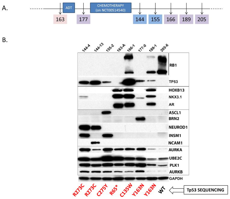
Experimental design: Fifty-nine prostate cancer samples from 40 clinical trial participants meeting AVPC criteria, and 8 patient-tumor derived xenografts (PDX) from 6 of them, were stained for markers aberrantly expressed in SCPC. DNA from 36 and 8 PDX was analyzed by Oncoscan for copy number gains (CNG) and losses (CNL). We used the AVPC PDX to expand observations and referenced publicly available datasets to arrive at a candidate molecular signature for the AVPC.

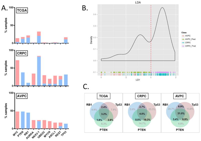
Results: Irrespective of morphology, Ki67 and Tp53 stained ≥10% cells in 80% and 41% of samples, respectively. RB1 stained <10% cells in 61% of samples and AR in 36%. MYC (surrogate for 8q) CNG and RB1 CNL showed in 54% of 44 samples each and PTEN CNL in 48%. All but 1 of 8 PDX bore Tp53 missense mutations. RB1 CNL was the strongest discriminator between unselected castration-resistant prostate cancer (CRPC) and the AVPC. Combined alterations in RB1, Tp53, and/or PTEN were more frequent in the AVPC than in unselected CRPC and in The Cancer Genome Atlas samples.

Conclusions: Clinically defined AVPC share molecular features with SCPC and are characterized by combined alterations in RB1, Tp53, and/or PTEN.


Conflict of interest statement

Disclosure of Potential Conflicts of Interest: None

Figures

Figure 1
Figure 1. Immunohistochemical analysis of NCT00514540 unique “Baseline” samples
(A) Labeling indices (% of cells staining positive for the marker listed on the y-axis, Li) for RB1, Tp53, AR, NKX3-1, AURKA, UBE2C and Ki67 in unique patient and PDX Baseline Samples (only one sample per patient is shown of the set obtained between −13 and +1 months from registration). Note, 177-0-XENO is included in the graph (n=32) but not in the analyses described in the text because the donor-patient tumor was not available for analysis. The BRN2 stains did not work and are not reported. Black bars depict nuclear staining. For AR, AURKA and UBE2C cytoplasmic stains are shown in light grey. Each individual sample is listed on the x-axis. Numbers indicate accession number on the protocol or PDX line. Letters indicate site of origin. Blue arrowheads indicate SCPC morphology. (B) Corrgram of selected tissue and serum markers among the 31 Baseline samples’ donors. Red represents positive values and blue represents negative values. The intensity of the color is proportional to the magnitude of the correlation. (*) indicate the IHC did not work. Abbreviations: RB, retinoblastoma 1; p53, tumor suppressor Tp53; NKX3-1, NK3 homeobox 1; AURKA, aurora kinase A; UBE2C, ubiquitin conjugating enzyme 2C; Ki67, antigen identified by monoclonal antibody Ki-67; RXPX, radical prostatectomy; BMBX, bone marrow biopsy; TURP, transurethral resection of prostate; CRAN, craniotomy; RCBX, rectal biopsy; XENO, xenograft; PVBX, pelvic mass biopsy; LNBX, lymph node biopsy; BOBX, bone biopsy; LIBX, liver biopsy; PAP, prostatic acid phosphatase; PSA, prostate specific antigen; -N, nuclear staining; -C, cytoplasmic staining; StdCHRA, standardized chromogranin A; CEA, carcinoembryonic antigen.
Figure 2
Figure 2. Copy number alterations at genes of interest and correlation with IHC results
(A) Segmental (values corresponding only to the gene of interest) vs regional (median values for the corresponding chromosome arm) copy number calls for PTEN, RB1, MYCN, AURKA, Tp53 and MYC. “n” in parenthesis following the gene name indicates the number of gene-specific probes present on the Oncoscan® chip. The blue and red dashed lines indicate the absolute cutoff values. The solid blue line represents the corresponding chromosome arm copy number. Red dots indicate samples with SCPC morphology. (B) Correlation between labeling indices (y-axis) and copy number (x-axis) for RB1.
Figure 3
Figure 3. Analyses in PDX
(A) PDX development relative to start of ADT and chemotherapy. Numbers indicate PDX line. The MDA-PCa 163 PDX line was derived from the prostate tumor of patient #80 on NCT00514540 in the non-castrate setting (pink shaded box). Blue shade indicates SCPC morphology. Purple shade indicates castration-resistant prostate tumor without SCPC morphology. (B) Western blot analyses of markers of interest and Tp53 mutations (shown in red) in PDX. Abbreviations: PDX, patient-tumor derived xenograft; ADT, androgen deprivation therapy; WT, wild-type.
Figure 4
Figure 4
AVPC data with reference to publicly available TCGA and unselected CRPC data sets. (A) Histograms showing percentage of samples with CNA (per absolute cutoffs) at segments corresponding to genes of interest in TCGA, unselected CRPC (middle) and AVPC 7. In blue are copy number losses and in red are copy number gains. (B) Linear Discriminant Analysis of AVPC and unselected CRPC samples using the 10-CNA-marker set. Green and purple dots at the bottom indicate predicted AVPC (AVPC_Pred) and non-AVPC (CRPC_Pred) predictions respectively while red and blue vertical lines indicate actual AVPC and unselected CRPC samples respectively. (C) Venn diagrams showing the rates of combined RB1, Tp53 and/or PTEN alterations in the TCGA (n=253), CRPC (n=150) and AVPC (n=29) samples.

References

    1. Jensen EV, Block GE, Smith S, Kyser K, DeSombre ER. Estrogen receptors and breast cancer response to adrenalectomy. National Cancer Institute monograph. 1971;34:55–70. - PubMed
    1. Slamon DJ, Clark GM, Wong SG, Levin WJ, Ullrich A, McGuire WL. Human breast cancer: correlation of relapse and survival with amplification of the HER-2/neu oncogene. Science. 1987;235(4785):177–82. - PubMed
    1. Lynch TJ, Bell DW, Sordella R, Gurubhagavatula S, Okimoto RA, Brannigan BW, et al. Activating mutations in the epidermal growth factor receptor underlying responsiveness of non-small-cell lung cancer to gefitinib. The New England journal of medicine. 2004;350(21):2129–39. - PubMed
    1. Kwak EL, Bang YJ, Camidge DR, Shaw AT, Solomon B, Maki RG, et al. Anaplastic lymphoma kinase inhibition in non-small-cell lung cancer. The New England journal of medicine. 2010;363(18):1693–703. - PMC - PubMed
    1. Flaherty KT, Puzanov I, Kim KB, Ribas A, McArthur GA, Sosman JA, et al. Inhibition of mutated, activated BRAF in metastatic melanoma. The New England journal of medicine. 2010;363(9):809–19. - PMC - PubMed

Publication types

MeSH terms