. 2015 Dec 25;290(52):31077-89.
doi: 10.1074/jbc.M115.677039. Epub 2015 Nov 6.

The Redox State Regulates the Conformation of Rv2466c to Activate the Antitubercular Prodrug TP053

Affiliations

The Redox State Regulates the Conformation of Rv2466c to Activate the Antitubercular Prodrug TP053

David Albesa-Jové et al. J Biol Chem. .

Abstract

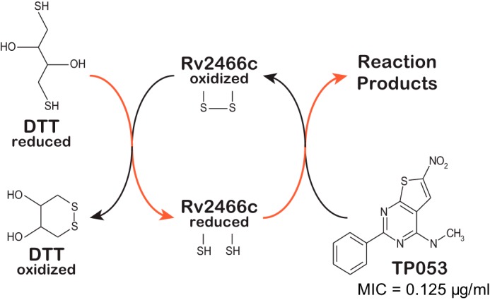
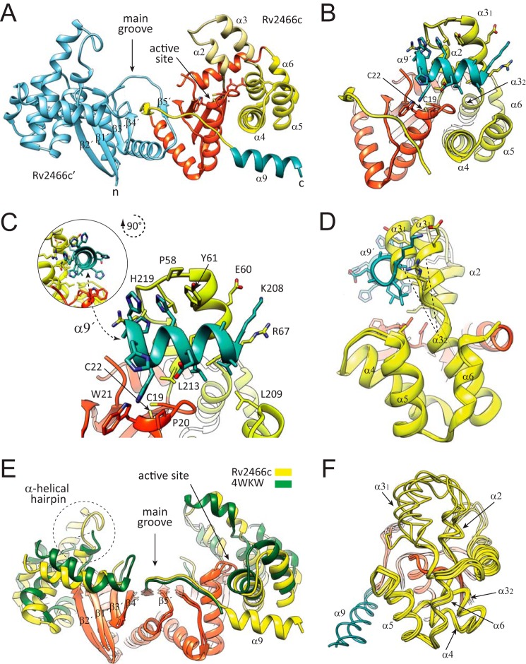
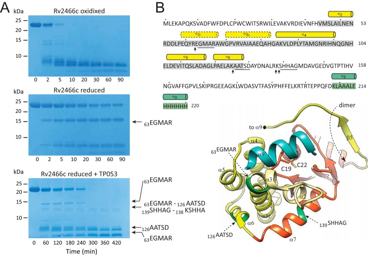
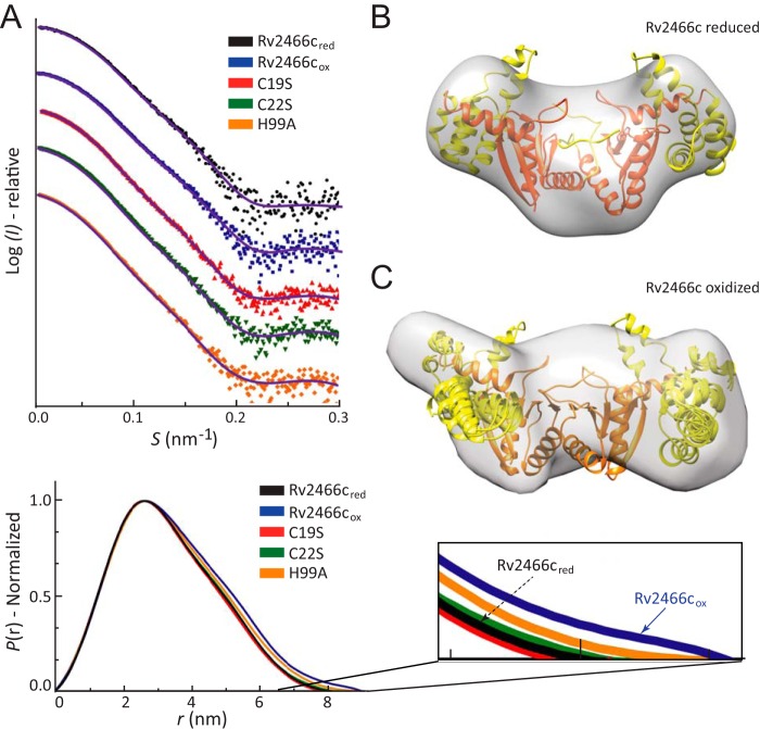
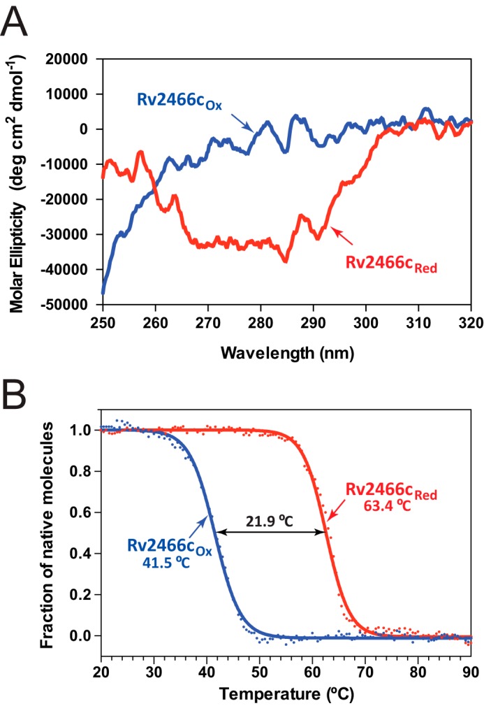
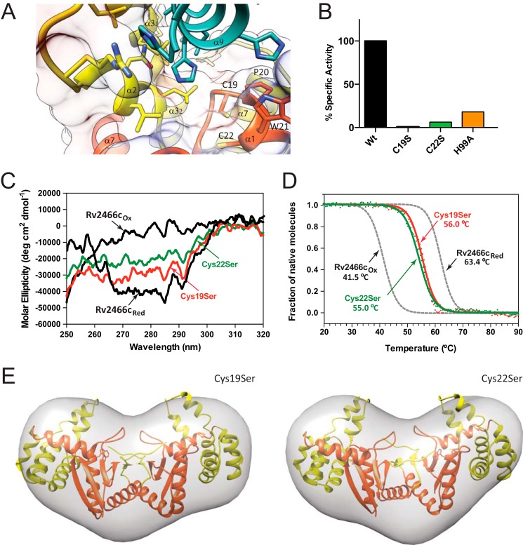
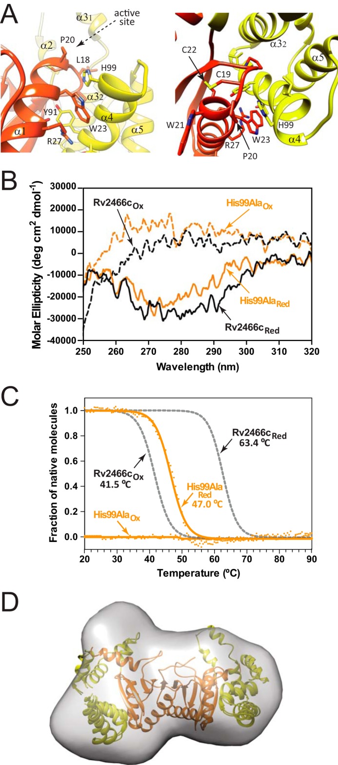
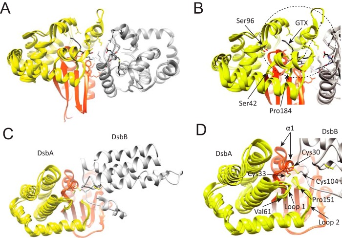
Rv2466c is a key oxidoreductase that mediates the reductive activation of TP053, a thienopyrimidine derivative that kills replicating and non-replicating Mycobacterium tuberculosis, but whose mode of action remains enigmatic. Rv2466c is a homodimer in which each subunit displays a modular architecture comprising a canonical thioredoxin-fold with a Cys(19)-Pro(20)-Trp(21)-Cys(22) motif, and an insertion consisting of a four α-helical bundle and a short α-helical hairpin. Strong evidence is provided for dramatic conformational changes during the Rv2466c redox cycle, which are essential for TP053 activity. Strikingly, a new crystal structure of the reduced form of Rv2466c revealed the binding of a C-terminal extension in α-helical conformation to a pocket next to the active site cysteine pair at the interface between the thioredoxin domain and the helical insertion domain. The ab initio low-resolution envelopes obtained from small angle x-ray scattering showed that the fully reduced form of Rv2466c adopts a "closed" compact conformation in solution, similar to that observed in the crystal structure. In contrast, the oxidized form of Rv2466c displays an "open" conformation, where tertiary structural changes in the α-helical subdomain suffice to account for the observed conformational transitions. Altogether our structural, biochemical, and biophysical data strongly support a model in which the formation of the catalytic disulfide bond upon TP053 reduction triggers local structural changes that open the substrate binding site of Rv2466c allowing the release of the activated, reduced form of TP053. Our studies suggest that similar structural changes might have a functional role in other members of the thioredoxin-fold superfamily.

Keywords: Mycobacterium tuberculosis; conformational change; enzyme; oxidation-reduction (redox); small-angle x-ray scattering (SAXS); thioredoxin; x-ray crystallography.


Figures

FIGURE 1.
FIGURE 1.
Rv2466c mediates the reduction of TP053 to kill M. tuberculosis. Model of the catalytic cycle of Rv2466c. Reduced Rv2466c reacts with TP053 to oxidized Rv2466c and reduced reaction products. Oxidized Rv2466c is then recycled to its reduced form by reduced DTT via disulfide exchange. The proposed electron flow during redox exchange is indicated in red. MIC, minimum inhibitory concentration.
FIGURE 2.
FIGURE 2.
Crystal structure of reduced Rv2466c-CT-His. A, schematic representation showing the structure of Rv2466c-CT-His. Monomer A (Rv2466c) is shown in orange (thioredoxin domain) and yellow (α-helical subdomain), whereas monomer B (Rv2466c′) is shown in cyan. The C-terminal peptide of 13 amino acids (KLAAALEHHHHHH) is shown in blue. B, schematic representation showing the catalytic motif (Cys19-Pro20-Trp21-Cys22) in close proximity to the α-helical subdomain, comprising a four α-helical bundle (α3″–α4–α5–α6) and a short α-helical hairpin (α2-α3′). The C terminus peptide of 13 amino acids binds to the active site and folds into an α-helix. C, close view of the active site of Rv2466c showing the molecular interactions with the peptide. D, conformational changes triggered by binding of the C-terminal peptide segment. Structural superposition of two selected monomers from the reduced Rv2466c (9) and Rv2466c-CT-His constructs, with the α9 in blue, suggesting that the α2–α3′ hairpin region displays structural variability. E, schematic representation showing the comparison between the crystal structure of Rv2466c-CT-His and the structural homologue from M. leprae (PDB code 4WKW). The canonical thioredoxin folds are shown in orange. The α-helical subdomains of Rv2466c-CT-His and the M. leprae homologue are shown in yellow and green, respectively. F, schematic representation showing the comparison of all monomers from the crystal structures of the Rv2466c (9), Rv2466c-CT-His, and the structural homologue from M. leprae (PDB code 4WKW).
FIGURE 3.
FIGURE 3.
Limited proteolysis experiments show significant conformational differences between the redox forms of Rv2466c. A, SDS-PAGE showing the trypsin cleavage profile for Rv2466cOX, Rv2466cRED, and Rv2466cRED in the presence of TP053. B, the peptide bonds that are cleaved specifically by trypsin are shown with arrows. The N-terminal sequences of selected proteolytic fragments are underlined in the amino acid sequence and labeled in dark green in the schematic representation of Rv2466c.
FIGURE 4.
FIGURE 4.
Conformation of oxidized and reduced Rv2466c, as characterized by SAXS. A, upper panel: scattering curves of Rv2466cRED and Rv2466cOX. Lower panel: P(r) function distributions of Rv2466cRED and Rv2466cOX. B and C, low resolution models of Rv2466cRED and Rv2466cOX in solution. Average low resolution structure of Rv2466cRED (B) and Rv2466cOX (C) with the high resolution crystal structure of Rv2466cRED (Protein Data Bank code 4NXI) fitted by rigid body docking, respectively.
FIGURE 5.
FIGURE 5.
Tertiary structure differences between the oxidized and reduced forms of Rv2466c analyzed by CD spectroscopy. A, near-UV CD spectra for Rv2466cRED and Rv2466cOX. B, thermal unfolding transitions of Rv2466cRED and Rv2466cOX monitored by the change in CD signal at 222 nm. The Tm values are given in the figures and represent the apparent melting temperature. The transition was tentatively fitted according to a two-state equilibrium (solid lines) and normalized.
FIGURE 6.
FIGURE 6.
The redox state of the CPWC motif regulates Rv2466c conformation and activity. A, localization of Cys19 and Cys22 in the active site of Rv2466c (top of α1 helix). B, activity measurements of Rv2466c WT and Rv2466c variants against TP053 (9). C, near-UV CD spectra of the C19S and C22S variants. D, thermal unfolding of the C19S and C22S variants as monitored by CD spectroscopy. E, low resolution models of C19S and C22S mutants in solution. Average low resolution structure of C19S and C22S mutants with the high resolution crystal structure of Rv2466cRED (Protein Data Bank code 4NXI) fitted by rigid body docking, respectively.
FIGURE 7.
FIGURE 7.
Conformational changes at the interface between the thioredoxin domain and the α-helical subdomain. A, two close views of the thioredoxin domain-α-helical subdomain interface, and the active site motif Cys19-Pro20-Trp21-Cys22. B, near-UV CD spectra of the oxidized and reduced H99A variant. C, normalized thermal unfolding transition of the oxidized and reduced H99A variant monitored by CD. D, low resolution model of the reduced H99A variant in solution deduced from SAXS data compared with the high-resolution crystal structure of Rv2466cRED (PDB code 4NXI).
FIGURE 8.
FIGURE 8.
Structural location of the α-helical subdomain in GST and DsbA. A, schematic representation of human κ class glutathione transferase (hGSTk). The transfer of GST reduced glutathione (GSH) to a variety of hydrophobic electrophiles for cellular detoxification. In this instance, the apo state of hGSTk (PDB code 3RPP) and when bound to a reaction product, S-hexylglutathione (GTX; PDB code 3RPN) is shown. B, close-up view of overlapping hGSTk structures where structural variability of the helical bundle can be observed; most evident is the variability within Ser42-Ser96 residues, which contains a region that structures upon substrate binding. C, schematic representation of disulfide bond oxidoreductases DsbA (orange-yellow) and DsbB (gray). DsbA is a periplasmic dithiol oxidase responsible for protein disulfide formation, DsbB is an integral membrane protein catalyzing the oxidation of DsbA by ubiquinone. Three crystal structures are overlapped, corresponding to the E. coli C33A DsbA mutant in complex with DsbB (PDB code 2HI7), oxidized DsbA (PDB code 1DSB), and C33A DsbA mutant (PDB code 1TI1). D. The canonical α1 of the thioredoxin-fold undergoes large conformational changes depending on its redox state. The helical bundle (yellow) shows significant structural variability between structures.

References

    1. World Health Organization (2014) Global Tuberculosis Report 2013, www.who.int/tb/publications/global_report/en
    1. Zumla A., Raviglione M., Hafner R., and von Reyn C. F. (2013) Tuberculosis. N. Engl. J. Med. 368, 745–755 - PubMed
    1. Andries K., Verhasselt P., Guillemont J., Göhlmann H. W., Neefs J. M., Winkler H., Van Gestel J., Timmerman P., Zhu M., Lee E., Williams P., de Chaffoy D., Huitric E., Hoffner S., Cambau E., Truffot-Pernot C., Lounis N., and Jarlier V. (2005) A diarylquinoline drug active on the ATP synthase of Mycobacterium tuberculosis. Science 307, 223–227 - PubMed
    1. Koul A., Dendouga N., Vergauwen K., Molenberghs B., Vranckx L., Willebrords R., Ristic Z., Lill H., Dorange I., Guillemont J., Bald D., and Andries K. (2007) Diarylquinolines target subunit c of mycobacterial ATP synthase. Nat. Chem. Biol. 3, 323–324 - PubMed
    1. Cohen J. (2013) Infectious disease: approval of novel TB drug celebrated with restraint. Science 339, 130. - PubMed

Publication types

MeSH terms

LinkOut - more resources