. 2015 Nov 7:12:62.
doi: 10.1186/s12950-015-0107-y. eCollection 2015.

CFTR Knockdown induces proinflammatory changes in intestinal epithelial cells

Affiliations

CFTR Knockdown induces proinflammatory changes in intestinal epithelial cells

Karoline St-Martin Crites et al. J Inflamm (Lond). .

Abstract

Background: Hyperinflammation is a hallmark feature of cystic fibrosis (CF) airways. However, inflammation has also been documented systemically and, more recently, in extrapulmonary CF-affected tissues such as the pancreas and intestine. The pathogenesis of CF-related inflammation and more specifically the role of the cystic fibrosis transmembrane conductance regulator (CFTR) in that respect are not entirely understood. We have tested the hypothesis that genetic depletion of CFTR will affect the inflammatory status of human intestinal epithelial cell lines.

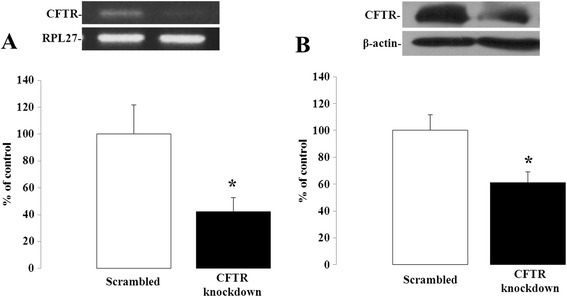
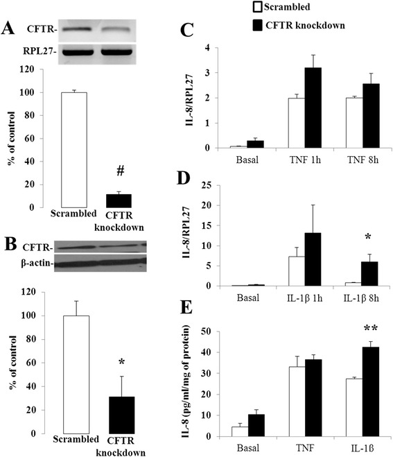
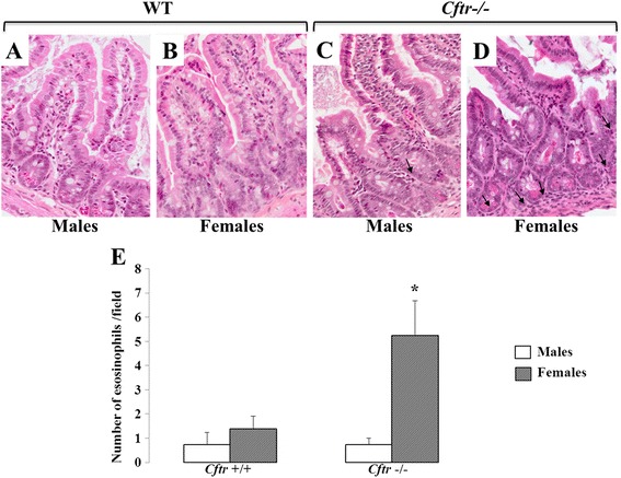
Methods: CFTR expression was genetically depleted from Caco-2/15 and HT-29 cells using short hairpin RNA interference (shRNAi). Inflammatory conditions were induced by the addition of human recombinant tumor necrosis factor (TNF) or Interleukin-1β (IL-1β) for various periods of time. Gene expression, mRNA stability and secreted levels of interleukin (IL)-6, -8 and 10 were assessed. Analysis of pro- and anti-inflammatory signaling pathways including mitogen-activated protein kinases (p38, ERK 1/2 and JNK), nuclear factor of kappa light polypeptide gene enhancer in B-cells inhibitor alpha (IκBα), and nuclear factor-kappa B (NF-κB) was also performed. Eosinophils were counted in the jejunal mucosa of Cftr-/- and Cftr+/+ mice.

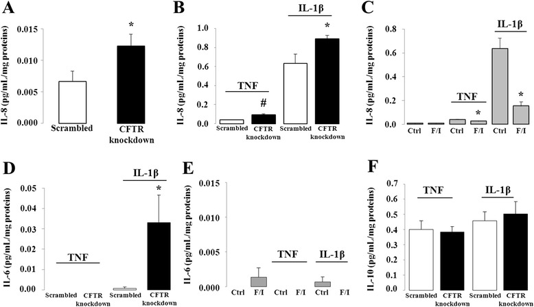
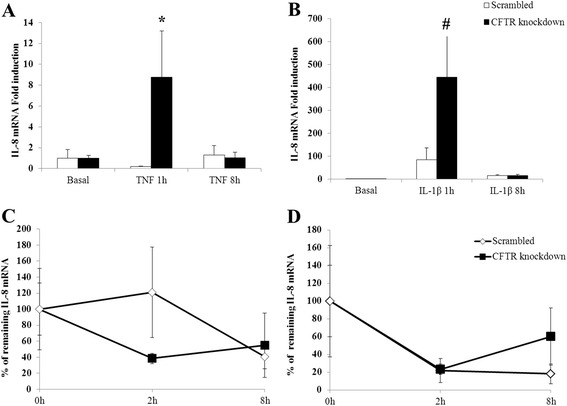
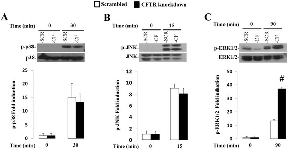
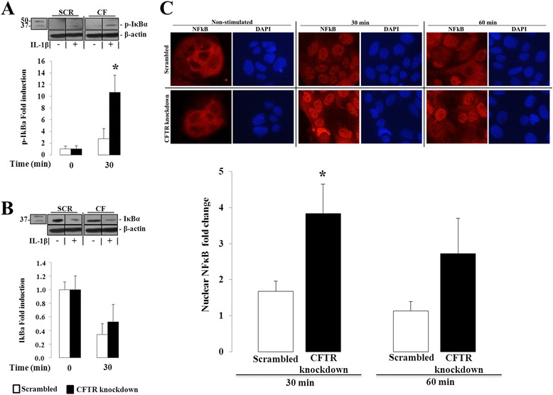
Results: CFTR gene and protein knockdown caused a significant increase in basal secretion of IL-8 as well as in IL-1β-induced secretion of IL-6 and -8. Release of the anti-inflammatory cytokine, IL-10, remained unaffected by CFTR depletion. The enhanced secretion of IL-8 stems in part from increased IL8 mRNA levels and greater activation of ERK1/2 MAPK, IκBα and NF-κB in the CFTR knockdown cells. By contrast, phosphorylation levels of p38 and JNK MAPK did not differ between control and knockdown cells. We also found a higher number of infiltrating eosinophils in the jejunal mucosa of Cftr -/- females, but not males, compared to Cftr +/+ mice, thus providing in vivo support to our in vitro findings.

Conclusion: Collectively, these data underscore the role played by CFTR in regulating the intestinal inflammatory responses. Such findings lend support to the theory that CFTR exerts functions that may go beyond its role as a chloride channel whereby its disruption may prevent cells to optimally respond to exogenous or endogenous challenges. These observations are of particular interest to CF patients who were found to display alterations in their intestinal microbiota, thus predisposing them to pathogens that may elicit exaggerated inflammatory responses.

Keywords: CFTR; Cystic fibrosis; Inflammation; Intestinal cell line.


Figures

Fig. 1
Fig. 1
CFTR knockdown in Caco-2/15 cells. Caco-2/15 cells were infected with a lentiviral vector carrying either a scrambled sequence or shRNAi against CFTR and analyzed for gene (a) and protein (b) expression of CFTR by Q-PCR and Western blotting when they reached 80 to 90 % of confluence. Results represent the means ± SEM of independent experiments and are illustrated as % of controls after calculating the data as densitometric ratios of CFTR to the housekeeping gene RPL27 for gene expression (n = 10) or CFTR to β-actin for protein expression (n = 4). *p < 0.05 vs. scrambled-infected cells
Fig. 2
Fig. 2
Effect of CFTR knockdown and activation on IL-6, −8 and −10 secretion in Caco-2/15 cells. CFTR knockdown or control (Scrambled) cells were incubated 24 h in the absence (a) or presence (b, d, f) of 25 ng/mL of either TNF or IL1-β. (c, e) TNF or IL1-β-stimulated interleukin secretion following pharmacological activation of CFTR. Prior to incubation with TNF or IL1-β, cells with intact CFTR were treated with a mixture of 10 μM forskolin and 100 μM IBMX for 24 h. Supernatants were then collected to quantify the secretion of IL-8 (a-c), −6 (d-e) and −10 (f) by ELISA. Results represent the means ± SEM of n = 3–8 independent experiments and are illustrated as pg/mL normalized to total protein concentration. *p <0.05 and #p <0.0001 vs. scrambled-infected cells
Fig. 3
Fig. 3
Effect of CFTR knockdown on IL-8 steady-state mRNA levels and IL-8 mRNA stability. CFTR knockdown or control (Scrambled) cells were incubated 1 and 8 h in the absence (a) or presence (b) of 25 ng/mL of either TNF or IL1-β. Total RNA was collected and subjected to Q-PCR using intron-spanning primers for the IL-8 gene. For mRNA stability, cells were incubated with 25 ng/mL of TNF (c) or IL1-β (d) for 1 h and then subjected to actinomycin D (5 μg/mL) to block transcription. Cells were incubated in the presence of actinomycin D for 2 and 8 h after which RNA was collected and analysed by Q-PCR for IL-8 mRNA expression. IL-8 mRNA was normalized to that of the housekeeping gene RPL27. Data represent the means ± SEM of n = 3-5 independent experiments. Steady state mRNA levels are reported as fold induction over basal levels. For mRNA stability, values represent percentages of remaining mRNA versus mRNA levels before the addition of actinomycin (time 0). *p <0.05 and #p <0.0001 vs. scrambled-infected cells
Fig. 4
Fig. 4
Effect of CFTR knockdown on p38MAPK, JNK, ERK1/2 pathways in Caco-2/15 cells. Cells were incubated from 0 to 120 min with 25 ng/mL of IL1-β. Protein expression of phosphorylated p38(a), JNK(b) and ERK1/2 MAPK(c) (p-p38, p-JNK and p-ERK1/2) and total p38, JNK and ERK1/2 MAPK was analyzed by Western blotting. Data represent the means ± SEM of n = 3-9 independent experiments and the phosphorylated form expression was normalized to that of total form and reported as fold induction over basal expression. #p <0.0001 vs. scrambled-infected cells
Fig. 5
Fig. 5
Effect of CFTR knockdown on IκB phosphorylation and NF-κBp65 nuclear translocation in Caco-2/15 cells. Cells were incubated from 0 to 60 min with 25 ng/mL of IL1-β. (a) Protein expression of phosphorylated IκB and (b) total IκB was analyzed by Western blotting. Data represent the means ± SEM of n = 4 independent experiments and both protein expressions were normalized to β-actin and reported as fold induction over basal expression. Western blot images are representative of the results obtained in the four independent cell culture experiments. (c) Ratio of nuclear to cytoplasmic fluorescence intensity of NF-κBp65 was calculated following the quantitative analysis of digitized immunofluorescence images. Data represent the means ± SEM of n = 5 independent experiments. Data are reported as fold induction over basal ratio of nuclear to cytoplasmic fluorescence intensity of NF-κB p65 after a 30 and 60 min-incubation with 25 ng/ml of IL1-β. Immunofluorescence images representing NF-κBp65 nuclear and cytoplasmic fluorescence intensity (in red) and DAPI nuclear staining (in blue) from scrambled and CFTR knockdown cells after 0, 30 and 60 min of IL1-β stimulation. Contrast and luminosity were standardised for all images. *p < 0.05 vs. scrambled-infected cells
Fig. 6
Fig. 6
Effect of CFTR knockdown on IL-8 mRNA and secretion levels in HT-29 cells. HT-29 cells were infected with a lentiviral vector carrying either a scrambled sequence or shRNAi against CFTR and analyzed for gene (a) and protein (b) expression of CFTR by Q-PCR and Western blotting. Results represent the means ± SEM of three independent experiments and are illustrated as % of controls after calculating the data as densitometric ratios of CFTR to the housekeeping gene RPL27 for gene expression (n = 3) or CFTR to β-actin for protein expression (n = 3). CFTR knockdown or control (Scrambled) cells were incubated 1 and 8 h in the absence or presence of 10 ng/mL of either TNF (c) or IL1-β (d). Total RNA was collected and subjected to Q-PCR using intron-spanning primers for the IL-8 gene. (e) CFTR knockdown or control (Scrambled) cells were incubated 24 h in the absence or presence of 10 ng/mL of either TNF or IL1-β and supernatants were collected to quantify the secretion of IL-8 by ELISA. Results represent the means ± SEM of n = 3 independent experiments and are illustrated as pg/mL normalized to total protein concentration. *p <0.05, **p <0.01 and #p <0.0001 vs. scrambled-infected cells
Fig. 7
Fig. 7
Eosinophil counts in the jejunal mucosa of Cftr −/− and Cftr +/+ mice. Hematoxylin-eosin staining of female mice jejunum showed the presence of eosinophils infiltrating the mucosa in the Cftr−/− specimen (d) compared to the Cftr+/+jejunum (b). In contrast, no such difference is observed in the male specimens (a and c). Black arrows indicate the presence of eosinophils. (e) Quantitation of eosinophils in the intercryptic lamina propria of Cftr−/− and Cftr+/+ mice from both genders. Results represent the means ± SEM of n = 4 and 5 Cftr+/+ males and females respectively, and 4 and 4 Cftr−/− males and females. 400 × magnification. *p <0.03 vs. Cftr+/+ females

References

    1. Cystic Fibrosis Foundation Patient Registry . 2013 Annual Data Report. Bethesda, Maryland: Cystic Fibrosis Foundation; 2014.
    1. Levy E, Gurbindo C, Lacaille F, Paradis K, Thibault L, Seidman E. Circulating tumor necrosis factor-alpha levels and lipid abnormalities in patients with cystic fibrosis. Pediatr Res. 1993;34:162–166. doi: 10.1203/00006450-199308000-00011. - DOI - PubMed
    1. Abu-El-Haija M, Ramachandran S, Meyerholz DK, Abu-El-Haija M, Griffin M, Giriyappa RL, Stoltz DA, Welsh MJ, McCray PB, Uc A. Pancreatic damage in fetal and newborn cystic fibrosis pigs involves the activation of inflammatory and remodeling pathways. Am J Pathol. 2012;181:499–507. doi: 10.1016/j.ajpath.2012.04.024. - DOI - PMC - PubMed
    1. Norkina O, Kaur S, Ziemer D, De Lisle RC. Inflammation of the cystic fibrosis mouse small intestine. Am J Physiol Gastrointest Liver Physiol. 2004;286:G1032–G1041. doi: 10.1152/ajpgi.00473.2003. - DOI - PubMed
    1. Raia V, Maiuri L, de Ritis G, de Vizia B, Vacca L, Conte R, Auricchio S, Londei M. Evidence of chronic inflammation in morphologically normal small intestine of cystic fibrosis patients. Pediatr Res. 2000;47:344–350. doi: 10.1203/00006450-200003000-00010. - DOI - PubMed