

Fusion of a supernumerary tooth to right mandibular second molar: a case report and literature review
- PMID: 26550101
- PMCID: PMC4612786
Fusion of a supernumerary tooth to right mandibular second molar: a case report and literature review
Abstract
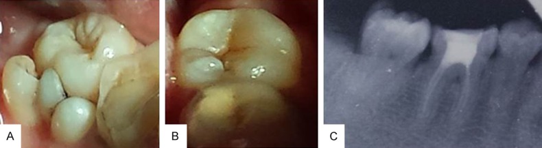
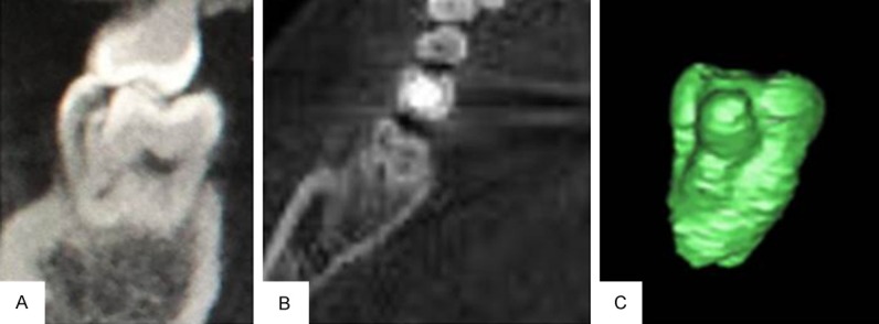
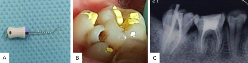
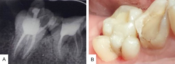
Gemination or fusion is a rare occurrence in the mandibular posterior teeth. The aim of this article is to describe the problems encountered and the strategy employed in treating such cases. A 34 years old patient came with the complaint of spontaneous and radiating pain in the right mandibular posterior region. The tooth in concern was an anomalous 'double' second mandibular molar diagnosed as having necrotic pulp with chronic apical abscess of endodontic origin. The present case emphasizes the importance of identifying anatomical anomalies during treatment of fused teeth with supernumerary tooth, and the need for the use of advanced imaging modalities like CBCT which is a critical aid in the diagnosis of such cases. Fused teeth can be managed quite efficiently by an overall combined treatment including both endodontic and periodontal therapy.
Keywords: Fusion; cone-beam computed tomography; endodontic treatment; supernumerary teeth.
Figures
Similar articles
-
Endodontic treatment of developmental anomalies in posterior teeth: treatment of geminated/fused teeth--report of two cases.Int Endod J. 2003 May;36(5):372-9. doi: 10.1046/j.1365-2591.2003.00666.x. Int Endod J. 2003. PMID: 12752652
-
Diagnosis and Endodontic Management of Fused Mandibular Second Molar and Paramolar with Concrescent Supernumerary Tooth Using Cone-beam CT and 3-D Printing Technology: A Case Report.Bull Tokyo Dent Coll. 2015;56(3):177-84. doi: 10.2209/tdcpublication.56.177. Bull Tokyo Dent Coll. 2015. PMID: 26370578
-
Rare case of gemination and fusion involving supernumerary teeth and second mandibular molar - Case report.J Clin Exp Dent. 2024 Feb 1;16(2):e236-e239. doi: 10.4317/jced.60865. eCollection 2024 Feb. J Clin Exp Dent. 2024. PMID: 38496816 Free PMC article.
-
Hemisection and vital treatment of a fused tooth--literature review and case report.Endod Dent Traumatol. 1997 Dec;13(6):253-8. doi: 10.1111/j.1600-9657.1997.tb00051.x. Endod Dent Traumatol. 1997. PMID: 9558505 Review.
-
The importance of cone-beam computed tomography in the management of endodontic problems: a review of the literature.J Endod. 2014 Dec;40(12):1895-901. doi: 10.1016/j.joen.2014.05.009. Epub 2014 Oct 3. J Endod. 2014. PMID: 25287321 Review.
Cited by
-
Fusion of a Tooth with a Supernumerary Tooth: A Case Report and Literature Review of 35 Cases.Children (Basel). 2023 Dec 20;11(1):6. doi: 10.3390/children11010006. Children (Basel). 2023. PMID: 38275427 Free PMC article.
-
Fusion of a maxillary third molar with a supernumerary fourth molar: A case report.Clin Case Rep. 2024 Feb 6;12(2):e8484. doi: 10.1002/ccr3.8484. eCollection 2024 Feb. Clin Case Rep. 2024. PMID: 38328490 Free PMC article.
-
Gemination of an Erupted Mandibular Third Molar: A Short Presentation of an Exceptionally Rare Clinical Occurrence.Eur J Dent. 2024 May;18(2):687-691. doi: 10.1055/s-0043-1772248. Epub 2023 Sep 20. Eur J Dent. 2024. PMID: 37729927 Free PMC article.
-
Hypertrophic olivary degeneration: a description of four cases of and a literature analysis.Quant Imaging Med Surg. 2022 Jun;12(6):3480-3488. doi: 10.21037/qims-21-1048. Quant Imaging Med Surg. 2022. PMID: 35655820 Free PMC article. No abstract available.
-
Clinical and Radiographic Features of Mandibular Third Molar Gemination: A Case Report and Literature Review.Case Rep Dent. 2025 May 20;2025:8934034. doi: 10.1155/crid/8934034. eCollection 2025. Case Rep Dent. 2025. PMID: 40433424 Free PMC article.
References
-
- Neville BW, Damm DD, Allen CM, Bouquot J. Oral and Maxillofacial Pathology. 3rd edition. Philadelphia: Saunders; 1993. Abnormalities of teeth; pp. 54–119.
-
- Yusof WZ. Non-syndromal multiple supernumerary teeth: Literature review. J Can Dent Assoc. 1990;56:147–149. - PubMed
-
- Rajab LD, Hamdan MA. Supernumerary teeth: Review of the literature and a survey of 152 cases. Int J Pediatr Dent. 2002;12:244–254. - PubMed
-
- Brook AH. Dental anomalies of number, form and size: Their prevalence in British school children. J Int Assoc Dent Child. 1974;5:37–53. - PubMed
Publication types
LinkOut - more resources
Full Text Sources