

Expansion of CD14(+)CD16(+) monocytes is related to acute leukemia
- PMID: 26550139
- PMCID: PMC4612824
Expansion of CD14(+)CD16(+) monocytes is related to acute leukemia
Abstract
Objective: Aim to investigate the proportion of CD14(+)CD16(+) monocytes and understand the pathogenesis of this monocyte subset in acute leukemia.
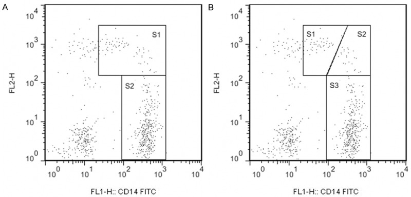
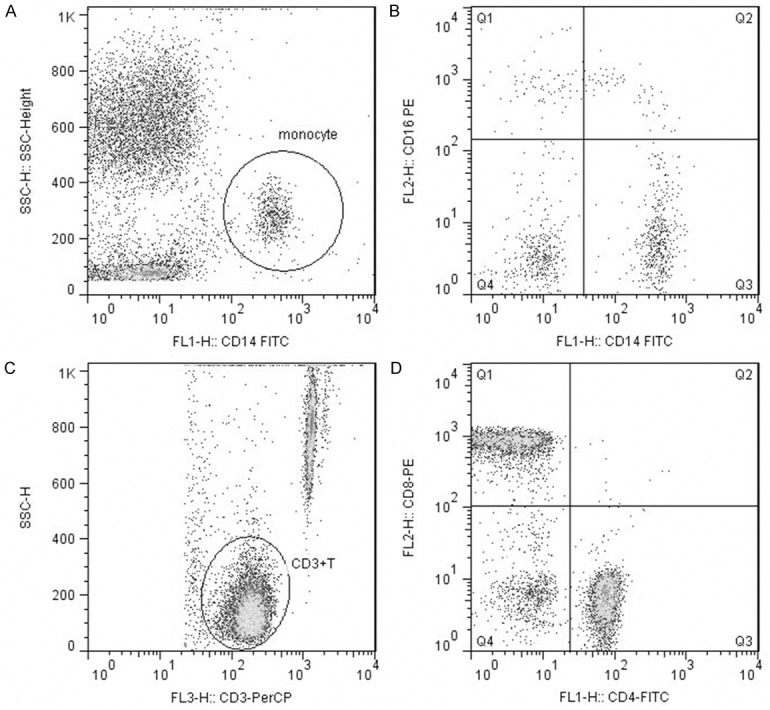
Methods: Flow cytometry was utilized to study the phenotype expression of CD14(+)CD16(+) monocytes and CD3(+) T lymphocytes in peripheral blood derived from patients with acute leukemia. All the data were analyzed by SPSS 13.0 software.
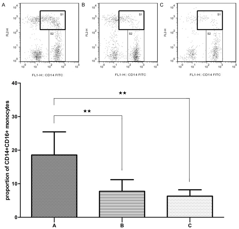
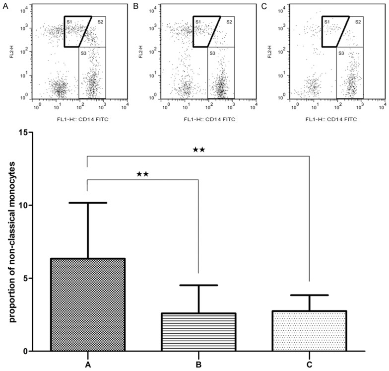
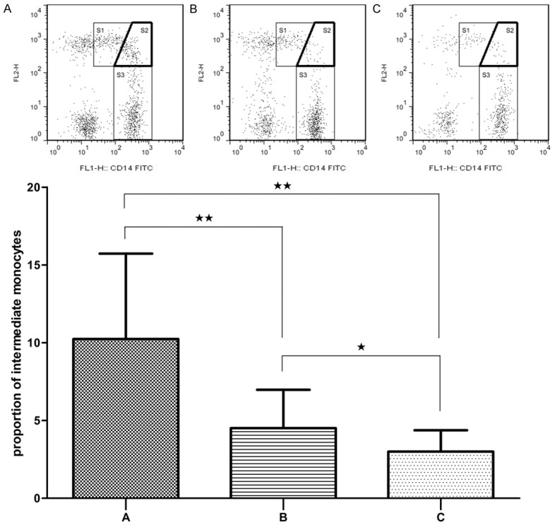
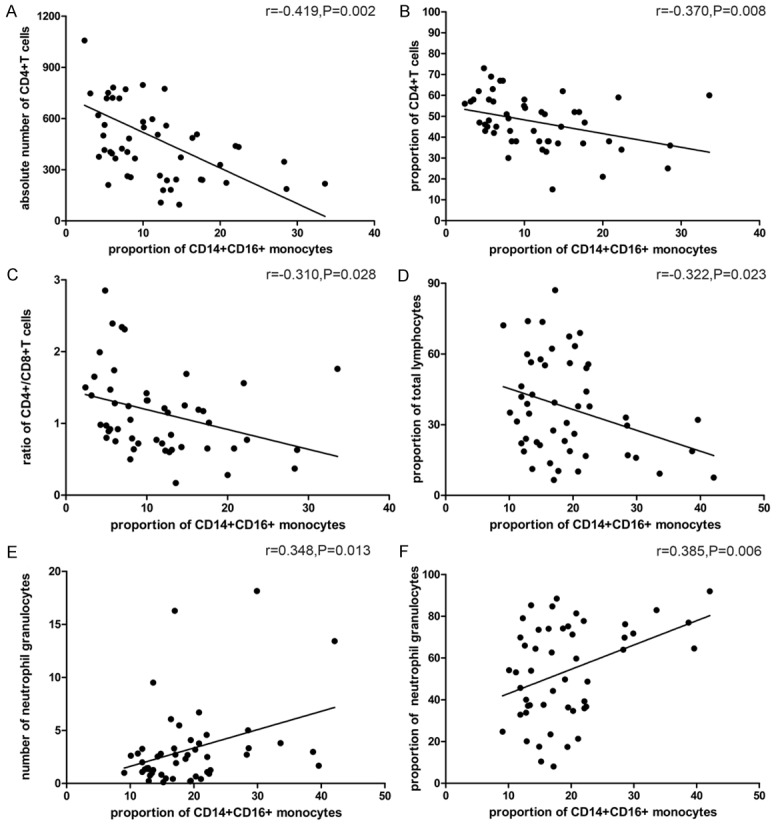
Results: The proportion of CD14(+)CD16(+) monocytes including both intermediate and non-classical monocytes, increased significantly in patients with acute leukemia and changed negatively or positively according to the disease process. Meanwhile, the proportion of CD14(+)CD16(+) monocytes was inversely correlated with absolute number of CD4(+) T lymphocytes, ratio of CD4(+)/CD8(+) T cells, and positively correlated with the proportion of neutrophil granulocytes.
Conclusions: The proportion of CD14(+)CD16(+) monocytes (especially the intermediate subpopulation) is related to the progression of acute leukemia, and the expansion of this monocyte subset could indicate the severity of the disease.
Keywords: Acute leukemia; T lymphocyte; inflammation; monocyte subset.
Figures
Similar articles
-
The CD14++CD16+ monocyte subset is expanded and controls Th1 cell development in Graves' disease.Clin Immunol. 2022 Dec;245:109160. doi: 10.1016/j.clim.2022.109160. Epub 2022 Oct 18. Clin Immunol. 2022. PMID: 36270470
-
CD14++ CD16+ HLA-DR+ Monocytes in Peripheral Blood are Quantitatively Correlated with the Severity of Pre-eclampsia.Am J Reprod Immunol. 2015 Aug;74(2):116-22. doi: 10.1111/aji.12389. Epub 2015 Apr 5. Am J Reprod Immunol. 2015. PMID: 25850575
-
Circulating intermediate CD14++CD16+monocytes are increased in patients with atrial fibrillation and reflect the functional remodelling of the left atrium.Europace. 2017 Jan;19(1):40-47. doi: 10.1093/europace/euv422. Epub 2016 Jan 29. Europace. 2017. PMID: 26826137
-
The CD14(bright) CD16+ monocyte subset is expanded in rheumatoid arthritis and promotes expansion of the Th17 cell population.Arthritis Rheum. 2012 Mar;64(3):671-7. doi: 10.1002/art.33418. Arthritis Rheum. 2012. PMID: 22006178
-
Type and Intensity as Key Variable of Exercise in Metainflammation Diseases: A Review.Int J Sports Med. 2022 Aug;43(9):743-767. doi: 10.1055/a-1720-0369. Epub 2022 Feb 18. Int J Sports Med. 2022. PMID: 34902867 Review.
Cited by
-
Causal association of circulating immune cells and lymphoma: A Mendelian randomization study.Open Med (Wars). 2024 Jul 15;19(1):20240984. doi: 10.1515/med-2024-0984. eCollection 2024. Open Med (Wars). 2024. PMID: 39015296 Free PMC article.
-
Cellular and Molecular Pathways of COVID-19 and Potential Points of Therapeutic Intervention.Front Pharmacol. 2020 Jul 29;11:1169. doi: 10.3389/fphar.2020.01169. eCollection 2020. Front Pharmacol. 2020. PMID: 32848776 Free PMC article.
-
Single-cell transcriptomic profiling reveals immune cell heterogeneity in acute myeloid leukaemia peripheral blood mononuclear cells after chemotherapy.Cell Oncol (Dordr). 2024 Feb;47(1):97-112. doi: 10.1007/s13402-023-00853-2. Epub 2023 Aug 24. Cell Oncol (Dordr). 2024. PMID: 37615858 Free PMC article.
-
Role of Antioxidants in Neonatal Hypoxic-Ischemic Brain Injury: New Therapeutic Approaches.Int J Mol Sci. 2017 Jan 28;18(2):265. doi: 10.3390/ijms18020265. Int J Mol Sci. 2017. PMID: 28134843 Free PMC article. Review.
-
Philadelphia-negative myeloproliferative neoplasms display alterations in monocyte subpopulations frequency and immunophenotype.Med Oncol. 2022 Sep 29;39(12):223. doi: 10.1007/s12032-022-01825-6. Med Oncol. 2022. PMID: 36175590 Free PMC article.
References
-
- Volk HD, Reinke P, Docke WD. Clinical aspects: from systemic inflammation to ‘immunoparalysis’. Chem Immunol. 2000;74:162–177. - PubMed
-
- Dayyani F, Belge KU, Frankenberger M, Mack M, Berki T, Ziegler-Heitbrock L. Mechanism of glucocorticoid-induced depletion of human CD14+CD16+ monocytes. J Leukoc Biol. 2003;74:33–39. - PubMed
-
- Passlick B, Flieger D, Ziegler-Heitbrock HW. Identification and characterization of a novel monocyte subpopulation in human peripheral blood. Blood. 1989;74:2527–2534. - PubMed
-
- Mobley JL, Leininger M, Madore S, Baginski TJ, Renkiewicz R. Genetic evidence of a functional monocyte dichotomy. Inflammation. 2007;30:189–197. - PubMed
LinkOut - more resources
Full Text Sources
Other Literature Sources
Research Materials