. 2015 Aug 15;8(8):12774-82.
eCollection 2015.

Bone mesenchymal stem cells overexpressing FGF4 contribute to liver regeneration in an animal model of liver cirrhosis

Affiliations

Bone mesenchymal stem cells overexpressing FGF4 contribute to liver regeneration in an animal model of liver cirrhosis

Jun Wang et al. Int J Clin Exp Med. .

Abstract

It is recognized that Fibroblast Growth Factor 4 (FGF-4) could not only increase the proliferation of bone marrow mesenchymal stem cells (BMSCs), but also induce BMSCs into hepatocyte-like cells in vitro. However, the role of FGF4 played in liver regeneration in vivo is unclear. This study constructed FGF4 overexpressing BMSCs and then transplanted them into cirrhotic rats to investigate the role of FGF4 played in liver regeneration. The results showed that FGF4 promoted the location of the BMSCs only at the early stage, and more proliferating cell nuclear antigen (PCNA), epithelial cell adhesion molecule (EpCAM) and Jagged-1 positive hepatocytes were found in the cirrhotic rats. This study indicated that FGF4 transduced BMSCs contributed to liver regeneration might by the transplanted microenvironment.

Keywords: BMSCs; microenvironment Introduction; migration; proliferation; transplant.


Figures
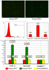
Figure 1
Figure 1
Expression of FGF4, EpCAM and Jagged-1 on FGF4 transduced BMSCs. A. Nearly 92% of FGF4-BMSCs were GFP-positive 96 hours after transduction, and 4.6-fold increase in FGF4 expression in FGF4-transduced BMSCs relative to the null-BMSCs evaluated by Q-PCR. B. Compared with the Group-GFP, FGF4 transduced BMSCs displayed a similar expression of Jagged-1, and a higher expression of EpCAM. Expression standard errors are expressed within brackets at the top of each bar. *means P < 0.001.
Figure 2
Figure 2
Effects of FGF4 on the homing of BMSCs to cirrhotic liver. Left: homing of BMSCs to the cirrhotic liver of different experimental groups was observed, 48 hours after transplantation. A brown nuclear signal in some cells indicated the presence of SRY-positive cells (200× magnification). Right: quantitative analysis shows that the proportion of SRY cells in the Group-BMSCs and the Group-GFP is not significantly different (P > 0.05). However, the proportion of SRY cells in the Group-GFP and the Group-FGF4 are significantly different (P < 0.05). Results are expressed as the mean ± SD.
Figure 3
Figure 3
Determination of the proliferation of hepatocytes. Left: PCNA expression in hepatocytes was observed in different experimental groups 48 hours after transplantation. A brown cytoplasmicsignal in some cells indicates the presence of PCNA positive cells (200× magnification). Right: quantitative analysis shows that the proportion of PCNA cells in the Group-LC and Group-BMSCs is significantly different (P < 0.05), whereas the proportion of PCNA cells in the Group-GFP and the Group-BMSCs are not significantly different (P > 0.05). A significant difference (P < 0.05) is observed in the Group-GFP and Group-FGF4. The results are expressed as the mean ± SD.
Figure 4
Figure 4
Effect of FGF4 on the proliferation of the HPCs of cirrhotic liver. A. Left: EpCAM expression in the liver tissues was observe in different experimental groups. A brown membranous and cytoplasmic signal in some cells indicates the presence of EpCAM-positive cells (200× magnification). Right: quantitative analysis shows that the proportion of EpCAM cells in the Group-LC and Group-BMSCs are significantly different (P < 0.05), whereas the proportion of EpCAM cells in the Group-GFP and the Group-BMSCs are not significantly different (P > 0.05). A significant difference (P < 0.05) was observed in the Group-GFP and Group-FGF4. The results are expressed as the mean ± SD. B. Left: Jagged-1 expression in the liver tissues was observed in different experimental groups 48 hours after transplantation. A brown membranous signal in some cells indicates the presence of Jagged-1-positive cells (200× magnification). Right: quantitative analysis shows that the proportion of Jagged-1 cells in the Group-LC and in the Group-BMSCs is significantly different (P < 0.05), whereas the proportion of Jagged-1 cells in the Group-GFP and the Group-BMSCs is not significantly different (P > 0.05). A significant difference (P < 0.05) is observed in the Group-GFP and Group-FGF4. The results are expressed as the mean ± SD.

References

    1. Liu J, Fan D. Hepatitis B in China. Lancet. 2007;369:1582–1583. - PubMed
    1. Fouraschen SM, Pan Q, de Ruiter PE, Farid WR, Kazemier G, Kwekkeboom J, Ijzermans JN, Metselaar HJ, Tilanus HW, de Jonge J, van der Laan LJ. Secreted factors of human liver-derived mesenchymal stem cells promote liver regeneration early after partial hepatectomy. Stem Cells Dev. 2012;21:2410–2419. - PubMed
    1. Stock P, Brückner S, Ebensing S, Hempel M, Dollinger MM, Christ B. The generation of hepatocytes from mesenchymal stem cells and engraftment into murine liver. Nat Protoc. 2010;5:617–627. - PubMed
    1. Li J, Zhang L, Xin J, Jiang L, Li J, Zhang T, Jin L, Li J, Zhou P, Hao S, Cao H, Li L. Immediate intraportal transplantation of human bone marrow mesenchymal stem cells prevents death from fulminant hepatic failure in pigs. Hepatology. 2012;56:1044–1052. - PubMed
    1. Puglisi MA, Tesori V, Lattanzi W, Piscaglia AC, Gasbarrini GB, D’Ugo DM, Gasbarrini A. Therapeutic implications of mesenchymal stem cells in liver injury. J Biomed Biotechnol. 2011;2011:860578. - PMC - PubMed

LinkOut - more resources