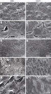
Osteogenic differentiation of CD271(+) cells from rabbit bone marrow cultured on three phase PCL/TZ-HA bioactive scaffolds: comparative study with mesenchymal stem cells (MSCs)
- PMID: 26550238
- PMCID: PMC4612923
Osteogenic differentiation of CD271(+) cells from rabbit bone marrow cultured on three phase PCL/TZ-HA bioactive scaffolds: comparative study with mesenchymal stem cells (MSCs)
Abstract
Tissue engineering is one of the major challenges of orthopedics and trauma surgery for bone regeneration. Biomaterials filled with mesenchymal stem cells (MSCs) are considered the most promising approach in bone tissue engineering. Furthermore, our previous study showed that the multi-phase poly [ε-caprolactone]/thermoplastic zein-hydroxyapatite (PCL/TZ-HA) biomaterials improved rabbit (r) MSCs adhesion and osteoblast differentiation, thus demonstrating high potential of this bioengineered scaffold for bone regeneration. In the recent past, CD271 has been applied as a specific selective marker for the enrichment of MSCs from bone marrow (BM-MSCs). In the present study, we aimed at establishing whether CD271-based enrichment could be an efficient method for the selection of rBM-MSCs, displaying higher ability in osteogenic differentiation than non-selected rBM-MSCs in an in vitro system. CD271(+) cells were isolated from rabbit bone marrow and were compared with rMSCs in their proliferation rate and osteogenic differentiation capability. Furthermore, rCD271(+) cells were tested in their ability to adhere, proliferate and differentiate into osteogenic lineage, while growing on PCL/TZ-HA scaffolds, in comparison to rMSCs. Our result demonstrate that rCD271(+) cells were able to adhere, proliferate and differentiate into osteoblasts when cultured on PCL/TZ-HA scaffolds in significantly higher levels as compared to rMSCs. Based on these findings, CD271 marker might serve as an optimal alternative MSCs selection method for the potential preclinical and clinical application of these cells in bone tissue regeneration.
Keywords: CD271; Rabbit bone marrow; biomaterials; bone tissue engineering; mesenchymal stem cells; osteoblast.
Figures


References
-
- Salerno A, Oliviero M, Di Maio E, Netti PA, Rofani C, Colosimo A, Guida V, Dallapiccola B, Palma P, Procaccini E, Berardi AC, Velardi F, Teti A, Iannace S. Design of novel three-phase PCL/TZ-HA biomaterials for use in bone regeneration applications. J Mater Sci Mater Med. 2010;21:2569–81. - PubMed
-
- Udehiya RK, Amarpal , Aithal HP, Kinjavdekar P, Pawde AM, Singh R, Taru Sharma G. Comparison of autogenic and allogenic bone marrow derived mesenchymal stem cells for repair of segmental bone defects in rabbits. Res Vet Sci. 2013;94:743–52. - PubMed
-
- Friedenstein AJ, Chailakhjan RK, Lalykina KS. The development of fibroblast colonies in monolayer cultures of guinea-pig bone marrow and spleen cells. Cell Tissue Kinet. 1970;3:393–403. - PubMed
-
- Eagan MJ, Zuk PA, Zhao KW, Bluth BE, Brinkmann EJ, Wu BM, McAllister DR. The suitability of human adipose-derived stem cells for the engineering of ligament tissue. J Tissue Eng Regen Med. 2012;6:702–9. - PubMed
LinkOut - more resources
Full Text Sources
Miscellaneous
