. 2015 Sep 15;7(9):1553-63.
eCollection 2015.

Low molecular weight fucoidan ameliorates diabetic nephropathy via inhibiting epithelial-mesenchymal transition and fibrotic processes

Affiliations

Low molecular weight fucoidan ameliorates diabetic nephropathy via inhibiting epithelial-mesenchymal transition and fibrotic processes

Jihui Chen et al. Am J Transl Res. .

Abstract

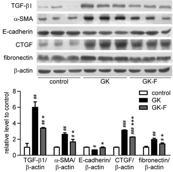
Diabetic nephropathy (DN) is one of the most serious microvascular complications of diabetes and may lead to end-stage renal disease (ESRD) and chronic renal failure. The aim of this study was to determine whether low-molecular-weight fucoidan (LMWF) can reduce harmful transforming growth factor-β (TGF-β)-mediated renal fibrosis in DN using in vitro and in vivo experimental models. The experimental results showed that LMWF significantly reversed TGF-β1-induced epithelial-mesenchymal transition and dose-dependently inhibited accumulation of extracellular matrix proteins, including connective tissue growth factor and fibronectin. It was found that LMWF significantly reduced blood urea nitrogen and blood creatinine in both type 1 and type 2 diabetic rat models. H&E, PAS and Masson's trichrome staining of kidney tissue showed LMWF significantly reduced renal interstitial fibrosis. Treatment with LMWF significantly increased E-cadherin expression and reduced α-SMA, CTGF and fibronectin expression in both type 1 and type 2 diabetic models. LMWF also decreased the phosphorylation of Akt, ERK1/2, p38 and Smad3 in vitro and in vivo. These data suggest that LMWF may protect kidney from dysfunction and fibrogenesis by inhibiting TGF-β pathway and have the potential benefit to slow down the progression of DN.

Keywords: Fucoidan; diabetic nephropathy; epithelialto-mesenchymal transition; extracellular matrix; transforming growth factor-β1; tubulointerstitial fibrosis.


Figures

Figure 1
Figure 1
LMWF reversed TGF-β1-induced epithelial-mesenchymal transition in HK2 cells. HK2 cells were incubated with 10 ng/ml of TGF-β1 and different concentrations of LMWF (5, 10, 20 μM) for 48 h. A. Morphology of the HK2 cells cultured in indicated conditions. B. Expression levels of E-cadherin, α-SMA, vimentin, CTGF, fibronectin, β-actin of HK2 cells incubated with 10 ng/ml of TGF-β1 and 0, 5, 10, 20 μg/ml LMWF for 48 h was detected by Western blot analysis. Representative blotting (left) and quantification of protein levels (right) are shown. Mean ± SEM. n=3; #P < 0.05, ##P < 0.01, ###P < 0.001 vs. control group; *P < 0.05, **P < 0.01 vs. TGF-β1 group.
Figure 2
Figure 2
LMWF inhibited downstream pathways of TGF-β1 in HK2 cells. Expression and phosphorylation levels of p-Akt, Akt, p-ERK1/2, ERK2, p-p38, p38, p-Smad3, Smad3, Snail, ZEB1 and β-actin of HK2 cells incubated with 10 ng/ml TGF-β1 and 0, 5, 10, 20 μg/ml LMWF for 48 h were detected by Western blot analysis. Representative blotting (left) and quantification of protein levels (right) are shown. Mean ± SEM. n=3; ##P < 0.01, ###P < 0.001 vs. control group; *P < 0.05 vs. TGF-β1 group.
Figure 3
Figure 3
LMWF attenuated renal functional defect and tissue damage in GK rats. GK rats were administered with vehicle or LMWF daily for 12 weeks from 12 weeks of age. Serum and kidneys were collected for renal function test and histological examination. A. Serum BUN. B. Serum creatinine concentration. C. Representative images of H&E, periodic acid-Schiff and Masson’s trichrome staining of kidney (original magnification ×400). Data are presented as mean ± SEM. n=5-8 for each group. *P < 0.05; **P < 0.01.
Figure 4
Figure 4
LMWF reduced renal interstitial fibrosis in GK rats. Expression of TGF-β1, E-cadherin, α-SMA, CTGF, fibronectin and β-actin of kidneys at 24 weeks of age was detected by Western blot analysis. Representative blotting (up) and quantification of protein levels (down) are shown. Mean ± SEM. n=3; #P < 0.05, ##P <0.01, ###P < 0.001 vs. control group; *P < 0.05, ***P < 0.001 vs. GK group.
Figure 5
Figure 5
LMWF inhibited Akt, ERK, p38, Smad signaling pathway in kidney of GK rats. Expression of p-Akt, p-ERK1/2, p-p38, p-Smad3 of kidneys at 24 weeks of age was detected by Western blotting, and then normalized versus nonphosphorylated forms (total forms). Representative blotting (up) and quantification of protein levels (down) are shown. Mean ± SEM. n=3; #P < 0.05, ##P < 0.01 vs. control group; *P < 0.05 vs. GK group.
Figure 6
Figure 6
LMWF alleviated DN of STZ rats. Rats were treated for 12 weeks with LMWF daily. A. Serum BUN. B. Serum creatinine. Data are presented as mean ± SEM. n=8 for each group. *P < 0.05, **P < 0.01. C. Representative images of H&E, periodic acid-Schiff (PAS) and Masson’s trichrome staining of kidney (original magnification ×400, scale bar: 100 mm). D. Expression of TGF-β1, α-SMA, E-cadherin, CTGF, fibronectin, β-actin, p-Akt, Akt, p-ERK1/2, ERK2, p-p38, p38, p-Smad3, Smad3 in kidneys treated for 12 weeks were detected by Western blot analysis. Representative blotting (left) and quantification of protein levels (right) are shown. Mean ± SEM, n=3; #P < 0.05, ##P < 0.01, ###P < 0.001 vs control group; *P < 0.05 vs STZ group.

References

    1. Gray SP, Cooper ME. Diabetic nephropathy in 2010: Alleviating the burden of diabetic nephropathy. Nat Rev Nephrol. 2011;7:71–73. - PubMed
    1. Eddy AA. Molecular basis of renal fibrosis. Pediatr Nephrol. 2000;15:290–301. - PubMed
    1. Fioretto P, Caramori ML, Mauer M. The kidney in diabetes: dynamic pathways of injury and repair. The Camillo Golgi Lecture 2007. Diabetologia. 2008;51:1347–1355. - PubMed
    1. Meran S, Steadman R. Fibroblasts and myofibroblasts in renal fibrosis. Int J Exp Pathol. 2011;92:158–167. - PMC - PubMed
    1. Zeisberg M, Kalluri R. The role of epithelialto-mesenchymal transition in renal fibrosis. J Mol Med (Berl) 2004;82:175–181. - PubMed

LinkOut - more resources