. 2015 Nov 5:4:51.
doi: 10.1186/s13742-015-0091-4. eCollection 2015.

Full-length single-cell RNA-seq applied to a viral human cancer: applications to HPV expression and splicing analysis in HeLa S3 cells

Affiliations

Full-length single-cell RNA-seq applied to a viral human cancer: applications to HPV expression and splicing analysis in HeLa S3 cells

Liang Wu et al. Gigascience. .

Abstract

Background: Viral infection causes multiple forms of human cancer, and HPV infection is the primary factor in cervical carcinomas. Recent single-cell RNA-seq studies highlight the tumor heterogeneity present in most cancers, but virally induced tumors have not been studied. HeLa is a well characterized HPV+ cervical cancer cell line.

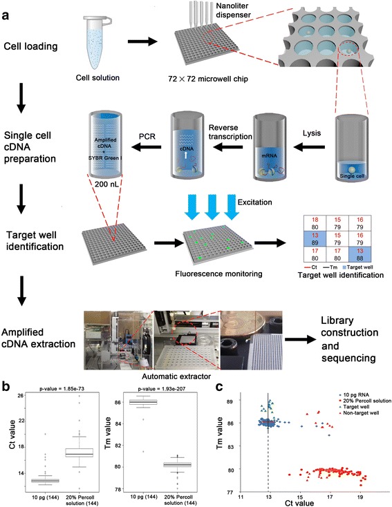
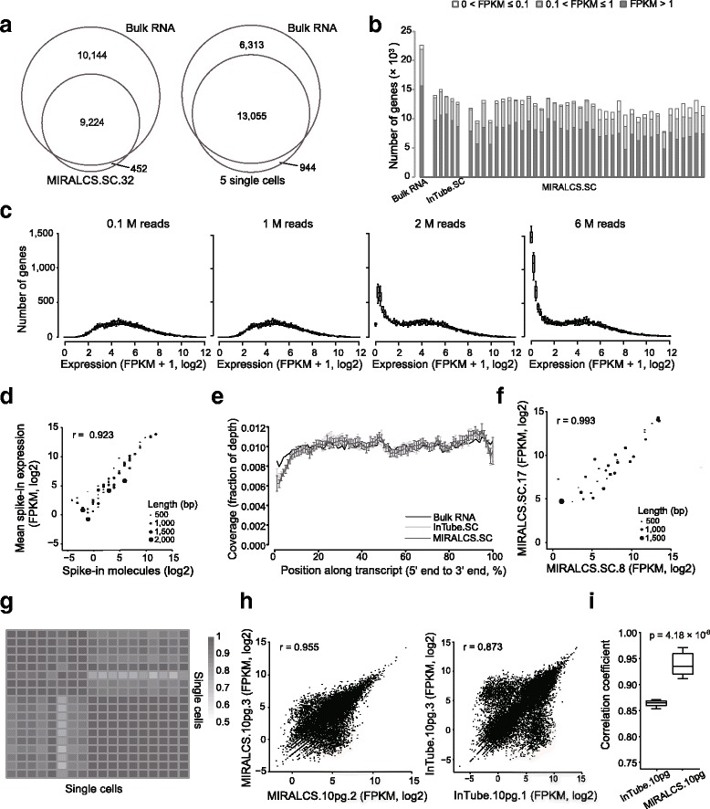
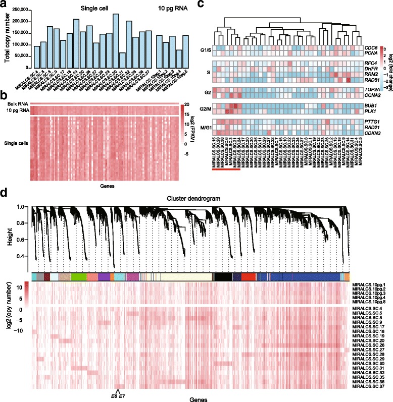
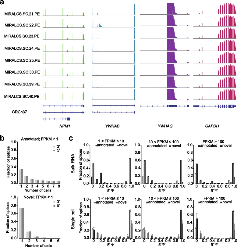
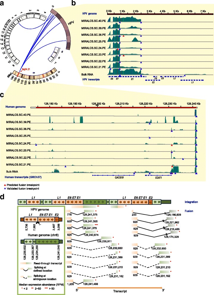
Result: We developed a new high throughput platform to prepare single-cell RNA on a nanoliter scale based on a customized microwell chip. Using this method, we successfully amplified full-length transcripts of 669 single HeLa S3 cells and 40 of them were randomly selected to perform single-cell RNA sequencing. Based on these data, we obtained a comprehensive understanding of the heterogeneity of HeLa S3 cells in gene expression, alternative splicing and fusions. Furthermore, we identified a high diversity of HPV-18 expression and splicing at the single-cell level. By co-expression analysis we identified 283 E6, E7 co-regulated genes, including CDC25, PCNA, PLK4, BUB1B and IRF1 known to interact with HPV viral proteins.

Conclusion: Our results reveal the heterogeneity of a virus-infected cell line. It not only provides a transcriptome characterization of HeLa S3 cells at the single cell level, but is a demonstration of the power of single cell RNA-seq analysis of virally infected cells and cancers.

Keywords: Cancer; HPV; HeLa; RNA splicing; Single-cell transcriptome; Tumor heterogeneity; Virus.


Figures

Fig. 1
Fig. 1
The schematic diagram of MIRALCS. a The flowchart of the MIRALCS. b The box plot of Ct (left) and Tm (right) value of the 20 % Percoll solution (negative control) and 10 pg total RNA (positive control), respectively. c The Ct value and Tm value distribution of 20 % Percoll solution, 10 pg total RNA, non-target well and target well during cDNA amplification process in microwells. The target wells (well with cell) and the non-target wells (without cells) were validated by Agilent 2100 Bioanlyzer. The line denotes Ct median. Horizontal bars denote ± 0.5
Fig. 2
Fig. 2
A high sensitivity, accuracy and reproducibility of MIRALCS. a Comparison of gene number between single cell (the smaller circle) and the 5 ng bulk sample (the larger circle). The left, a typical cell; the right, 5 randomly selected cells (randomly sampling 0.4 million reads per cell) vs. the 5 ng bulk sample (2 million reads). b Gene detection in MIRALCS single-cells, regular tube-based single cells and 5 ng bulk RNA sample. c The distribution of gene number on gene expression along sequencing depths. d The correlation of the mean expression (FPKM) and the number of input molecules of spike-ins of all MIRALCS single-cell libraries. e The reads coverage along the transcript position from 5′ to 3′end. Error bar stands for the standard deviation. f The correlation of spike-ins expression (FPKM) between two randomly selected MIRALCS single cells. g Heat map of correlation coefficients of spike-ins expression levels with input molecules >1 for each library (n = 19). h The correlation of gene expression (FPKM) between technical replicates. Left: two randomly selected MIRALCS 10 pg replicates. Right: two randomly selected tube-based 10 pg replicates. i The pair-wise correlation in MIRALCS 10 pg RNA replicates and tube-based 10 pg RNA replicates
Fig. 3
Fig. 3
Heterogeneity of gene expression in HeLa S3 single cells. a The mRNA molecular number in single cells and 10 pg RNA replicates. b The heat map of the FPKM values of extremely highly expressed genes (FPKM > 500 in bulk RNA) in single cells and 10 pg replicates. c Single-cell subpopulations identification based on cell cycle relative genes. The cells with underline are in G2/M phase. d Gene co-expression modules derived from 19 single cells based on RNA molecular number (modules are distinguished by colors). The detailed of each module stands for were shown on Additional file 4: Table S7. The weighted gene correlation network was constructed using the WCGNA R package [38]
Fig. 4
Fig. 4
Heterogeneity of alternative splicing and distributions of splices in in single cells. a The sequencing depth for genes of NPM1, YWHAB, YWHAQ and GAPDH in single cells. b The frequency distribution of detected annotated and novel spliced junctions. c The distributions of the ψ scores of annotated and novel spliced junctions in the bulk RNA (upper) and single cells (lower)
Fig. 5
Fig. 5
The landscape of the HPV-18/cellular fusion and diversity of HPV-host splicing and expression in HeLa S3 cells. a The overview of the HPV-18 cellular fusion based on HeLa cell transcriptome. Blue lines denote fusion events. b The read coverage of HPV-18 genome in single cells and the bulk RNA. Colored vertical lines denote nucleotides of SNPs detected in the transcriptome. Light green, A; red, T; orange, G; blue, C. c The read coverage of the host region on chromosome 8 in single cells and the bulk RNA. d The schematic diagram of the inferred HPV integration structure (upper) and splicing forms (lower). RPM stands for reads per million

References

    1. Bouvard V, Baan R, Straif K, Grosse Y, Secretan B, El Ghissassi F, et al. A review of human carcinogens–part B: biological agents. Lancet Oncol. 2009;10(4):321–322. doi: 10.1016/S1470-2045(09)70096-8. - DOI - PubMed
    1. Boyle P, Levin B. World cancer report 2008. Lyon: International Agency for Research on Cancer and World Health Organization Press; 2008.
    1. de Martel C, Ferlay J, Franceschi S, Vignat J, Bray F, Forman D, et al. Global burden of cancers attributable to infections in 2008: a review and synthetic analysis. Lancet Oncol. 2012;13(6):607–615. doi: 10.1016/S1470-2045(12)70137-7. - DOI - PubMed
    1. Mesri EA, Feitelson MA, Munger K. Human viral oncogenesis: a cancer hallmarks analysis. Cell Host Microbe. 2014;15(3):266–282. doi: 10.1016/j.chom.2014.02.011. - DOI - PMC - PubMed
    1. Hourdequin KC, Lefferts JA, Brennick JB, Ernstoff MS, Tsongalis GJ, Pipas JM. Merkel cell polyomavirus and extrapulmonary small cell carcinoma. Oncol Lett. 2013;6(4):1049–1052. - PMC - PubMed

Publication types