

Comparison of (18)F-FDG PET/CT and PET/MRI in patients with multiple myeloma
- PMID: 26550538
- PMCID: PMC4620174
Comparison of (18)F-FDG PET/CT and PET/MRI in patients with multiple myeloma
Abstract
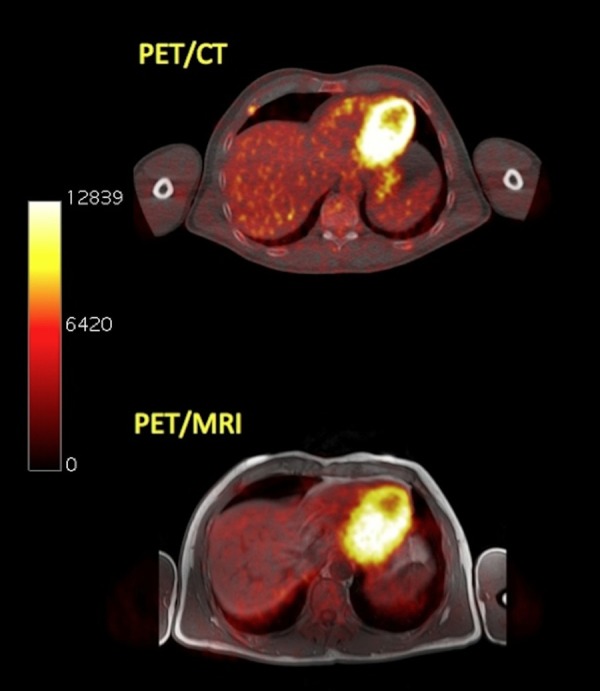
PET/MRI represents a promising hybrid imaging modality with several potential clinical applications. Although PET/MRI seems highly attractive in the diagnostic approach of multiple myeloma (MM), its role has not yet been evaluated. The aims of this prospective study are to evaluate the feasibility of (18)F-FDG PET/MRI in detection of MM lesions, and to investigate the reproducibility of bone marrow lesions detection and quantitative data of (18)F-FDG uptake between the functional (PET) component of PET/CT and PET/MRI in MM patients. The study includes 30 MM patients. All patients initially underwent (18)F-FDG PET/CT (60 min p.i.), followed by PET/MRI (120 min p.i.). PET/CT and PET/MRI data were assessed and compared based on qualitative (lesion detection) and quantitative (SUV) evaluation. The hybrid PET/MRI system provided good image quality in all cases without artefacts. PET/MRI identified 65 of the 69 lesions, which were detectable with PET/CT (94.2%). Quantitative PET evaluations showed the following mean values in MM lesions: SUVaverage=5.5 and SUVmax=7.9 for PET/CT; SUVaverage=3.9 and SUVmax=5.8 for PET/MRI. Both SUVaverage and SUVmax were significantly higher on PET/CT than on PET/MRI. Spearman correlation analysis demonstrated a strong correlation between both lesional SUVaverage (r=0.744) and lesional SUVmax (r=0.855) values derived from PET/CT and PET/MRI. Regarding detection of myeloma skeletal lesions, PET/MRI exhibited equivalent performance to PET/CT. In terms of tracer uptake quantitation, a significant correlation between the two techniques was demonstrated, despite the statistically significant differences in lesional SUVs between PET/CT and PET/MRI.
Keywords: Multiple myeloma; PET/CT; PET/MRI; SUV.
Figures


Similar articles
-
PET/CT studies of multiple myeloma using (18) F-FDG and (18) F-NaF: comparison of distribution patterns and tracers' pharmacokinetics.Eur J Nucl Med Mol Imaging. 2014 Jul;41(7):1343-53. doi: 10.1007/s00259-014-2721-y. Epub 2014 Feb 22. Eur J Nucl Med Mol Imaging. 2014. PMID: 24562650
-
(18)F-FDG dynamic PET/CT in patients with multiple myeloma: patterns of tracer uptake and correlation with bone marrow plasma cell infiltration rate.Clin Nucl Med. 2015 Jun;40(6):e300-7. doi: 10.1097/RLU.0000000000000773. Clin Nucl Med. 2015. PMID: 25783508
-
More advantages in detecting bone and soft tissue metastases from prostate cancer using 18F-PSMA PET/CT.Hell J Nucl Med. 2019 Jan-Apr;22(1):6-9. doi: 10.1967/s002449910952. Epub 2019 Mar 7. Hell J Nucl Med. 2019. PMID: 30843003
-
Comparison of whole-body PET/CT and PET/MRI in breast cancer patients: lesion detection and quantitation of 18F-deoxyglucose uptake in lesions and in normal organ tissues.Eur J Radiol. 2014 Feb;83(2):289-96. doi: 10.1016/j.ejrad.2013.11.002. Epub 2013 Nov 23. Eur J Radiol. 2014. PMID: 24331845
-
Quantitative analysis of 18F-NaF dynamic PET/CT cannot differentiate malignant from benign lesions in multiple myeloma.Am J Nucl Med Mol Imaging. 2017 Sep 1;7(4):148-156. eCollection 2017. Am J Nucl Med Mol Imaging. 2017. PMID: 28913153 Free PMC article.
Cited by
-
Diagnostic Innovations: Advances in imaging techniques for diagnosis and follow-up of multiple myeloma.J Bone Oncol. 2025 Feb 28;51:100669. doi: 10.1016/j.jbo.2025.100669. eCollection 2025 Apr. J Bone Oncol. 2025. PMID: 40124904 Free PMC article. Review.
-
Prognostic value of [18F]fluorodeoxyglucose-PET/MRI(CT) novel interpretation criteria (IMPeTUs) in multiple myeloma.Eur J Nucl Med Mol Imaging. 2025 Aug;52(10):3781-3791. doi: 10.1007/s00259-025-07219-w. Epub 2025 Apr 3. Eur J Nucl Med Mol Imaging. 2025. PMID: 40175849
-
Application of (18)F-FDG PET and diffusion weighted imaging (DWI) in multiple myeloma: comparison of functional imaging modalities.Am J Nucl Med Mol Imaging. 2015 Oct 12;5(5):479-92. eCollection 2015. Am J Nucl Med Mol Imaging. 2015. PMID: 26550539 Free PMC article.
-
Positron Emission Tomography (PET) Radiopharmaceuticals in Multiple Myeloma.Molecules. 2019 Dec 29;25(1):134. doi: 10.3390/molecules25010134. Molecules. 2019. PMID: 31905752 Free PMC article. Review.
-
Improving MR sequence of 18F-FDG PET/MR for diagnosing and staging gastric Cancer: a comparison study to 18F-FDG PET/CT.Cancer Imaging. 2020 Jun 16;20(1):39. doi: 10.1186/s40644-020-00317-y. Cancer Imaging. 2020. PMID: 32546207 Free PMC article.
References
-
- Kyle RA, Gertz MA, Witzig TE, Lust JA, Lacy MQ, Dispenzieri A, Fonseca R, Rajkumar SV, Offord JR, Larson DR, Plevak ME, Therneau TM, Greipp PR. Review of 1027 patients with newly diagnosed multiple myeloma. Mayo Clin Proc. 2003;78:21–33. - PubMed
-
- International Myeloma Working Group. Criteria for the classification of monoclonal gammopathies, multiple myeloma and related disorders: a report of the International Myeloma Working Group. Br J Haematol. 2003;121:749–757. - PubMed
-
- Zamagni E, Cavo M. The role of imaging techniques in the management of multiple myeloma. Br J Haematol. 2012;159:499–513. - PubMed
-
- Palumbo A, Anderson K. Multiple myeloma. N Engl J Med. 2011;364:1046–1060. - PubMed
-
- Rajkumar SV, Dimopoulos MA, Palumbo A, Blade J, Merlini G, Mateos MV, Kumar S, Hillengass J, Kastritis E, Richardson P, Landgren O, Paiva B, Dispenzieri A, Weiss B, LeLeu X, Zweegman S, Lonial S, Rosinol L, Zamagni E, Jagannath S, Sezer O, Kristinsson SY, Caers J, Usmani SZ, Lahuerta JJ, Johnsen HE, Beksac M, Cavo M, Goldschmidt H, Terpos E, Kyle RA, Anderson KC, Durie BG, Miguel JF. International Myeloma Working Group updated criteria for the diagnosis of multiple myeloma. Lancet Oncol. 2014;15:e538–e548. - PubMed
LinkOut - more resources
Full Text Sources