The use of dynamic nuclear polarization (13)C-pyruvate MRS in cancer
- PMID: 26550544
- PMCID: PMC4620180
The use of dynamic nuclear polarization (13)C-pyruvate MRS in cancer
Abstract
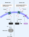
In recent years there has been an immense development of new targeted anti-cancer drugs. For practicing precision medicine, a sensitive method imaging for non-invasive, assessment of early treatment response and for assisting in developing new drugs is warranted. Magnetic Resonance Spectroscopy (MRS) is a potent technique for non-invasive in vivo investigation of tissue chemistry and cellular metabolism. Hyperpolarization by Dynamic Nuclear Polarization (DNP) is capable of creating solutions of molecules with polarized nuclear spins in a range of biological molecules and has enabled the real-time investigation of in vivo metabolism. The development of this new method has been demonstrated to enhance the nuclear polarization more than 10,000-fold, thereby significantly increasing the sensitivity of the MRS with a spatial resolution to the millimeters and a temporal resolution at the subsecond range. Furthermore, the method enables measuring kinetics of conversion of substrates into cell metabolites and can be integrated with anatomical proton magnetic resonance imaging (MRI). Many nuclei and substrates have been hyperpolarized using the DNP method. Currently, the most widely used compound is (13)C-pyruvate due to favoring technicalities. Intravenous injection of the hyperpolarized (13)C-pyruvate results in appearance of (13)C-lactate, (13)C-alanine and (13)C-bicarbonate resonance peaks depending on the tissue, disease and the metabolic state probed. In cancer, the lactate level is increased due to increased glycolysis. The use of DNP enhanced (13)C-pyruvate has in preclinical studies shown to be a sensitive method for detecting cancer and for assessment of early treatment response in a variety of cancers. Recently, a first-in-man 31-patient study was conducted with the primary objective to assess the safety of hyperpolarized (13)C-pyruvate in healthy subjects and prostate cancer patients. The study showed an elevated (13)C-lactate/(13)C-pyruvate ratio in regions of biopsy-proven prostate cancer compared to noncancerous tissue. However, more studies are needed in order to establish use of hyperpolarized (13)C MRS imaging of cancer.
Keywords: 13C-pyruvate; Dynamic nuclear polarization; MR; MRS; cancer; response monitoring.
Figures


References
-
- Gambhir SS. Molecular imaging of cancer with positron emission tomography. Nat Rev Cancer. 2002;2:683–93. - PubMed
-
- Adams MC, Turkington TG, Wilson JM, Wong TZ. A Systematic Review of the Factors Affecting Accuracy of SUV Measurements. AJR. 2010;195:310–20. - PubMed
-
- Patterson DM, Padhani AR, Collins DJ. Technology Insight: water diffusion MRI-a potential new biomarker of response to cancer therapy. Nat Clin Pract Oncol. 2008;5:220–33. - PubMed
-
- McKnight TR, Bussche von dem MH, Vigneron DB, Lu Y, Berger MS, McDermott MW, Dillon WP, Graves EE, Pirzkall A, Nelson SJ. Histopathological validation of a three-dimensional magnetic resonance spectroscopy index as a predictor of tumor presence. J Neurosurg. 2002;97:794–802. - PubMed
Publication types
LinkOut - more resources
Full Text Sources
Other Literature Sources
