Review
. 2015 Dec;1(4):201-19.
doi: 10.1159/000441252. Epub 2015 Oct 28.

Evidence of Mitochondrial Dysfunction within the Complex Genetic Etiology of Schizophrenia

Affiliations
Review

Evidence of Mitochondrial Dysfunction within the Complex Genetic Etiology of Schizophrenia

Brooke E Hjelm et al. Mol Neuropsychiatry. 2015 Dec.

Erratum in

  • Erratum.
    [No authors listed] [No authors listed] Mol Neuropsychiatry. 2016 May;2(1):60. doi: 10.1159/000444029. Epub 2016 May 17. Mol Neuropsychiatry. 2016. PMID: 27602361 Free PMC article.

Abstract

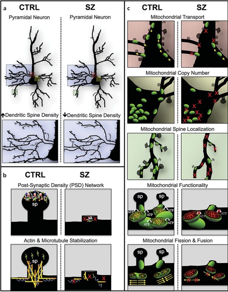
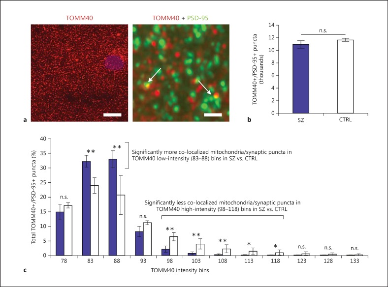
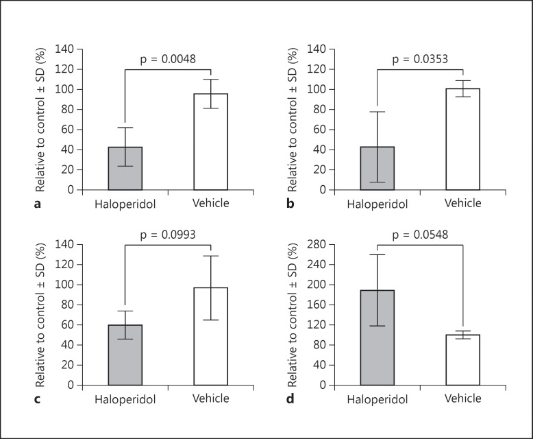
Genetic evidence has supported the hypothesis that schizophrenia (SZ) is a polygenic disorder caused by the disruption in function of several or many genes. The most common and reproducible cellular phenotype associated with SZ is a reduction in dendritic spines within the neocortex, suggesting alterations in dendritic architecture may cause aberrant cortical circuitry and SZ symptoms. Here, we review evidence supporting a multifactorial model of mitochondrial dysfunction in SZ etiology and discuss how these multiple paths to mitochondrial dysfunction may contribute to dendritic spine loss and/or underdevelopment in some SZ subjects. The pathophysiological role of mitochondrial dysfunction in SZ is based upon genomic analyses of both the mitochondrial genome and nuclear genes involved in mitochondrial function. Previous studies and preliminary data suggest SZ is associated with specific alleles and haplogroups of the mitochondrial genome, and also correlates with a reduction in mitochondrial copy number and an increase in synonymous and nonsynonymous substitutions of mitochondrial DNA. Mitochondrial dysfunction has also been widely implicated in SZ by genome-wide association, exome sequencing, altered gene expression, proteomics, microscopy analyses, and induced pluripotent stem cell studies. Together, these data support the hypothesis that SZ is a polygenic disorder with an enrichment of mitochondrial targets.

Keywords: Antipsychotic drug; Dendritic spines; Fluorescence deconvolution tomography; Genome; Induced pluripotent stem cell; Mitochondria; Polygenic disorder; Proteome; Schizophrenia; Transcriptome.


Figures

Fig. 1
Fig. 1
Multifactorial model of mitochondrial dysfunction in SZ. Cartoon illustration of our hypothesis of a multifactorial model where alterations in gene networks specific to PSD, cytoskeleton organization, and an array of mitochondrial processes may independently (or additively) lead to dendritic spine deficits, SZ symptom onset and polygenic disease risk. a Pyramidal neurons in control (CTRL) and SZ brains displaying a reduction in dendritic spine density in SZ pathology (blue box) and neuronal regions potentially affected by mitochondrial dysfunction (pink, yellow and green boxes). Colors refer to the online version only. b Dendritic spines (sp) in CTRL brains and spine loss in SZ brains due to polygenic variants in nonmitochondrial networks that have been implicated in SZ etiology (i.e., postsynaptic density and cytoskeleton organization). c Neuronal regions potentially affected by a variety of mitochondrial processes (i.e., transport, copy number, spine localization, functionality, and fusion/fission) and their possible role in dendritic spine loss in SZ brains compared to CTRL. We hypothesize that a signature of mitochondrial dysfunction can occur in SZ independently or additively in b and c. In b, mitochondrial deficits are a direct result of spine loss due to the polygenic burden in nonmitochondrial pathways like the PSD and cytoskeleton networks (i.e., PSD/cytoskeleton gene aberrations → spine loss ← → mitochondrial dysfunction); in c, mitochondrial deficits are a direct result of aberrations in nuclear genes with mitochondrial function and/or in the mitochondrial genome, and there is also spine loss-induced mitochondria loss (i.e., mitochondrial gene aberrations → mitochondrial dysfunction ← → spine loss); in c the mitochondria loss of function is primary, while in b the spine loss is the primary causative event.
Fig. 2
Fig. 2
FDT morphometric analysis of mitochondria and synaptic puncta in SZ brains. FDT from dorsolateral PFC layer I of SZ (n = 6) compared to controls (CTRL; n = 2) suggests synaptic puncta of SZ subjects have lower density of (aggregated) mitochondria or abnormal mitochondrial morphology. a Fluorescence images from mitochondria and synaptic puncta immunohistochemistry co-localization used for FDT analysis. Left: Low-magnification image of TOMM40 immunofluorescence (i.e., mitochondria label). Scale bar = 10 μm. Right: High-magnification image of TOMM40 (mitochondria = red) and PSD-95 (synaptic puncta = green) immunofluorescence. Colors refer to the online version only. Scale bar = 1 μm. Arrows point to co-localization of both markers used for morphometric analysis between SZ and CTRL. b Mean and standard deviation of co-localized puncta (TOMM40+/PSD-95+) across all subjects suggests no difference between SZ and CTRL (i.e., the same number of co-localized puncta were observed in both groups). c Mean and standard deviation of co-localized puncta (TOMM40+/PSD-95+) across all subjects, binned according to TOMM40 fluorescence intensity and normalized to the total number of puncta counts per subject, demonstrates significant differences between SZ and CTRL and suggests SZ synapses have lower density of (aggregated) mitochondria or abnormal mitochondrial morphology. n.s. = Not significant, * p < 0.05, ** p < 0.01.
Fig. 3
Fig. 3
Haloperidol induces hypofunctional effects on mitochondria in vitro. LCLs treated with 30 μm haloperidol for 24-48 h exhibit mitochondrial hypofunction. Results display mean and standard deviation (SD) across 4 independent experiments analyzed with the XF Cell Mito Stress Test Kit and XF Analyzer (Seahorse Bioscience), comparing 30 μm haloperidol with vehicle control treatments. a Adjusted basal respiration (i.e., basal respiration - nonmitochondrial respiration). b Coupling efficiency. c Maximum respiratory capacity. d ATP turnover. Statistical comparisons (p values) displayed are from Welch's t tests between both treatment groups.

References

    1. Glausier JR, Lewis DA. Dendritic spine pathology in schizophrenia. Neuroscience. 2013;251:90–107. - PMC - PubMed
    1. Harrison PJ, Weinberger DR. Schizophrenia genes, gene expression, and neuropathology: on the matter of their convergence. Mol Psychiatry. 2005;10:40–68. - PubMed
    1. Katsel P, Davis KL, Gorman JM, Haroutunian V. Variations in differential gene expression patterns across multiple brain regions in schizophrenia. Schizophr Res. 2005;77:241–252. - PubMed
    1. Vawter MP, Barrett T, Cheadle C, Sokolov BP, Wood WH 3rd, Donovan DM, Webster M, Freed WJ, Becker KG. Application of cDNA microarrays to examine gene expression differences in schizophrenia. Brain Res Bull. 2001;55:641–650. - PubMed
    1. Arion D, Corradi JP, Tang S, Datta D, Boothe F, He A, Cacace AM, Zaczek R, Albright CF, Tseng G, Lewis DA. Distinctive transcriptome alterations of prefrontal pyramidal neurons in schizophrenia and schizoaffective disorder. Mol Psychiatry. DOI: 10.1038/mp.2014.171. - PMC - PubMed