. 2016 Jan:487:230-41.
doi: 10.1016/j.virol.2015.09.023. Epub 2015 Nov 6.

Both RIG-I and MDA5 detect alphavirus replication in concentration-dependent mode

Affiliations

Both RIG-I and MDA5 detect alphavirus replication in concentration-dependent mode

Ivan Akhrymuk et al. Virology. 2016 Jan.

Abstract

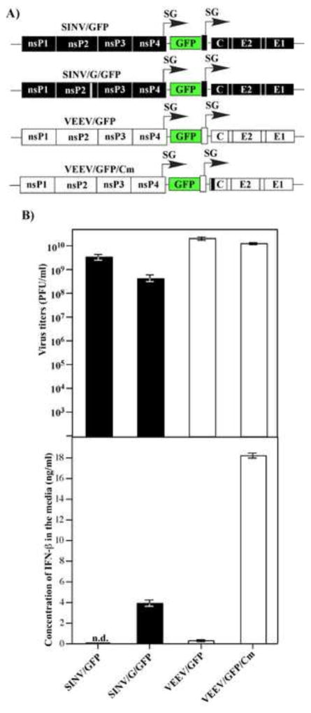
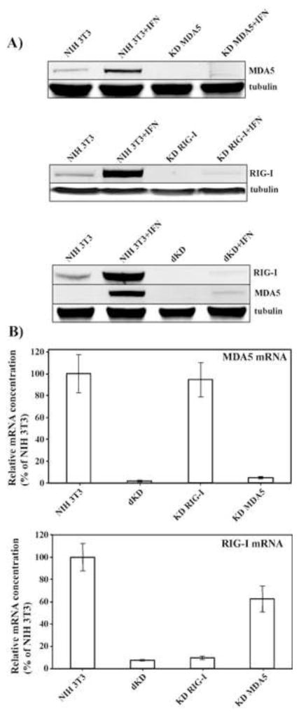
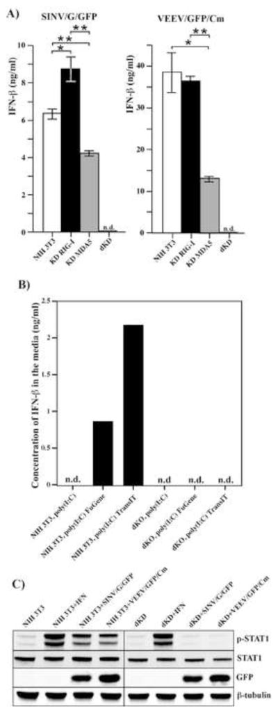
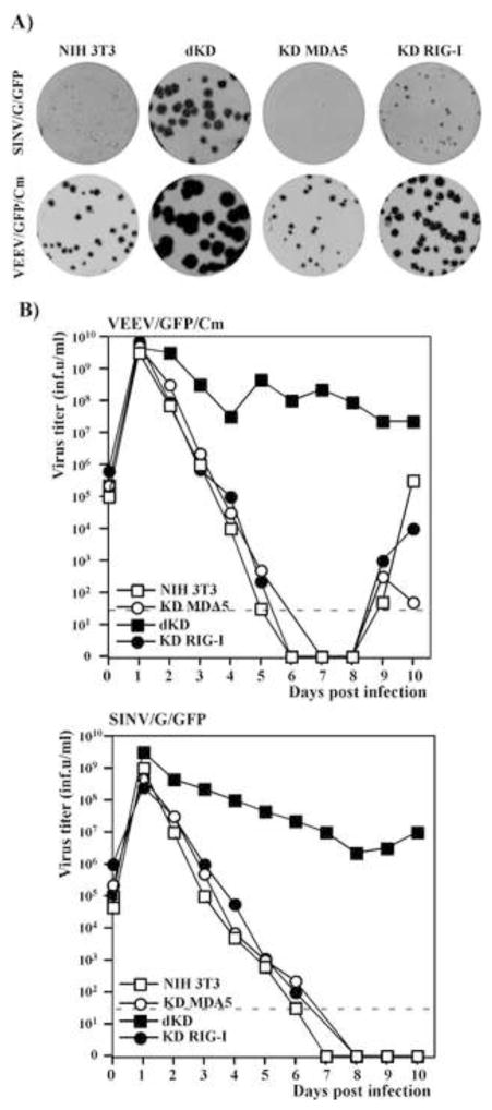
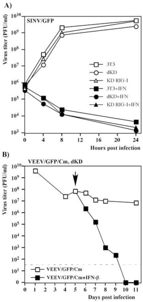
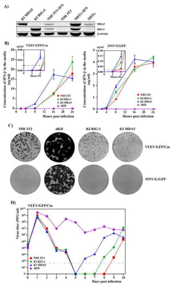
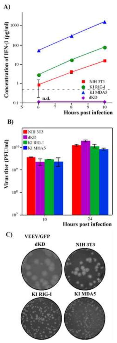
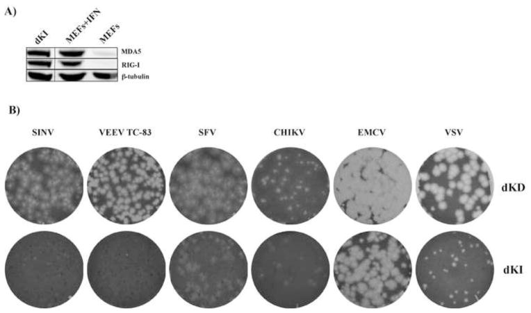
Alphaviruses are a family of positive-strand RNA viruses that circulate on all continents between mosquito vectors and vertebrate hosts. Despite a significant public health threat, their biology is not sufficiently investigated, and the mechanisms of alphavirus replication and virus-host interaction are insufficiently understood. In this study, we have applied a variety of experimental systems to further understand the mechanism by which infected cells detect replicating alphaviruses. Our new data strongly suggest that activation of the antiviral response by alphavirus-infected cells is determined by the integrity of viral genes encoding proteins with nuclear functions, and by the presence of two cellular pattern recognition receptors (PRRs), RIG-I and MDA5. No type I IFN response is induced in their absence. The presence of either of these PRRs is sufficient for detecting virus replication. However, type I IFN activation in response to pathogenic alphaviruses depends on the basal levels of RIG-I or MDA5.

Keywords: Alphaviruses; Innate immunity; MDA5; Pattern recognition receptors; RIG-I; Type I interferon; Virus–host interactions.


Figures

Fig. 1
Fig. 1
Alphaviruses deficient in nuclear functions, but not their wt variants, induce high levels of IFN-β. (A) The schematic representation of alphavirus genomes encoding wt and mutated nsP2- and capsid-coding genes (SINV/G/GFP and VEEV/GFP/Cm, respectively). (B) NIH 3T3 cells were infected with the indicated viruses at an MOI of 20 PFU/cell. Media were harvested at 16 h post infection and used for assessment of both virus titers and IFN-β levels. Means of three biological repeats with SD are presented.
Fig. 2
Fig. 2
RIG-I and MDA5 expression is strongly inhibited in the selected KD cell lines. (A) Equal numbers of NIH 3T3 cells and indicated clone-derived cell lines were treated with mouse IFN-β (500 IU/ml) for 24 h or remained untreated. Cell lysates, corresponding to equal numbers of cells were analyzed by SDS-PAGE, followed by Western blot using MDA5- and RIG-I-specific, and tubulin-specific antibodies in conjunction with infrared dye-labeled secondary antibodies. Membranes were scanned on a LI-COR imager. (B) The indicated clone-derived cell lines were treated with mouse IFN-β (500 IU/ml) for 24 h. RNAs were isolated, and relative concentrations of RIG-I- and MDA5-specific mRNAs were determined by RT-qPCR as described in the Materials and Methods.
Fig. 3
Fig. 3
Single PRR KD, but not dKD, cells respond to alphavirus replication by IFN-β release. (A) Parental NIH 3T3, KD RIG-I, KD MDA5 and dKD cells were infected with SINV/G/GFP and VEEV/GFP/Cm viruses at an MOI of 20 PFU/cell. Media was replaced at 24 h post infection and harvested again at 48 h post infection. Concentration of IFN-β was assessed as described in the Materials and methods. Sensitivity of the assay was ~1 pg/ml. Means of three biological repeats with SD are presented. (B) NIH 3T3 and dKD cells were transfected with poly(I:C) using the indicated transfection reagents or treated with poly(I:C)-containing media as described in the Materials and Methods. Media were harvested at 18 h post transfection, and concentration of IFN-β was determined as indicated above. This experiment was repeated twice with reproducible results. (C) NIH 3T3 and dKD cells were infected with SINV/G/GFP and VEEV/GFP/Cm viruses at an MOI of 20 PFU/cell for 8 h or treated with IFN-β at a concentration of 500 IU/ml for 30 min. Cell lysates were analyzed by Western blot using p-STAT1-, STAT1-, GFP- and β-tubulin-specific antibodies.
Fig. 4
Fig. 4
Single PRR KD cells are capable of both downregulating the spread of mutant alphaviruses and inhibiting already established virus replication. (A) NIH 3T3, KD RIG-I, KD MDA5 and dKD cells were seeded into 6-well Costar plates at a concentration of 5×10⁵ cells per well. SINV/G/GFP and VEEV/GFP/Cm virus stocks were serially diluted and used for infection of the indicated cells with different numbers of infectious units. After 1-h-long incubation at 37°C, the virus-containing media were replaced with 2 ml of media supplemented with 0.6% agarose. After incubation at 37°C for 48 h, cells were fixed, and plates were scanned on a Typhoon imager to enumerate and assess foci of GFP-expressing cells. Images represent wells infected with the same numbers of infectious units. (B) Subconfluent monolayers of NIH 3T3, KD RIG-I, KD MDA5 and dKD cells were infected with SINV/G/GFP and VEEV/GFP/Cm at an MOI of 20 PFU/cell. Media were replaced every day and cells were split upon reaching confluency, usually every 24 hours. Virus titers in harvested media were determined by plaque assay on BHK-21 cells. Dashed lines represent the limits of detection. This experiment was repeated twice with similar results.
Fig. 5
Fig. 5
Single KD and dKD cells remain fully capable of responding to IFN-β treatment and activating the antiviral state. (A) NIH 3T3, KD RIG-I and dKD cells were treated with IFN-β at a concentration of 500 IU/ml for 20 h or mock-treated and then infected with SINV/GFP at an MOI of 5 PFU/cell. Media were replaced at the indicated time points and virus titers were determined by plaque assay on BHK-21 cells. (B) dKD cells were infected with the VEEV/GFP/Cm mutant at an MOI of 5 PFU/cell and incubated for 5 days with periodic splitting. After 5 days, the persistently infected cells were either treated with IFN-β at concentration of 1000 IU/ml or mock treated. Titers of released virus were assessed by plaque assay on BHK-21 cells. The arrow indicates the beginning of IFN treatment. The dashed line indicates the limit of detection.
Fig. 6
Fig. 6
Ectopic expression of RIG-I or MDA5 in dKD cells restores their ability to rapidly respond to replication of alphavirus mutants. (A) Comparative levels of RIG-I and MDA5 expression in the parental NIH 3T3 cells and MEFs, treated with IFN-β at a concentration of 500 IU/ml for 24 h, and in selected clones of KI cells. (B) NIH 3T3, dKD, KI RIG-I and KI MDA5 cells were infected with VEEV/GFP/Cm and SINV/G/GFP viruses at an MOI of 20 PFU/cell. Concentrations of released IFN-β were assessed at the indicated times post infection. Means of three biological repeats with SD are presented. (C) The NIH 3T3, KI RIG-I, KI MDA5 and dKD cells were seeded into 6-well Costar plates at a concentration of 5×10⁵ cells per well. SINV/G/GFP and VEEV/GFP/Cm virus stocks were serially diluted and used for infection of indicated cells with different numbers of infectious units. After 1 h incubation at 37°C the virus-containing media were replaced with 2 ml of media supplemented with 0.6% agarose. After incubation at 37°C for 48 h, cells were fixed and plates were scanned on a Typhoon imager to enumerate and assess foci of GFP-expressing cells. Images represent wells infected with the same numbers of infectious units. (D) NIH 3T3, dKD, KI RIG-I and KI MDA5 cells were infected with VEEV/GFP/Cm at an MOI of 20 PFU/cell. Media were replaced every 24 h and cells were split upon reaching confluency in a 1:2 ratio. Virus titers were determined by plaque assay on BHK-21 cells. Dashed line represents the limit of detection. This experiment was repeated twice with similar results.
Fig. 7
Fig. 7
Ectopic expression of RIG-I or MDA5 in dKD cells leads to efficient IFN induction in response to replication of VEEV/GFP, which encodes wt capsid protein. (A) NIH 3T3, dKD, KI RIG-I and KI MDA5 cells were infected with VEEV/GFP at an MOI of 20 PFU/cell, and accumulation of IFN-β in the media was assessed at the indicated time points. Means of three biological repeats with SD are presented. (B) NIH 3T3, KI RIG-I, KI MDA5 and dKD cells were infected with VEEV/GFP at an MOI of 20 PFU/cell. Media were replaced at the indicated time points and titers were determined by plaque assay on BHK-21 cells. Means of three biological repeats with SD are presented. (C) NIH 3T3, KI RIG-I, KI MDA5 and dKD cells were seeded into 6-well Costar plates at a concentration of 5×10⁵ cells per well. VEEV/GFP virus stock was serially diluted and used for infection of indicated cells with different numbers of PFUs. After 1 h incubation at 37°C the virus-containing media were replaced with 2 ml of media supplemented with 0.6% agarose. After incubation at 37°C for 48 h, cells were fixed and stained with Crystal Violet. Images represent wells infected with the same numbers of PFUs.
Fig. 8
Fig. 8
Ectopic, simultaneous expression of both RIG-I and MDA5 in dKD cells (dKI cell line) differentially affects replication of alpha- and other viruses. (A) Comparative levels of RIG-I and MDA5 expression in MEFs, mock-treated or treated with IFN-β at a concentration of 500 IU/ml for 24 h, and in stable dKI cells. (B) The original dKD and stable dKI cells were seeded into 6-well Costar plates at a concentration of 5×10⁵ cells per well. Stocks of the indicated viruses were serially diluted and used for infection of cells with different numbers of PFUs. After 1 h incubation at 37°C the virus-containing media were replaced with 2 ml of media supplemented with 0.6% agarose. After incubation at 37°C for 48 h, cells were fixed and stained with Crystal Violet. Images represent wells infected with the same numbers of PFUs.

References

    1. Aguilar PV, Paessler S, Carrara AS, Baron S, Poast J, Wang E, Moncayo AC, Anishchenko M, Watts D, Tesh RB, Weaver SC. Variation in interferon sensitivity and induction among strains of eastern equine encephalitis virus. J Virol. 2005;79:11300–11310. - PMC - PubMed
    1. Akhrymuk I, Kulemzin SV, Frolova EI. Evasion of the innate immune response: the Old World alphavirus nsP2 protein induces rapid degradation of Rpb1, a catalytic subunit of RNA polymerase II. J Virol. 2012;86:7180–7191. - PMC - PubMed
    1. Atasheva S, Akhrymuk M, Frolova EI, Frolov I. New PARP gene with an anti-alphavirus function. J Virol. 2012;86:8147–8160. - PMC - PubMed
    1. Atasheva S, Fish A, Fornerod M, Frolova EI. Venezuelan equine Encephalitis virus capsid protein forms a tetrameric complex with CRM1 and importin alpha/beta that obstructs nuclear pore complex function. J Virol. 2010a;84:4158–4171. - PMC - PubMed
    1. Atasheva S, Garmashova N, Frolov I, Frolova E. Venezuelan equine encephalitis virus capsid protein inhibits nuclear import in Mammalian but not in mosquito cells. J Virol. 2008;82:4028–4041. - PMC - PubMed

Publication types

LinkOut - more resources