. 2015 Dec;18(12):1756-62.
doi: 10.1038/nn.4162. Epub 2015 Nov 9.

Piezo2 is the principal mechanotransduction channel for proprioception

Affiliations

Piezo2 is the principal mechanotransduction channel for proprioception

Seung-Hyun Woo et al. Nat Neurosci. 2015 Dec.

Abstract

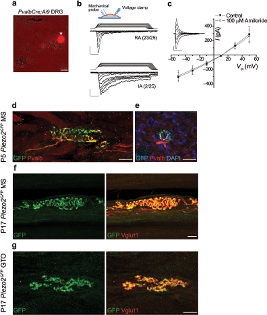
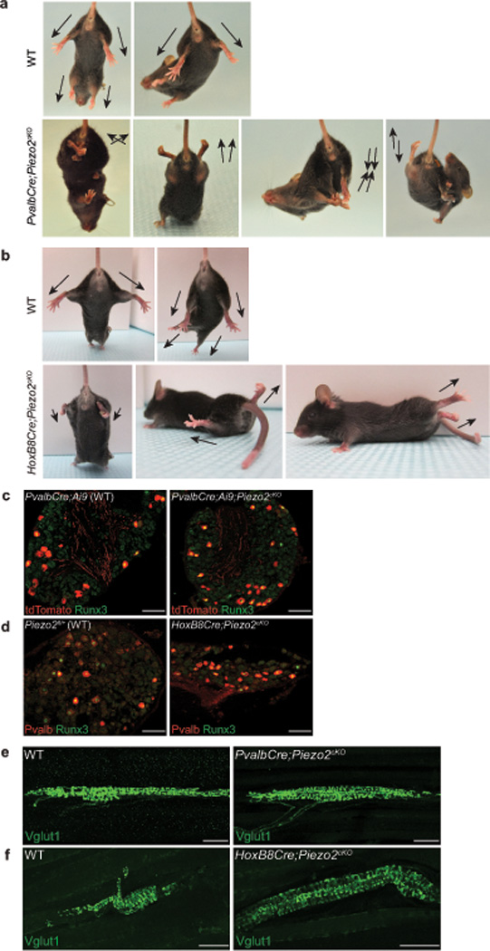
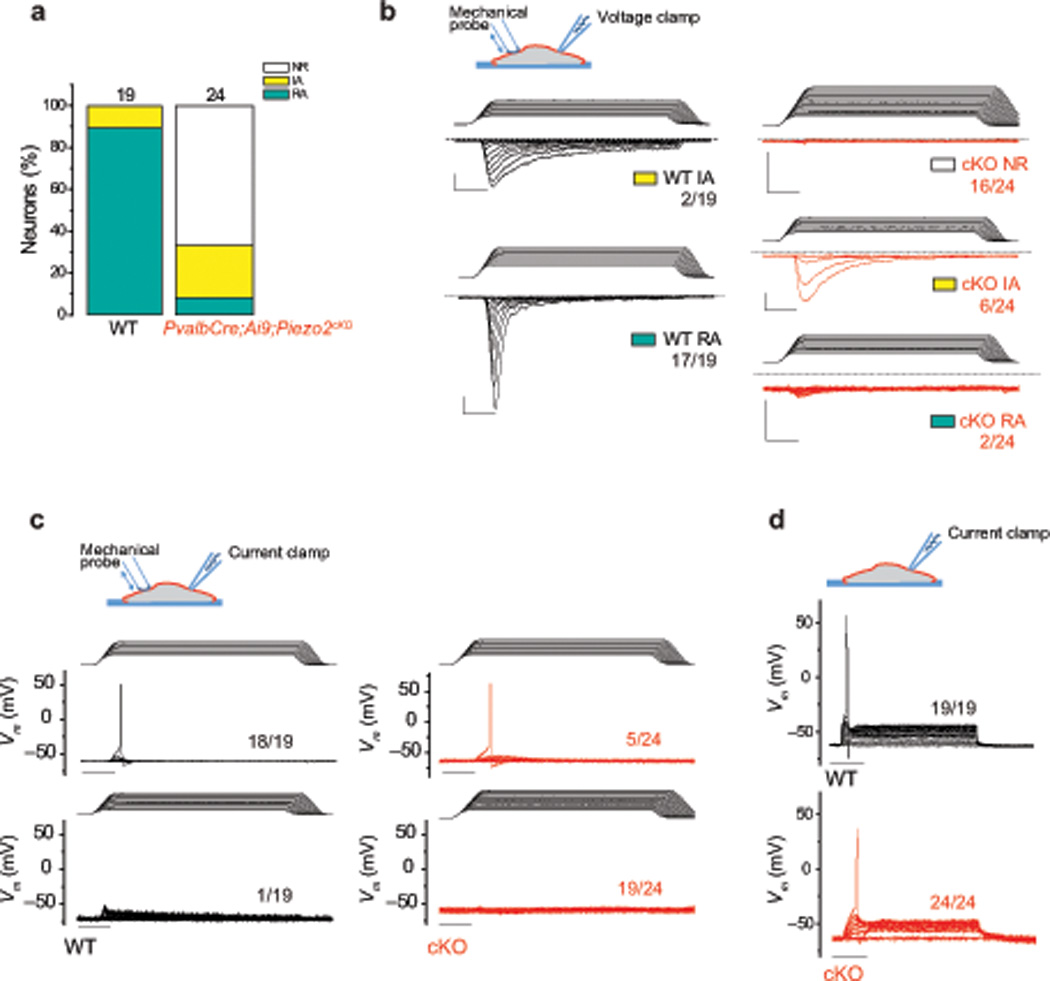
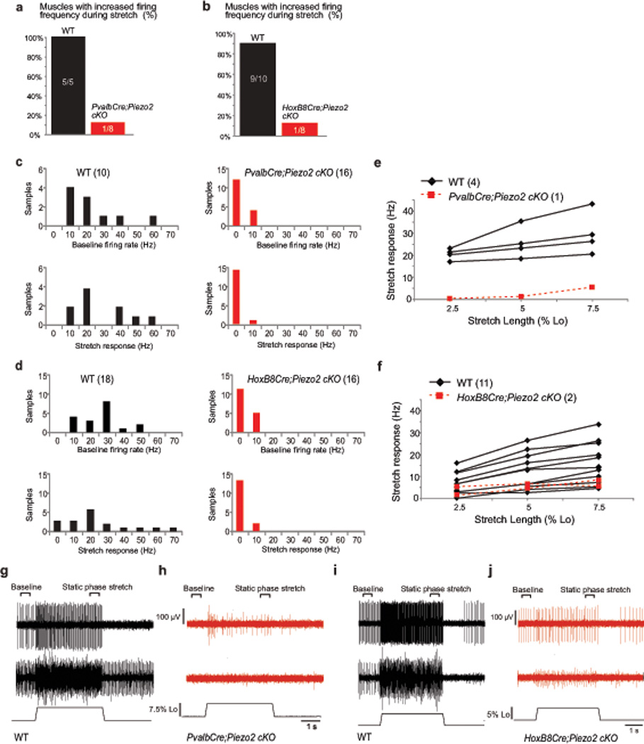
Proprioception, the perception of body and limb position, is mediated by proprioceptors, specialized mechanosensory neurons that convey information about the stretch and tension experienced by muscles, tendons, skin and joints. In mammals, the molecular identity of the stretch-sensitive channel that mediates proprioception is unknown. We found that the mechanically activated nonselective cation channel Piezo2 was expressed in sensory endings of proprioceptors innervating muscle spindles and Golgi tendon organs in mice. Two independent mouse lines that lack Piezo2 in proprioceptive neurons showed severely uncoordinated body movements and abnormal limb positions. Moreover, the mechanosensitivity of parvalbumin-expressing neurons that predominantly mark proprioceptors was dependent on Piezo2 expression in vitro, and the stretch-induced firing of proprioceptors in muscle-nerve recordings was markedly reduced in Piezo2-deficient mice. Together, our results indicate that Piezo2 is the major mechanotransducer of mammalian proprioceptors.


Figures

Figure 1
Figure 1. Characterization of mechanically activated currents and Piezo2 expression in proprioceptive neurons
a, tdTomato+ neuron (asterisk) in isolated DRG cultures from adult PvalbCre;Ai9 mice. b, Whole-cell voltage-clamp recordings from tdTomato+ DRG neurons using mechanical stimuli by displacement of a blunt glass probe in 1 µm increments. Example traces of tdTomato+ DRG neurons responding with rapidly (RA, τinact < 10 ms) and intermediately (IA, 10 ms < τinact < 30 ms) adapting mechanically activated currents from PvalbCre;Ai9 mice are shown. n=25 neurons total. Inset depicts experimental setting; ramp-and-hold traces on top of current recordings show displacement of glass probe. Vertical scale bars, 100 pA. Horizontal scale bars, 25 ms. Holding potential, –60 mV. c, Current (I)-voltage (V) relationship of RA responses at 4–5 µm past mechanical threshold before (closed squares) and after (open squares) the application of 100 uM amiloride. n=11 neurons in each group. Representative background-subtracted control trace shown in inset. Vertical scale bar, 200 pA. Horizontal scale bar, 25 ms. d, Immunofluorescence for GFP and Pvalb in MS from P5 Piezo2GFP hind leg muscle. e, Immunofluorescence for GFP and Pvalb in MS in a transverse section of P5 Piezo2GFP intercostal muscle. f, g, Immunofluorescence for GFP and Vglut1 in MS (f) and in GTO (g) from P17 Piezo2GFP hind leg muscle. Scale bars: a, 30 µm; d–g, 20 µm.
Figure 2
Figure 2. Characterization of two tissue-specific Piezo2 conditional knockout mice
a, b, Representative images showing limb positions of 4–5 week old WT littermate and PvalbCre;Piezo2cKO mice (a) and WT littermate and HoxB8Cre;Piezo2cKO mice (b). Arrows mark the direction of the limbs. c, Runx3 immunostaining with tdTomato epifluorescence in lumbar DRG from 4–5 week old PvalbCre;Ai9 (WT, left) and PvalbCre;Ai9;Piezo2cKO (right) mice. In WT, tdTomato+ neurons: 15.4% (154/1003), tdTomato+ Runx3+ neurons: 6.8% (68/1003); in cKO, tdTomato+ neurons: 15.4% (204/1327), tdTomato+ Runx3+ neurons: 8.7% (116/1327). d, Runx3 and Pvalb immunostaining in lumbar DRG from 4–5 week old WT (left) and HoxB8Cre;Piezo2cKO (right) mice. In WT, Pvalb+ neurons: 8.5% (87/1029), Pvalb+ Runx3+ neurons: 6.2% (64/1029); in cKO, Pvalb+ neurons: 8.8% (83/938), Pvalb+ Runx3+ neurons: 4.6% (43/938). Note that we detected a higher percentage of Pvalb+ neurons in the PvalbCre;Ai9 line (15.4%) compared to the HoxB8Cre line (8.5%). tdTomato epifluorescence is very strong compared to Pvalb immunostaining in adult DRG, thus a difference in the detection method could explain the higher % of Pvalb+ neurons in the PvalbCre;Ai9 line. e, f, Vglut1 immunostaining in MS endings of 4–5 week old WT littermate and PvalbCre;Piezo2cKO hind leg muscles (e) and WT littermate and HoxB8Cre;Piezo2cKO hind leg muscles (f). Scale bars: c, d, 100 µm; e, f, 50 µm.
Figure 3
Figure 3. Characterization of mechanically activated currents in proprioceptive neurons of Piezo2-deficient mice
Whole-cell patch clamp recordings were conducted using mechanical stimulation with a blunt-end glass probe in 1 µm increments. a, The proportion of tdTomato+ DRG neurons responding with rapidly (RA, τinact < 10 ms) and intermediately (IA, 10 ms < τinact < 30 ms) adapting mechanically activated currents from 4–6 week old PvalbCre;Ai9 (WT) and PvalbCre;Ai9;Piezo2cKO mice in voltage-clamp experiments. NR, non-responsive to mechanical displacements. b, Example traces of mechanically induced currents in WT (black traces) and cKO (red traces) tdTomato+ neurons. Holding potential: −60 mV. c, Current-clamp recordings of WT (black traces) and cKO (red traces) tdTomato+ neurons with mechanical stimulation. d, Current-clamp recordings of WT (black traces) and cKO tdTomato+ neurons (red traces). Depolarizing currents were injected in 50 pA increments from a holding current of 0 pA. All vertical scale bars, 200 pA; all horizontal scale bars, 25 ms. Numbers adjacent to traces indicate the number of neurons represented by the trace over the total number of neurons tested.
Figure 4
Figure 4. Ex vivo recordings of stretch-sensitive muscle afferent activities in two Piezo2 conditional knockout mice
(PvalbCre;Piezo2cKO mice = 4, WT mice = 3; HoxB8Cre;Piezo2cKO mice = 4, WT mice = 5; both EDL muscles were used for analysis except for 1 WT animal for PvalbCre). a, b, Percentage of muscles with stretch-sensitive MS afferent activity in adult Piezo2fl/+ (WT) and PvalbCre;Piezo2cKO mice (a: Pearson Chi-Square, X2 = 9.479, df = 1, p < 0.05) and in Piezo2fl/+ (WT) and HoxB8Cre;Piezo2cKO mice (b: Pearson Chi-Square, X2 = 10.811, df = 1, p < 0.05). c, d, Average baseline instantaneous firing rate (Hz, top) and stretch response (bottom: firing rate during static phase of stretch – baseline firing rate; sample area labeled on g) from WT and PvalbCre;Piezo2cKO mice (c) and WT and HoxB8Cre;Piezo2cKO mice (d). e, f, Calculated stretch responses from 4 identified MS afferents from WT (black) and 1 MS afferent from PvalbCre;Piezo2cKO mice (red) (e) and from 11 identified MS afferents from WT (black) and 2 MS afferents from HoxB8Cre;Piezo2cKO mice (red) (f). Stretch response calculated by subtracting baseline instantaneous firing frequency (Hz) from instantaneous firing frequency during static phase of stretch. g, Two representative responses to stretch from WT muscles (control littermates of PvalbCre;Piezo2cKO mice). Top trace includes a single identified MS afferent. Middle trace is an example of multiple units firing during baseline and stretch. Individual unit responses could not be determined from this sample, although all units paused during twitch contraction. Length of the muscle shown on bottom. h, Top trace: only stretch responsive sample recorded in PvalbCre;Piezo2cKO muscle. Middle trace: representative of a non-responsive sample site. i, Two representative responses to stretch from WT muscles (control littermates of HoxB8Cre;Piezo2cKO mice) similar to (g). j, Two stretch responsive samples recorded in HoxB8Cre;Piezo2cKO muscle.

References

    1. Sherrington C. On the proprio-ceptive system, especially in its reflex aspect. Brain. 1907;29:467–482.
    1. Proske U, Gandevia SC. The proprioceptive senses: their roles in signaling body shape, body position and movement, and muscle force. Physiological reviews. 2012;92:1651–1697. - PubMed
    1. Akay T, Tourtellotte WG, Arber S, Jessell TM. Degradation of mouse locomotor pattern in the absence of proprioceptive sensory feedback. Proceedings of the National Academy of Sciences of the United States of America. 2014;111:16877–16882. - PMC - PubMed
    1. Bewick GS, Banks RW. Mechanotransduction in the muscle spindle. Pflugers Archiv : European journal of physiology. 2015;467:175–190. - PMC - PubMed
    1. Walker RG, Willingham AT, Zuker CSA. Drosophila mechanosensory transduction channel. Science. 2000;287:2229–2234. - PubMed

Publication types