
Development of autoantibodies against muscle-specific FHL1 in severe inflammatory myopathies

Inka Albrecht et al. J Clin Invest. 2015 Dec.

Abstract

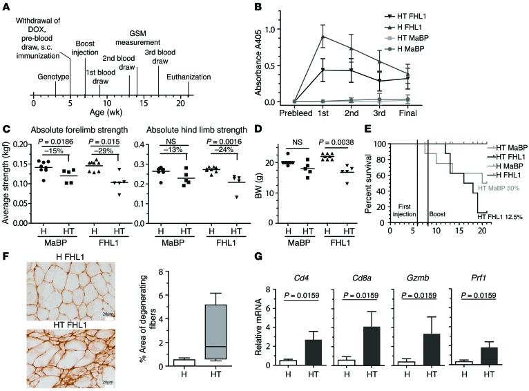
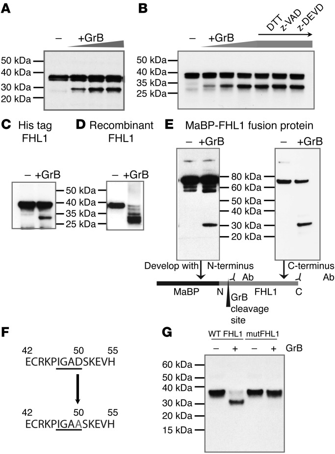
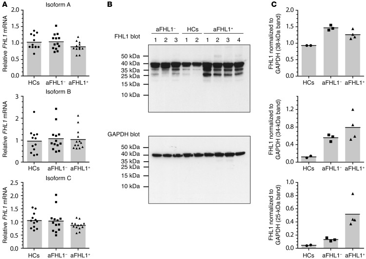
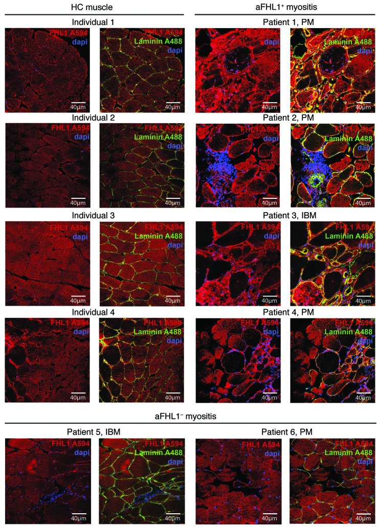
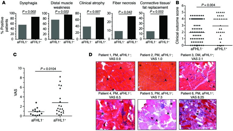
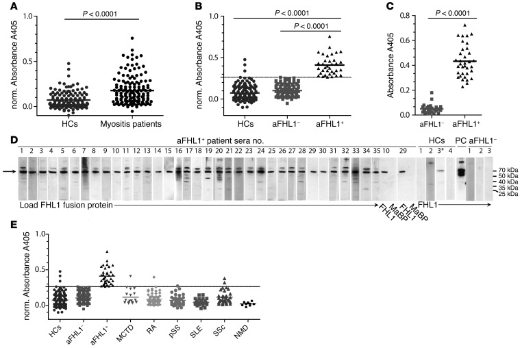
Mutations of the gene encoding four-and-a-half LIM domain 1 (FHL1) are the causative factor of several X-linked hereditary myopathies that are collectively termed FHL1-related myopathies. These disorders are characterized by severe muscle dysfunction and damage. Here, we have shown that patients with idiopathic inflammatory myopathies (IIMs) develop autoimmunity to FHL1, which is a muscle-specific protein. Anti-FHL1 autoantibodies were detected in 25% of IIM patients, while patients with other autoimmune diseases or muscular dystrophies were largely anti-FHL1 negative. Anti-FHL1 reactivity was predictive for muscle atrophy, dysphagia, pronounced muscle fiber damage, and vasculitis. FHL1 showed an altered expression pattern, with focal accumulation in the muscle fibers of autoantibody-positive patients compared with a homogeneous expression in anti-FHL1-negative patients and healthy controls. We determined that FHL1 is a target of the cytotoxic protease granzyme B, indicating that the generation of FHL1 fragments may initiate FHL1 autoimmunity. Moreover, immunization of myositis-prone mice with FHL1 aggravated muscle weakness and increased mortality, suggesting a direct link between anti-FHL1 responses and muscle damage. Together, our findings provide evidence that FHL1 may be involved in the pathogenesis not only of genetic FHL1-related myopathies but also of autoimmune IIM. Importantly, these results indicate that anti-FHL1 autoantibodies in peripheral blood have promising potential as a biomarker to identify a subset of severe IIM.


Figures

Figure 6
Figure 6. Immunization of HT mice with FHL1 aggravates muscle pathology.
At the time points indicated in the animal experimental scheme (A), serum was taken and analyzed by ELISA for the presence of anti-FHL1 autoantibodies, with recombinant His-tagged FHL1 as an antigen (B). DOX, doxycycline; GSM, grip strength measurement. At the age of 16 weeks, 9 weeks after the mice were immunized, forelimbs and hind limb grip strength was measured using a grip strength meter (C), and BW was determined (D). H MaBP mice and H FHL1 mice, n = 8; HT MaBP mice and HT FHL1 mice, n = 5. P = 0.0038, 2-tailed Mann-Whitney U test. (E) The hazard ratio of the Kaplan-Meier survival plot is 2.2, indicating that mice in the HT-FHL1 group died 2.2 times faster than did mice in the matching MaBP control group. (F) Muscle injury was determined by staining of IgM depositions in HT FHL1 (n = 5) versus H FHL1 (n = 4) mice. Scale bars: 20 μm. (G) Hind-limb muscle tissue from HT (n = 5) and H mice (n = 4) immunized with FHL1 was analyzed by qPCR for relative mRNA expression of Cd4, Cd8a, Gzmb, and Prf1. P values in G were calculated by a 2-tailed Mann-Whitney U test.
Figure 5
Figure 5. FHL1 is a target of granzyme B.
(A) Cleavage of FHL1 by granzyme B was demonstrated by performing a cleavage assay using skeletal muscle cell lysates and increasing concentrations of recombinant granzyme B (50, 100, and 250 U) and (B), as indicated, in lanes 5–7, DTT (5 mM), z-VAD (500 nM), or z-DEVD (500 nM) was added. The results for the cleavage assay are representative of 5 independent experiments. In addition, recombinant His-tagged FHL1 (C), commercially available FHL1 (OriGene) (D), and FHL1-MaBP fusion protein (E) were cleaved with granzyme B. For the latter, the immunoblot was developed with either an antibody binding to the N-terminus or an antibody binding to the C-terminus to identify the location of the cleavage site. (F) The granzyme B cleavage site in FHL1 was predicted to be IGAD, with D at amino acid position 50. (G) Using site-directed mutagenesis, the cleavage site was mutated and IVTT-expressed WT or mutated FHL1 (mutFHL1) was incubated with granzyme B or left untreated and analyzed by immunoblotting.
Figure 4
Figure 4. A higher amount of lower-molecular-weight protein bands can be detected in anti-FHL1+ muscle biopsy lysates.
(A) mRNA was extracted from muscle biopsies from anti-FHL1+ (n = 13) and anti-FHL1 (n = 13) patients as well as from HCs (n = 12) and transcribed into cDNA. FHL1 mRNA expression was analyzed by TaqMan PCR using specific primers for amplification of the A, B, and C isoforms. Each data point represents 1 individual, and horizontal bars indicate the mean values. (B) Protein lysates were generated from muscle biopsy material from anti-FHL1+ (n = 4) and anti-FHL1 (n = 3) patients as well as from healthy muscle tissue (n = 2) and immunoblotted with commercially available anti-FHL1 antibody. GAPDH was used as a loading control for immunoblotting. (C) The 3 major bands detected by immunoblotting were quantified using Quantity One 1-D Analysis software and normalized to the GAPDH loading control by calculating as follows: FHL1 bandMean value intensity/GAPDH bandMean value intensity.
Figure 3
Figure 3. FHL1 protein has an altered expression pattern in muscles of anti-FHL1+ IIM patients.
Muscle tissue sections from HCs (left 2 panels) were compared by confocal microscopy with muscle tissue sections from anti-FHL1+ patients (right 2 panels) and from anti-FHL1 patients (lower panels). FHL1 staining is shown in red (secondary antibody coupled to Alexa Fluor 594), and nuclei were visualized using DAPI (blue). In addition, laminin staining was done to visualize the sarcolemma (green, Alexa Fluor 488), and an overlay was done with FHL1 staining and DAPI. Scale bars: 40 μm.
Figure 2
Figure 2. The presence of anti-FHL1 autoantibodies is associated with pronounced muscle damage.
(A) Statistical analysis revealed that anti-FHL1 positivity was associated with dysphagia (P = 0.002, Fisher’s exact test), distal muscle weakness (P = 0.022, Fisher’s exact test), clinical atrophy (P = 0.007, Fisher’s exact test), fiber necrosis (P = 0.042, Fisher’s exact test), and connective tissue/fat replacement (P = 0.002, Fisher’s exact test). (B) Clinical disease severity score for anti-FHL1+ compared with anti-FHL1 IIM patients. Scoring for disease severity was done by examining disease outcome determined at the patient’s last clinic visit. (C) H&E- and Gomori trichrome–stained muscle tissue sections of 20 patients with anti-FHL1 autoantibodies were examined for histopathology and compared with stained muscle tissue sections from sex-, age-, and diagnosis-matched (PM, DM, and IBM; n = 13) anti-FHL1 patients. Scoring was done using a 0–10 cm VAS. (D) Representative H&E-stained images of muscle tissue from 2 anti-FHL1 patients with low histopathological VAS scores (patients 1 and 2); 1 anti FHL1+ patient with a medium VAS score (patient 3); and 3 anti-FHL1+ patients with high VAS scores (patients 4, 5, and 6). Images show inflammatory infiltrates, connective tissue/fat replacement (indicated by asterisks), internal nuclei (indicated by arrows), and massive fiber size variation. Scoring in B and C was done blindly with regard to anti-FHL1 autoantibody status. Statistics for B and C were calculated by 2-tailed Mann-Whitney U test; each data point represents 1 individual, and horizontal bars indicate the mean values.
Figure 1
Figure 1. IIM patients have anti-FHL1 autoantibodies that are specific to this disease.
(A) Sera from patients with IIM (PM, DM, or IBM; n = 141) were analyzed by ELISA for reactivity to recombinant FHL1-MaBP fusion protein and compared with sera from sex- and age-matched HCs (n = 126). (B) A cutoff value was calculated, allowing subdivision of the patients into anti-FHL1 (aFHL1–) and anti-FHL1+ patients (cutoff = mean [norm. absorbance HC] + 2 × SD = 0.26228). (C) Anti-FHL1 positivity was confirmed by another ELISA using recombinant His-tagged FHL1 and by comparing anti-FHL1+ (n = 35) with sex- and age-matched anti-FHL1 patients (n = 30) as well as by Western blotting (D) using recombinant FHL1-MaBP fusion protein. All 35 of the anti-FHL1+ patients were analyzed (lanes 1–35). Lanes show reactivity of sera from anti-FHL1+ patients 10 and 29 to MaBP loaded next to FHL1-MaBP, of sera from 4 HCs (sera 3* with positive reactivity detected by ELISA in B), of sera from a positive control (PC) using a commercial anti-FHL1 antibody, and of sera from 3 anti-FHL1 patients. (E) Reactivity to FHL1 in sera from HCs and IIM patients was compared by ELISA with anti-FHL1 reactivity in sera from patients with MCTD (n = 19), RA (n = 67), pSS (n = 35), SLE (n = 33), and SSc (n = 32), as well as in sera from patients with neuromuscular disease (NMD; n = 9). Statistical analysis for AC was performed using a 2-tailed Mann Whitney U test; each data point represents 1 individual, and horizontal bars indicate the mean values. For A, B, and E, normalized A405 values (norm. A405 = FHL1-MaBP-A405 minus MaBP-A405) are shown.

Comment in

References

    1. Dalakas MC, Sivakumar K. The immunopathologic and inflammatory differences between dermatomyositis, polymyositis and sporadic inclusion body myositis. Curr Opin Neurol. 1996;9(3):235–239. doi: 10.1097/00019052-199606000-00015. - DOI - PubMed
    1. Ernste FC, Reed AM. Idiopathic inflammatory myopathies: current trends in pathogenesis, clinical features, and up-to-date treatment recommendations. Mayo Clin Proc. 2013;88(1):83–105. doi: 10.1016/j.mayocp.2012.10.017. - DOI - PubMed
    1. Emslie-Smith AM, Engel AG. Necrotizing myopathy with pipestem capillaries, microvascular deposition of the complement membrane attack complex (MAC), and minimal cellular infiltration. Neurology. 1991;41(6):936–939. doi: 10.1212/WNL.41.6.936. - DOI - PubMed
    1. Christopher-Stine L, Casciola-Rosen LA, Hong G, Chung T, Corse AM, Mammen AL. A novel autoantibody recognizing 200-kd and 100-kd proteins is associated with an immune-mediated necrotizing myopathy. Arthritis Rheum. 2010;62(9):2757–2766. doi: 10.1002/art.27572. - DOI - PMC - PubMed
    1. Salajegheh M, Lam T, Greenberg SA. Autoantibodies against a 43 KDa muscle protein in inclusion body myositis. PLoS One. 2011;6(5): doi: 10.1371/journal.pone.0020266. - DOI - PMC - PubMed

Publication types

MeSH terms