Platelet-derived HMGB1 is a critical mediator of thrombosis

Sebastian Vogel et al. J Clin Invest. 2015 Dec.

Abstract

Thrombosis and inflammation are intricately linked in several major clinical disorders, including disseminated intravascular coagulation and acute ischemic events. The damage-associated molecular pattern molecule high-mobility group box 1 (HMGB1) is upregulated by activated platelets in multiple inflammatory diseases; however, the contribution of platelet-derived HMGB1 in thrombosis remains unexplored. Here, we generated transgenic mice with platelet-specific ablation of HMGB1 and determined that platelet-derived HMGB1 is a critical mediator of thrombosis. Mice lacking HMGB1 in platelets exhibited increased bleeding times as well as reduced thrombus formation, platelet aggregation, inflammation, and organ damage during experimental trauma/hemorrhagic shock. Platelets were the major source of HMGB1 within thrombi. In trauma patients, HMGB1 expression on the surface of circulating platelets was markedly upregulated. Moreover, evaluation of isolated platelets revealed that HMGB1 is critical for regulating platelet activation, granule secretion, adhesion, and spreading. These effects were mediated via TLR4- and MyD88-dependent recruitment of platelet guanylyl cyclase (GC) toward the plasma membrane, followed by MyD88/GC complex formation and activation of the cGMP-dependent protein kinase I (cGKI). Thus, we establish platelet-derived HMGB1 as an important mediator of thrombosis and identify a HMGB1-driven link between MyD88 and GC/cGKI in platelets. Additionally, these findings suggest a potential therapeutic target for patients sustaining trauma and other inflammatory disorders associated with abnormal coagulation.


Figures

Figure 8
Figure 8. Schematic overview of the molecular mechanism underlying HMGB1-driven effects in platelets.
Platelet-derived HMGB1 promotes platelet activation, aggregation, and thrombus formation via TLR4- and MyD88-dependent recruitment of GC toward the platelet plasma membrane, followed by MyD88/GC complex formation, GC activation, and cGMP-dependent activation of cGKI in platelets. sGC, soluble GC.
Figure 7
Figure 7. HMGB1 induces MyD88-dependent recruitment of GC to the platelet plasma membrane, MyD88/GC complex formation, and cGMP production in platelets.
Detection of intracellular (A) MyD88 and (A and B) GC in isolated (A) human and (B) murine (WT, Myd88–/–) platelets by immunofluorescence staining and confocal laser scanning microscopy. (A) Treatment of human platelets with HMGB1 or LPS induces translocation of MyD88 and GC toward the plasma membrane in platelets, whereas untreated and DEA/NO-treated platelets show a uniform intracellular distribution of MyD88 and GC. Scale bar: 5 μm (top 3 rows), 1 μm (bottom row). (B) HMGB1 also induces translocation of GC toward the plasma membrane in murine WT platelets, which does not occur in Myd88–/– platelets. Scale bar: 5 μm (top row), 1 μm (bottom row). (CF) Coimmunoprecipitation studies reveal HMGB1-dependent complex formation of MyD88 and GC in murine platelets. (G) HMGB1 treatment induces cGMP production in WT platelets, which does not occur in Myd88–/– platelets. Data show mean ± SD of the results from at least 3 separate experiments and n ≥ 3 mice per group.
Figure 6
Figure 6. HMGB1 enhances agonist-induced platelet activation and granule secretion via TLR4/MyD88/cGKI.
Incubation of CRP-activated platelets (0.5–2.0 μg/ml) with (A) rHMGB1 or (B) LPS enhances the expression of CD62P on the platelet surface, as evaluated by flow cytometry. (C and D) HMGB1-induced enhanced expression of CD62P on CRP-activated WT platelets does not occur on Tlr4–/–, Myd88–/–, or CGKI–/– platelets. (E) Collagen-induced CD62P expression on the surface of HMGB1-deficient platelets is reduced, as compared with Flox control platelets. The addition of rHMGB1 enhances CD62P expression on platelets derived both from Hmgb1 Pf4 mice and Flox control mice, with stronger effects in the latter. (F and G) Incubation of CRP-activated WT platelets with rHMGB1 enhances ATP release, which does not occur in Tlr4–/–, Myd88–/–, or CGKI–/– platelets. Data show mean ± SD of the results from at least 3 separate experiments and n ≥ 4 mice per group. *P < 0.05, **P < 0.01, ***P < 0.001 (Student’s t test in A and B; 1-way ANOVA with Tukey’s post-hoc test in CG).
Figure 5
Figure 5. HMGB1 induces TLR4/MyD88-dependent cGKI activation in platelets, promoting thrombus formation and platelet aggregation.
(A) Incubation of WT platelets with rHMGB1 (H) induces activation of cGKI in platelets, as validated by Western blot analysis of VASP phosphorylation (P-VASP). LPS and 8-Br-cGMP were used as positive controls. HMGB1 fails to upregulate VASP phosphorylation in (B) Tlr4–/– and (C) Myd88–/– platelets. (D) The HMGB1-induced prothrombotic effect is reversed in CGKI–/– blood. Scale bar: 70 μm. (E) The HMGB1-induced proaggregatory effect is reversed in CGKI–/– platelets. (F) HMGB1-mediated thrombus formation is inhibited in the presence of the cGKI inhibitor DT-2 (tail vein injections in FeCl3 model), as quantified by time to vessel occlusion. Data show mean ± SD of the results from at least 3 separate experiments and n ≥ 3 mice per group. *P < 0.05, **P < 0.01, ***P < 0.001 (Student’s t test in E and F; 1-way ANOVA with Tukey’s post-hoc test in D).
Figure 4
Figure 4. HMGB1 promotes thrombus formation and platelet aggregation via TLR4/MyD88.
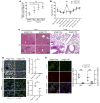
(A) Tail vein injection of rHMGB1 in C57BL/6 mice results in enhanced clot formation in a FeCl3 model (quantified as time to vessel occlusion). (B) Injection of rHMGB1 in C57BL/6 mice increases platelet sequestration in lungs and livers. Scale bar: 100 μm (top rows), 30 μm (bottom rows). This is quantified in C. (D) In a FeCl3 model, HMGB1 decreases the time to vessel occlusion in Tlr4 Flox control mice; this does not occur in Tlr4 Pf4 mice. (E) HMGB1 treatment of blood from WT mice induces a strong prothrombotic effect, which is reversed when repeated with blood from Tlr4–/– and Myd88–/– mice. Scale bar: 70 μm. (F) HMGB1 treatment of blood from WT mice enhances CRP-induced platelet aggregation; this is reversed when repeated with platelets from Tlr4–/– and Myd88–/– mice. Data show mean ± SD of the results from at least 3 separate experiments and n ≥ 3 mice per group. *P < 0.05, **P < 0.01, ***P < 0.001 (Student’s t test in A, C, D, and F; 1-way ANOVA with Tukey’s post-hoc test in E).
Figure 3
Figure 3. HMGB1 enhances platelet adhesion and spreading.
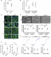
(A) The addition of rHMGB1 to isolated human platelets spreading on collagen or vWF increases final platelet surface area (after 30 minutes), as investigated with SICM at single-cell level. Scale bar: 2 μm. (B) HMGB1 increases the speed of platelet spreading (quantified by area growth exponent) on collagen or vWF within a 9-minute investigation period. Scale bar: 3 μm. Data show mean ± SD for (A) n ≥ 27 or (B) n ≥ 6 from at least 3 separate experiments in all studies. **P < 0.01, ***P < 0.001 (Student’s t test).
Figure 2
Figure 2. Platelet-derived HMGB1 is critical for platelet aggregation, small vessel thrombosis, NET formation, and recruitment of innate immune cells during trauma/hemorrhagic shock.
(A) Expression of HMGB1 on the surface of circulating platelets is increased in trauma patients as compared with healthy subjects. (B) Systemic aggregation of circulating platelets is upregulated in Hmgb1 Flox mice 30 minutes after induction of experimental trauma/hemorrhagic shock as compared with Hmgb1 Pf4 mice. (C) Small vessel thrombi (H&E staining) and (D) CD41-positive platelet aggregates (immunofluorescence staining and quantification of platelets) are detected in lungs and livers of Hmgb1 Flox mice but not in or only very little in Hmgb1 Pf4 mice after trauma/hemorrhagic shock. Arrows indicate small vessel thrombus formation and vascular congestion. Scale bar: 200 μm (C, top row), 100 μm (C, bottom row, and D, top rows), 50 μm (D, bottom rows). (E) CitH3-positive NETs and Ly6G-positive immune cell infiltrates are decreased in lungs of Hmgb1 Pf4 animals subjected to trauma/hemorrhagic shock compared with Hmgb1 Flox mice subjected to trauma/hemorrhagic shock (immunofluorescence staining and quantification). Scale bar: 10 μm. Data show mean ± SD for (A) n = 4 patients or (BE) n ≥ 4 mice per group from at least 3 separate experiments in all studies. *P < 0.05, **P < 0.01, ***P < 0.001 (Student’s t test in B, D, and E; 1-way ANOVA with Tukey’s post-hoc test in A).
Figure 1
Figure 1. Platelet-derived HMGB1 promotes platelet aggregation and thrombus formation.
(A) Hmgb1 Pf4 mice have prolonged bleeding time compared with Hmgb1 Flox control mice. (B) PT, (C) aPTT, and (D) TT are not altered in Hmgb1 Pf4 mice as compared with control mice. (E and F) Reduction in collagen-induced platelet aggregation in Hmgb1 Pf4 mice. (G) Blood derived from Hmgb1 Pf4 mice is less thrombogenic in a flow chamber system. Exogenous HMGB1 increases thrombus formation in blood from both Hmgb1 Pf4 and Hmgb1 Flox mice. (HJ) FeCl3-induced thrombus formation is inhibited in Hmgb1 Pf4 mice, with (H) improved blood flow and (I) prolonged time to vessel occlusion (as measured by laser Doppler imaging, quantified in J). (K) Immunofluorescence staining of thrombi from Hmgb1 Flox control mice demonstrates large clusters of CD41-positive platelets and surrounding deposition of HMGB1 overlying a fibrinogen network. Thrombi from Hmgb1 Pf4 animals show smaller clusters without HMGB1 expression. Scale bar: 100 μm (top row), 40 μm (bottom row). (L) Ly6G-positive immune cell infiltrates and citH3-positive NETs detected in FeCl3-induced thrombi from Hmgb1 Flox mice but not Hmgb1 Pf4 mice. Scale bar: 100 μm (top row), 40 μm (bottom row). (M) Western blot of clots isolated from Hmgb1 Pf4 mice reveals almost no expression of HMGB1 as compared with control. Data show mean ± SD from at least 3 separate experiments and (BD) n = 3, (G, H, and J) n = 4, (F) n = 5, and (A) n = 12 mice per group. (E, I, and KM) Representative images from at least 4 mice per group. *P < 0.05, **P < 0.01, ***P < 0.001 (Student’s t test in AD, F, H, and J; 1-way ANOVA with Tukey’s post-hoc test in G).

References

    1. Jackson SP. Arterial thrombosis — insidious, unpredictable and deadly. Nat Med. 2011;17(11):1423–1436. doi: 10.1038/nm.2515. - DOI - PubMed
    1. Ruggeri ZM. Platelets in atherothrombosis. Nat Med. 2002;8(11):1227–1234. doi: 10.1038/nm1102-1227. - DOI - PubMed
    1. Gawaz M, Langer H, May AE. Platelets in inflammation and atherogenesis. J Clin Invest. 2005;115(12):3378–3384. doi: 10.1172/JCI27196. - DOI - PMC - PubMed
    1. Gawaz M, Vogel S. Platelets in tissue repair: control of apoptosis and interactions with regenerative cells. Blood. 2013;122(15):2550–2554. doi: 10.1182/blood-2013-05-468694. - DOI - PubMed
    1. Morrell CN, Aggrey AA, Chapman LM, Modjeski KL. Emerging roles for platelets as immune and inflammatory cells. Blood. 2014;123(18):2759–2767. doi: 10.1182/blood-2013-11-462432. - DOI - PMC - PubMed

Publication types

MeSH terms