

[Rare parotid gland tumors: enhanced computed tomography and pathological correlation]
- PMID: 26552248
- PMCID: PMC7030452
- DOI: 10.7518/hxkq.2015.04.019
[Rare parotid gland tumors: enhanced computed tomography and pathological correlation]
Abstract
Objective: To investigate the correlation between enhanced computed tomography (CT) findings and pathological results of rare parotid gland tumors, and improve diagnosis accuracy.
Methods: The enhanced CT manifestations of 22 cases with pathologically documented rare parotid gland tumors, which included 6 cases of basal cell tumor, 5 cases of myoepithelioma, 4 cases of vascular invasion, 3 cases of lymphatic cyst, 3 cases of lipoma, and 1 case of chondrosarcoma, were retrospectively analyzed. The location, size, shape, density, and relationship with surrounding structure were evaluated on CT images.
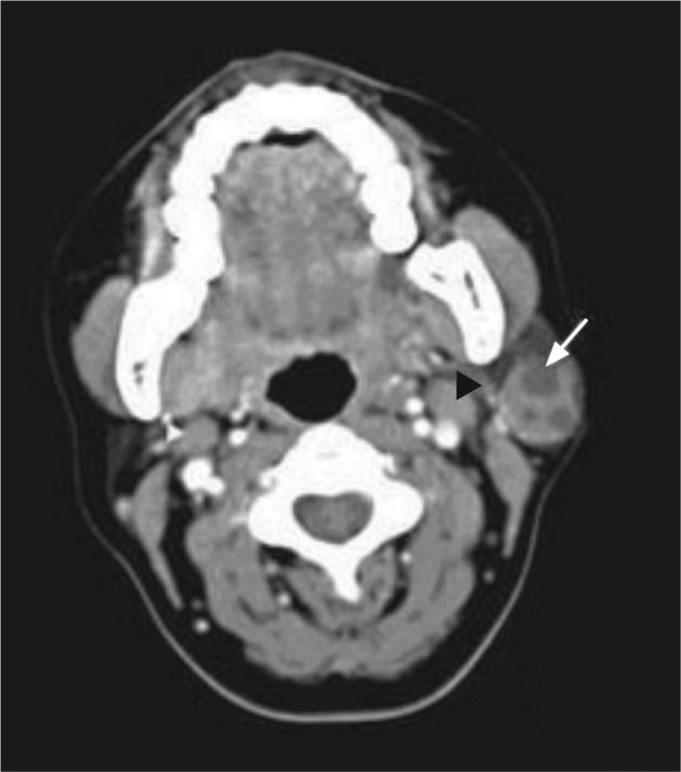
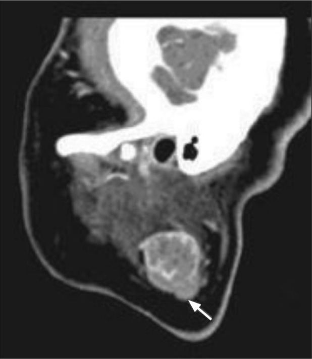
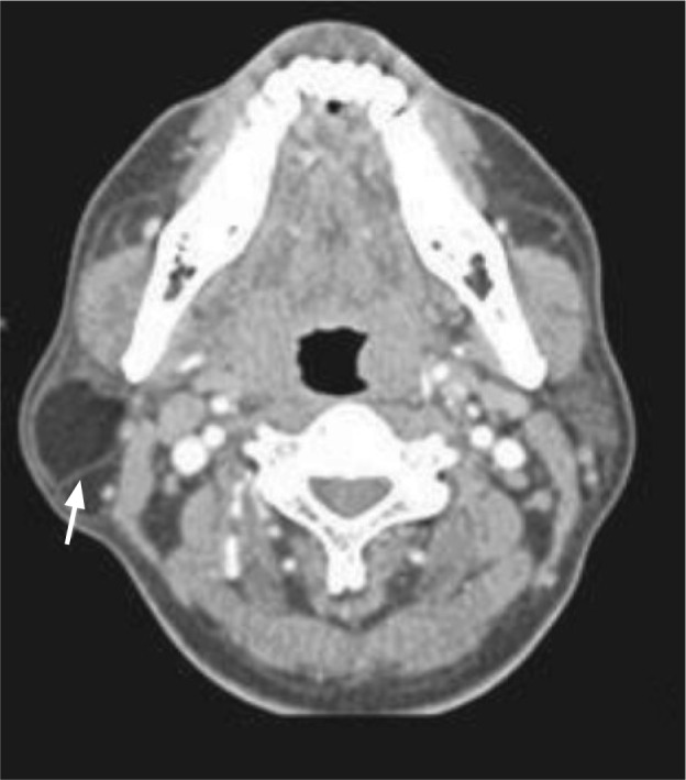
Results: The enhanced CT showed that basal cell tumors occurred in the superficial lobe of the parotid gland, with clear boundary, within the cystic lesion. The lesions were moderate to obviously enhanced, which may be accompanied by enlarged lymph nodes. Myoepithelial tumors were located in the superficial lobe of the parotid gland, with a small cystic prone and microcalcification within a few cases. The lesions were moderate to obviously enhanced. Hemangiomas of soft tissue mass prominent in the parotid gland surface were mild to significantly enhanced. Larger lesions may occupy the entire parotid gland, with uneven density and visible vein stone. The CT density values of the lymphatic cyst were usually higher. Chondrosarcoma mainly manifested cystic mass at the calcification edge. Lipoma with fat density mass exhibited clear boundary without enhancement. Fiber separation could be observed in the lesion.
Conclusion: CT can reflect the pathological features of rare parotid gland tumors by demonstrating their corresponding imaging features. Enhanced CT is the most effective means of imaging to identify the nature of rare tumor of the parotid gland lesions.
目的: 探讨腮腺少见肿瘤增强CT表现特征与病理的相关性,以提高影像诊断水平。
方法: 对22例经手术病理证实的少见腮腺肿瘤进行回顾性分析,包括基底细胞瘤6例,肌细胞上皮瘤5例,脉管瘤4例,淋巴上皮囊肿及腮腺脂肪瘤各3例,软骨肉瘤1例。在增强CT图像上,从病变所在腮腺的位置、病变大小和形态、病变密度以及其与周围结构的关系进行总结分析。
结果: 基底细胞瘤发生在腮腺浅叶,边界清楚,病变内易囊变。增强扫描呈不均匀中度-明显强化肿块,边缘或内部可见结节状强化,可伴有增大的淋巴结。肌细胞上皮瘤多发于腮腺浅叶,易发生小囊变,少数病变内可有小点状钙化。实性部分增强后中度-明显强化,多有典型的动脉期强化结节和边缘显著强化。血管瘤为软组织肿块,病变较大可占据整个腮腺,密度均匀或不均匀,可见静脉石,突出于腮腺表面,增强后轻度-明显强化。淋巴上皮囊肿囊液较黏稠,CT值密度较高。软骨肉瘤以囊性为主的肿块,含边缘钙化、骨化成分的肿块。腮腺脂肪瘤可见脂肪密度肿块,边界清晰,无强化,病变内可见纤维分隔。
结论: 腮腺少见肿瘤的增强CT表现具有一定的影像学特征,可揭示其病理基础;增强CT是诊断腮腺少见肿瘤病变性质有效的影像检查手段。
Figures


Similar articles
-
[Parotid adenolymphoma: the enhanced MSCT manifestations and clinical pathological analysis].Lin Chuang Er Bi Yan Hou Tou Jing Wai Ke Za Zhi. 2015 Dec;29(24):2129-32. Lin Chuang Er Bi Yan Hou Tou Jing Wai Ke Za Zhi. 2015. PMID: 27093811 Chinese.
-
MRI and CT imaging characteristics of myoepithelioma of the parotid gland.Acta Radiol. 2016 Jul;57(7):837-43. doi: 10.1177/0284185115609364. Epub 2015 Oct 26. Acta Radiol. 2016. PMID: 26508793
-
Myoepithelioma of the parotid gland: CT imaging findings.AJNR Am J Neuroradiol. 2008 Aug;29(7):1372-5. doi: 10.3174/ajnr.A1109. Epub 2008 May 8. AJNR Am J Neuroradiol. 2008. PMID: 18467518 Free PMC article.
-
Huge lipoma of the right parotid gland: Case report and review of 42 cases.Ear Nose Throat J. 2016 Jan;95(1):E8-E13. doi: 10.1177/014556131609500103. Ear Nose Throat J. 2016. PMID: 26829698 Review.
-
Wide excision of accessory parotid gland with anterior approach.J Craniofac Surg. 2012 Jan;23(1):165-8. doi: 10.1097/SCS.0b013e3182413f19. J Craniofac Surg. 2012. PMID: 22337398 Review.
Cited by
-
[Clinical value of quantitative parameters by spectral CT in parotid gland tumor].Hua Xi Kou Qiang Yi Xue Za Zhi. 2019 Dec 1;37(6):631-635. doi: 10.7518/hxkq.2019.06.011. Hua Xi Kou Qiang Yi Xue Za Zhi. 2019. PMID: 31875442 Free PMC article. Chinese.
References
-
- 张 启禄, 钱 斌, 刘 良. 腮腺基底细胞瘤的CT影像诊断及鉴别[J] 临床放射学杂志. 2012;31(11):1547–1550.
-
- 刘 春玲, 黄 飚, 周 正根, et al. 腮腺基底细胞腺瘤的CT和MRI特点[J] 中华放射学杂志. 2009;43(6):600–603. - PubMed
-
- Yaman H, Gerek M, Tosun F, et al. Myoepithelioma of the parotid gland in a child: a case report[J] J Pediatr Surg. 2010;45(7):E5–E7. - PubMed
-
- Astarci HM, Celik A, Sungu N, et al. Cystic clear cell myoepithelioma of the parotid gland. A case report[J] Oral Maxillofac Surg. 2009;13(1):45–48. - PubMed
MeSH terms
LinkOut - more resources
Full Text Sources